1644
Characterization of biomechanical properties of cerebral vessels using magnetic resonance elastography1Shanghai Jiao Tong University, Shanghai, China, 2Department of Radiology, The first affiliated hospital of Soochow University, Suzhou, China, 3Mayo Clinic, Phoenix, AZ, United States, 4Washington University in St. Louis, St. Louis, MO, United States, 5University of Delaware, Newark, DE, United States
Synopsis
Measurements of the biomechanical properties of the cerebral vessels could help diagnosis and prognosis of diseases such as lacunar infarction. In this study, shear moduli of the cerebral vessels were measured using both magnetic resonance elastography (MRE) and MR Angiography (MRA). The magnitude images from MRE were registered to MRA images to obtain the shear moduli of the cerebral vessels. Results showed significant correlations between age and gender in terms of the storage modulus. Significant differences between lacunar infarction and healthy control group were also observed in terms of the absolution shear modulus.
Introduction
Studies have shown that biomechanical properties of the vasculature are closely related to vascular diseases 1, 2. Decreased elastic modulus of the vessel probably contributed to lumen infarction 3. Therefore, in vivo measurement of biomechanical properties of the cerebral vasculature could help the diagnosis and prognosis of the related diseases such as lacunar infarction. In this study, we proposed a method to measure the shear modulus of the cerebral vessels with MRE. The relationships between the cerebral vessel elasticity, gender, age as well as cerebral diseases were investigated.Methods
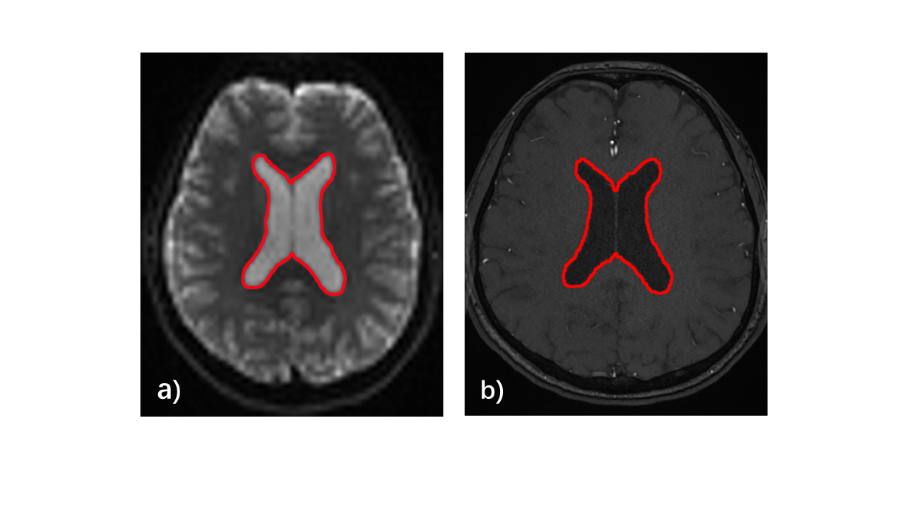
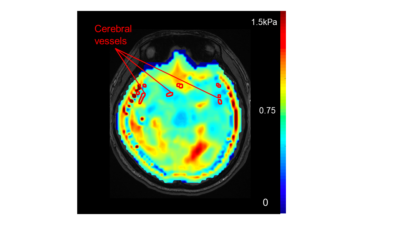
Both MRA and MRE images were acquired using a 3.0T scanner (SKYRA, Siemens, Germany). MRA images were obtained using a 3D time-of-flight (TOF) MRA sequence. MRE images were obtained using a 3D multislab, multishot spiral sequence 4. Lateral ventricles were segmented from both MRA and magnitude images of MRE (Figure 1). The target vessel region was delineated in the MRA images. By registering the MRE and MRA images based on the ventricle region, the shear modulus map of the vessels was obtained (Figure 2).A total of 23 patients and 27 volunteers were recruited between 2018 and 2019. Both MRA and MRE images were acquired for each sample. The 27 healthy volunteers included 12 males and 15 females, with an age range between 12 and 77. The 23 cases of vascular diseases included 13 lacunar infarction and 10 arterial dysplasia. To analyze the relationship between the biomechanical properties of the cerebral vessels and the age, Pearson correlation analyses were carried out. Gender differences in the biomechanical properties were also analyzed using student t-tests. Potentials of using elastic modulus to diagnose cerebral vessel disease were also investigated.
Results
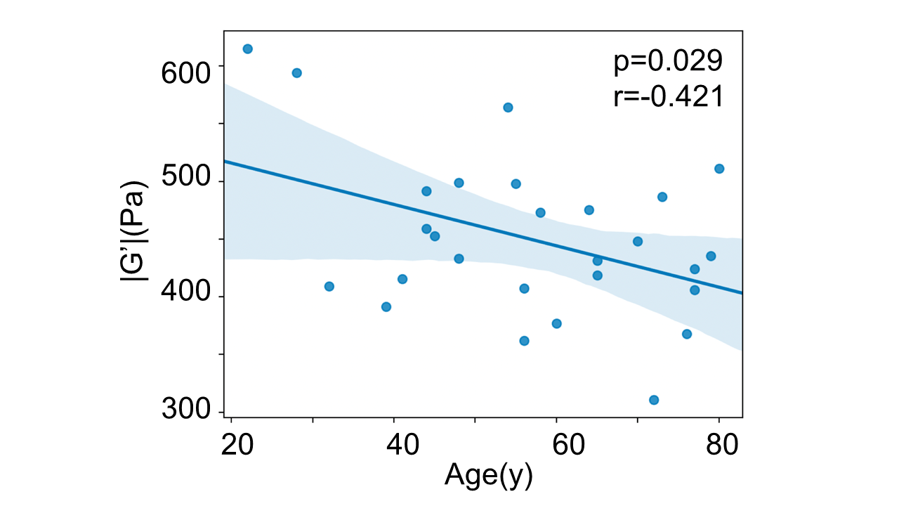
No significant correlation was found between the age and absolute shear modulus |G*| (Figure 3a). In addition, no significant correlations were found between gender and vessel elasticity of the healthy group (p=0.812) (Figure 3b). We observed that the mean |G*| of the lacunar infarction group was lower than that of the control group (Figure 3c). A significant difference was found between the lacunar infarction group and the control group (p=0.002) in terms of |G*|, whereas no significant difference was found between the arterial dysplasia group and the control group (p=0.175). However, in terms of storage modulus G’, a negative correlation was found between G’ and the age (Figure 4).Discussion
In this study, a procedure to measure the biomechanical properties of cerebral vessels using MRE was proposed. Results showed that the storage moduli of the vessels were negatively correlated to age, and the absolute shear moduli of the vessels were significantly lower for the lacunar infarction group. These complied with the current hypothesis that biomechanical properties of the cerebral vessels were closely related to the diseases and age. Future studies will improve the current procedure and expand the patient pool.Acknowledgements
National Natural Science Foundation of China (NSFC) grant 31870941, Shanghai Science and Technology Committee (STCSM) grant 19441907700.References
[1] K. S. Cheng, C. R. Baker, G. Hamilton, A. P. G. Hoeks, and A. M. Seifalian, “Arterial elastic properties and cardiovascular risk/event,” European Journal of Vascular and Endovascular Surgery, vol. 24, no. 5, pp. 383-397, Nov, 2002.
[2] J. C. Lasheras, “The Biomechanics of Arterial Aneurysms,” Annual Review of Fluid Mechanics, vol. 39, no. 1, pp. 293-319, 2007/01/01, 2006.
[3] P. Lacolley, V. Regnault, P. Segers, and S. Laurent, “VASCULAR SMOOTH MUSCLE CELLS AND ARTERIAL STIFFENING: RELEVANCE IN DEVELOPMENT, AGING, AND DISEASE,” Physiological Reviews, vol. 97, no. 4, pp. 1555-1617, Oct, 2017.
[4] C. L. Johnson, J. L. Holtrop, M. D. J. McGarry, J. B. Weaver, K. D. Paulsen, J. G. Georgiadis, and B. P. Sutton, “3D Multislab, Multishot Acquisition for Fast, Whole-Brain MR Elastography with High SNR Efficiency,” Magnetic resonance in medicine: official journal of the Society of Magnetic Resonance in Medicine / Society of Magnetic Resonance in Medicine, vol. 71, no. 2, pp. 477-485, 2014.
[5] H. Kandil, A. Soliman, L. Fraiwan, A. Shalaby, A. Mahmoud, A. ElTanboly, A. Elmaghraby, G. Giridharan, A. El-Baz, “A NOVEL MRA FRAMEWORK BASED ON INTEGRATED GLOBAL AND LOCAL ANALYSIS FOR ACCURATE SEGMENTATION OF THE CEREBRAL VASCULAR SYSTEM,” 2018 IEEE15th International Symposium on Biomedical Imaging (ISBI2018), pp. 1365-1368, 2018.
Figures