1643
Altered Spontaneous Brain Activity in Patients with MELAS at Attack and Remission Stages: Evidence from Resting-state fMRI1Huashan Hospital, Shanghai, China
Synopsis
This is the first study that used resting-state fMRI (rs-fMRI) to assess the alterations of spontaneous neuronal activity in patients with mitochondrial encephalomyopathy, lactic acidosis and stroke-like episodes (MELAS) at attack and remission stages, which may provide a new insight into the mechanisms of cerebral cortex damage in MELAS.
Background and purpose
Stroke-like episodes (SLEs) are one of the cardinal features of MELAS, which occur in 84–99% of affected individuals.1 These episodes present with protean manifestations including headaches, seizures, hearing loss, cortical blindness, aphasia, motor weakness and dementia. SLEs are characterized by recurrent attack followed by symptomatic remission, which eventually lead to the progressive accumulation of neurological deficits.2 Resting-state functional MRI (rs-fMRI) is a powerful tool for evaluating spontaneous neural activity of human brain.3 Until now, no study has reported the changes of intrinsic brain activity in MELAS. In this study, we aimed to determine the altered spontaneous brain activity patterns of MELAS patients at attack and remission stages, so as to investigate the underlying mechanisms of brain impairment during these two different stages. Additionally, we also explore a neuroimaging biomarker for distinguishing MELAS patients at attack stage from remission stage.Methods
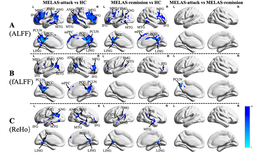
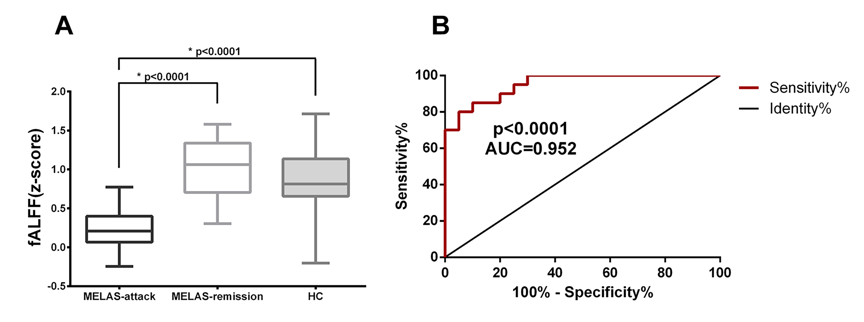
Resting state functional magnetic resonance imaging (rs-fMRI) data were acquired from 40 patients with MELAS at attack stage (MELAS-attack, n=20) and remission stage (MELAS-remission, n=20) and 22 healthy controls (HC). Amplitude of low frequency fluctuations (ALFF), fractional ALFF (fALFF) and regional homogeneity (ReHo) were used to investigate spontaneous brain activity over the whole brain. A receiver operating characteristics (ROC) curve was analyzed to distinguish MELAS-attack from MELAS-remission.Results
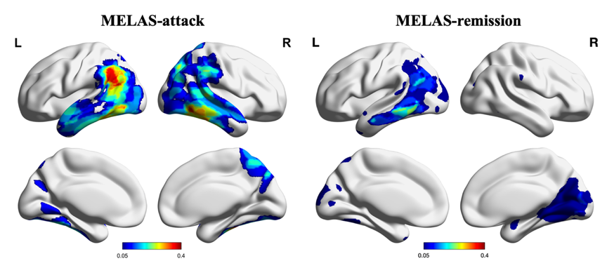
Compared with HC, MELAS-attack and MELAS-remission groups showed reduced ALFF, fALFF and ReHo values in regions mainly involving the default mode network (DMN), language, visual and auditory networks, extending beyond the distribution areas of stroke-like lesions (SLLs). These altered spontaneous brain activities were much more widespread in MELAS-attack group than MELAS-remission group. In comparison with MELAS-remission group, MELAS-attack group exhibited significantly decreased fALFF values in left precuneus. The ROC analysis demonstrated that fALFF values in the left precuneus could efficiently distinguish MELAS patients at attack stage from remission stage (area under the curve (AUC) = 0.952, P < 0.0001).Conclusion
MELAS patients presented decreased spontaneous brain activities beyond the SLLs, with a downward trend from attack stage to remission stage. Additionally, fALFF value in left precuneus could be used as an objective neuroimaging biomarker formonitoring the disease status of MELAS.Acknowledgements
This study was supported by the Science and Technology Commission of Shanghai Municipality (17411953700, 19ZR1407900).References
1. El-Hattab AW, Adesina AM, Jones J, Scaglia F. MELAS syndrome: Clinical manifestations, pathogenesis, and treatment options. Molecular genetics and metabolism 2015;116:4-12.
2. Kim HS, Kim DI, Lee BI, et al. Diffusion-weighted image and MR spectroscopic analysis of a case of MELAS with repeated attacks. Yonsei medical journal 2001;42:128-133.
3. Biswal B, Yetkin FZ, Haughton VM, Hyde JS. Functional connectivity in the motor cortex of resting human brain using echo-planar MRI. Magnetic resonance in medicine 1995;34:537-541.
Figures