1621
Rapid Brain Arterial Spin Labeling (ASL) using pseudo-Continuous ASL Echo Planar Imaging with Compressed SENSE (pCASL-EPICS)1Kumamoto University Hospital, Kumamoto, Japan, 2Philips Japan, Tokyo, Japan, 3Graduate School of Medical Sciences, Kumamoto University, Kumamoto, Japan
Synopsis
Multi-slice single-shot 2D echo-planar imaging (EPI) readout can be used effectively for pseudo-continuous arterial spin labeling (pCASL) perfusion imaging. In this study we tried to utilize the Compressed SENSE reconstruction for pCASL-EPI without further optimization of EPI sampling scheme. pCASL-EPICS clearly reduces noise-like artifacts and significantly improves the robustness of perfusion images in a short scan time less than 3 minutes compared with conventional pCASL SENSE, without any penalty for scan parameters. This technique may be helpful to further assess the many brain diseases.
PURPOSE
Multi-slice single-shot 2D echo-planar imaging (EPI) readout can be used effectively for pseudo-continuous arterial spin labeling (pCASL) perfusion imaging and it should be considered a viable alternative to segmented 3D sequences1, because they are available on all systems and are insensitive to image artifacts from motion. However, 2D imaging basically requires many signal averages to gain signal-to-noise ratio, which leads to longer scan time. In addition, typical clinical brain EPI-based ASL imaging has limited spatial resolution due to its high sensitivity to B0 inhomogeneities as well as diffusion-weighted imaging (DWI). EPI with sensitivity encoding (SENSE) helps to reduce the voxel size without increasing of image distortion, but it often suffers from increased noise-like artifacts on the center of the images due to the high geometry factor2,3. Recently, a combination of parallel imaging and compressed sensing technique (Compressed SENSE, C-SENSE) has been developed to accelerate the acquisition time without increasing the image artifacts4,5. Although C-SENSE is basically applied for non-EPI scans, it has been demonstrated that the C-SENSE reconstruction could clearly reduce noise-like artifacts and significantly improve the image quality of EPI based DWI without further optimization of EPI sampling scheme6,7. We hypothesized that EPI-based ASL can also be applied the C-SENSE reconstruction framework as well as DW-EPI. The purpose of this study was to demonstrate the feasibility of a combination of EPI-based ASL and C-SENSE (pCASL-EPICS) for improving multi-slice perfusion imaging.MATERIALS AND METHODS
Experimental data was collected from 6 healthy volunteers. Written informed consent was obtained from each volunteer and the protocol was approved by the ethics committee. All studies were performed with clinical 3.0T MR scanner (Philips, Ingenia 3.0T CX) and 32-channel dS-head coil.EPICS is based single-shot EPI. We did not modify its sampling pattern of single-shot EPI and we applied it into the C-SENSE framework. Scan parameters of pCASL-EPICS were as follows: Transverse plane acquisition, TR/TE = 4550/7.9ms, slices thickness = 6mm, voxel size = 3.75 × 3.93 × 6.0mm (similar setting with commercially available 3D pCASL Gradient spin-echo (GraSE) protocol), number of slices = 14, 18 dynamic scans, EPI factor = 21, CS factor or SENSE factor = 3.0, pCASL label duration =1800ms, post label delay =2000ms, two-inversion pulses background suppression and acquisition time = 2:52.
pCASL-EPICS were compared to conventional 2D SENSE-EPI images and 3D GraSE-pCASL images for image quality, especially for the reduction of image noise. ROIs were placed on left and right gray matter (GM). We calculated the Coefficient of Variation (CV) of signal intensities (SI) of GM on perfusion image in respective volunteers as follows: CV = SDSI / AverageSI, to compare the consistency of perfusion related signal intensities among pCASL-EPICS, SENSE 2D EPI and 3D GraSE-pCASL.
RESULTS
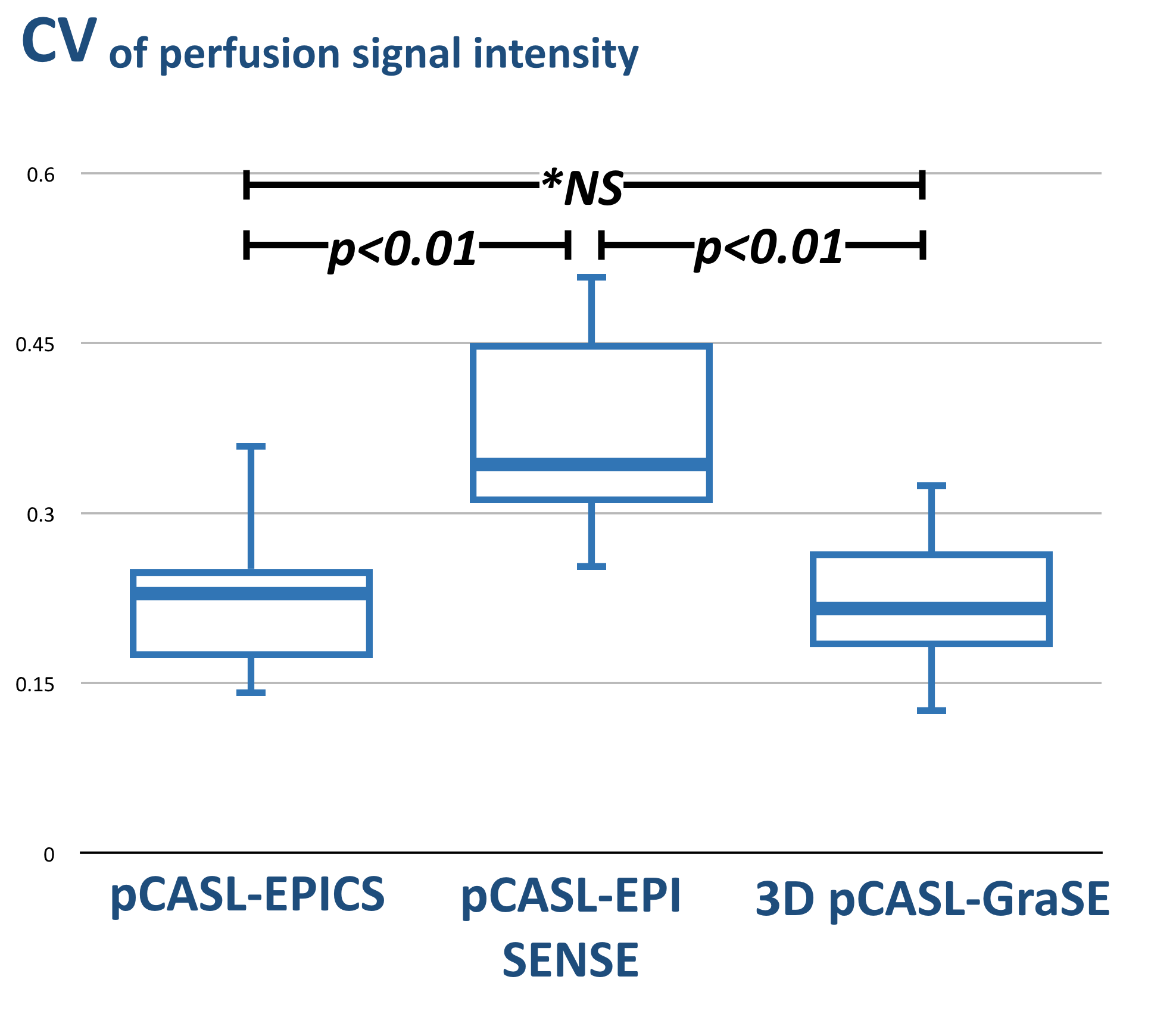
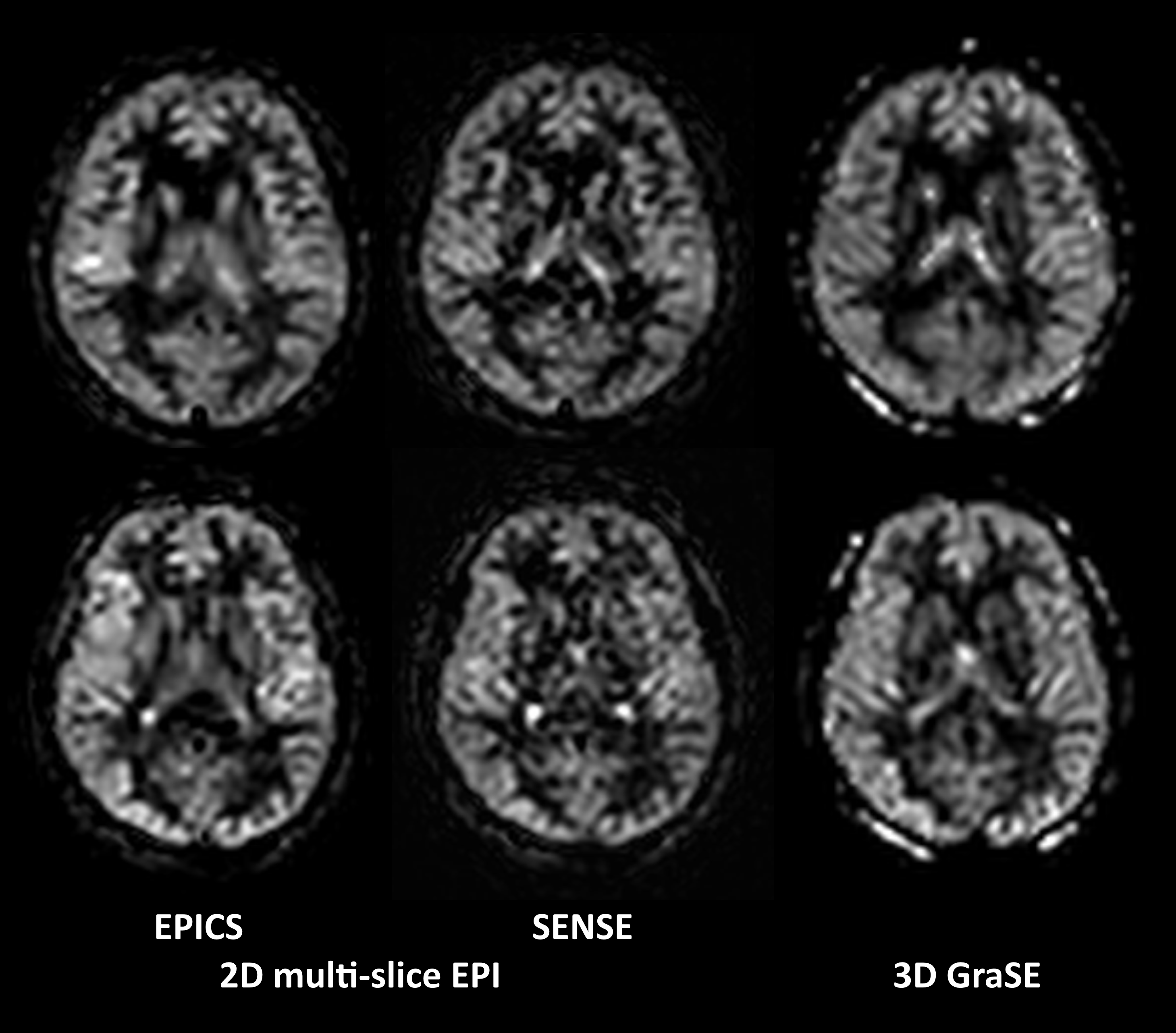
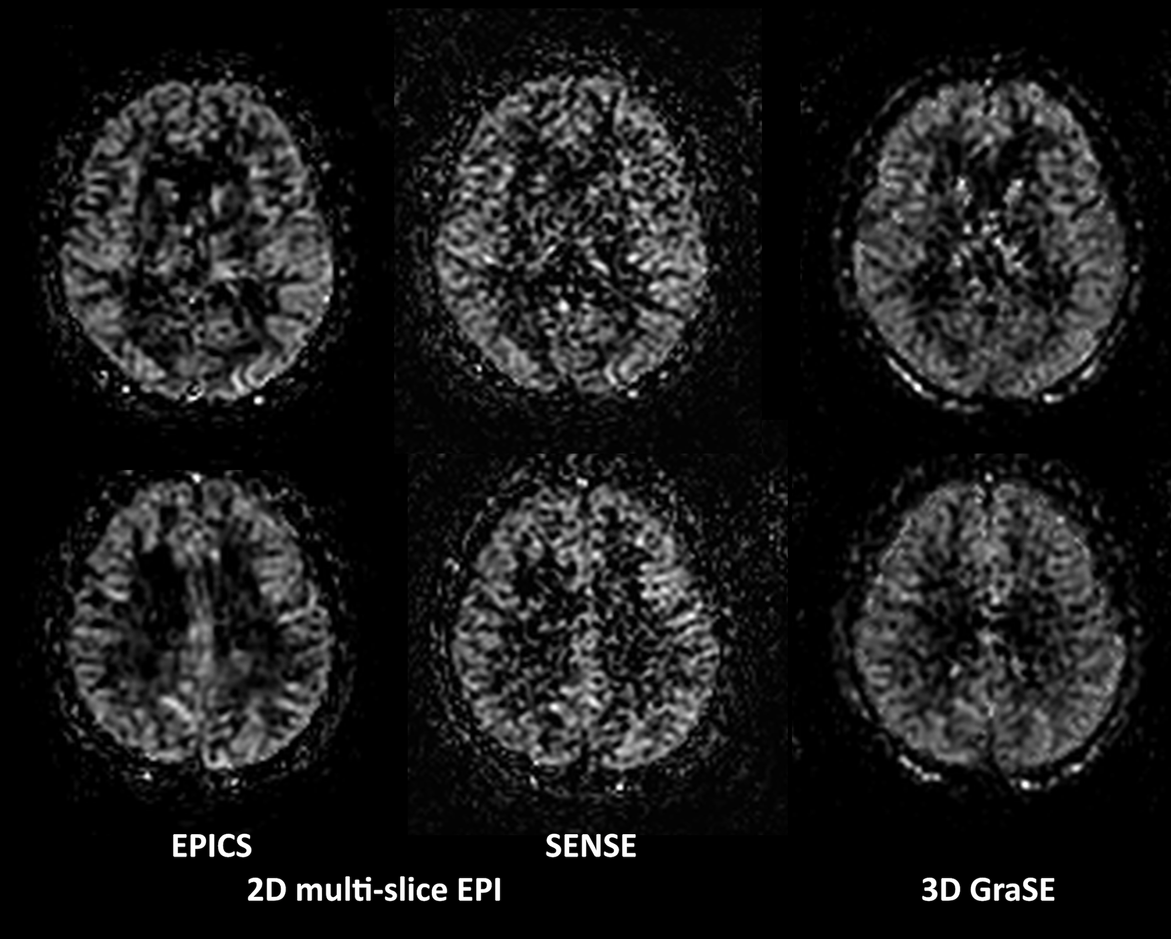
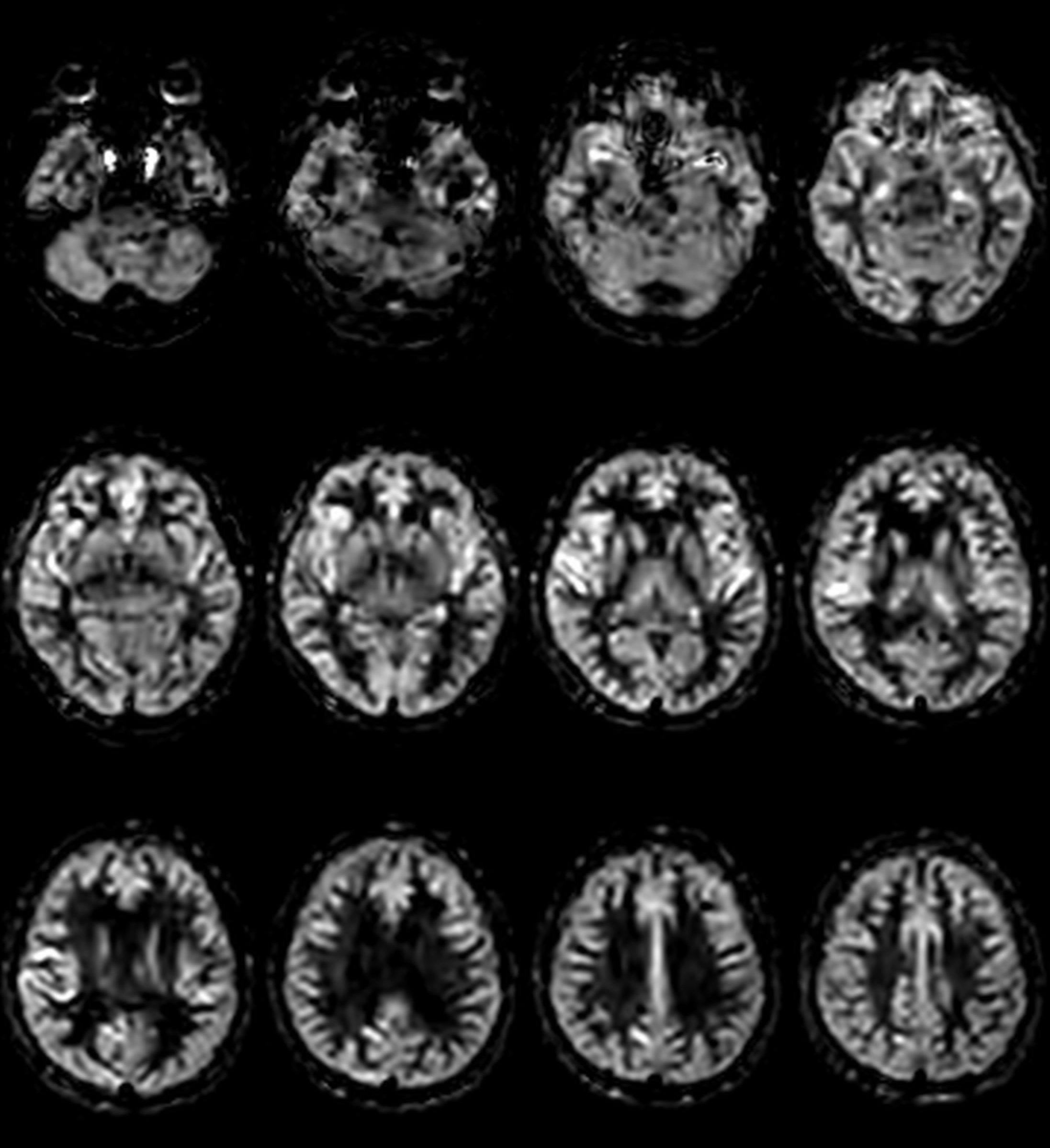
Figure 1 shows the results of CV values on the perfusion images b among pCASL-EPICS, SENSE 2D EPI and 3D GraSE-pCASL. CV values of pCASL-EPICS (0.23±0.07) was significantly lower than that of SENSE 2D EPI (0.37±0.08) (p < 0.01). The cause of high CV values of SENSE was most likely due to the presence of severe noise across the images. On the other hand, there were no significant differences between CV values of pCASL-EPICS and 3D GraSE-pCASL(0.24±0.10) (p=0.47). Figure 2 shows representative perfusion images using pCASL-EPICS, SENSE 2D EPI and 3D GraSE-pCASL in two volunteers. pCASL-EPICS clearly reduced the noise which exists in the center of the SENSE images and almost similar SNR compared with 3D GraSE-pCASL. It indicated that pCASL-EPICS can provide more accurate perfusion information with high reproducibility and robustness. Figure 3 also shows representative perfusion images using pCASL-EPICS, SENSE 2D EPI and 3D GraSE-pCASL with increased voxel size (2.5mm2) from same setting of Figure 2 images. Although further optimization to obtain more sufficient SNR is needed, pCASL-EPICS still maintained higher SNR compared with other two methods. Figure 4 shows the representative multi-slice perfusion images using optimized pCASL-EPICS.CONCLUSION
pCASL-EPICS clearly reduces noise-like artifacts and significantly improves the robustness of perfusion images in a short scan time less than 3 minutes compared with conventional pCASL SENSE, without any penalty for scan parameters. This technique may be helpful to further assess the many brain diseases.Acknowledgements
No acknowledgement found.References
1. Alsop DC, et al. Recommended implementation of arterial spin-labeled perfusion MRI for clinical applications: A consensus of the ISMRM perfusion study group and the European consortium for ASL in dementia. Magn Reson Med. 2015;73(1):102-16.
2. Patricia N, et al. Parallel Imaging Artifacts in Body Magnetic Resonance Imaging. Can Assoc Radiol J. 2009;60: 91–98.
3. Yanasak NE, et al. MR imaging artifacts and parallel imaging techniques with calibration scanning: a new twist on old problems. Radiographics. 2014;34:532-48.
4. Geerts-Ossevoort L, et al. Compressed SENSE Speed done right. Every time. The Netherlands: Philips Healthcare; 2018 Jan. Report No: 4522 991 31821. https://www.philips.de/content/dam/b2bhc/de/resourcecatalog/landingpages/ingeniaelition/White_Paper_Compressed_SENSE-opt.pdf
5. Mönch S, et al. Magnetic Resonance Imaging of the Brain Using Compressed Sensing - Quality Assessment in Daily Clinical Routine. Clin Neuroradiol. 2019 May 16. doi: 10.1007/s00062-019-00789-x. [Epub ahead of print]
6. Morita, et al. Pseudo-3D Diffusion-Weighted Imaging of the Brain using Echo Planar Imaging with Compressed SENSE (EPICS). Proc. ISMRM. 2019:3355.
7. Yoneyama, et al. Noise Reduction in Prostate Single-Shot DW-EPI utilizing Compressed SENSE Framework. Proc. ISMRM. 2019:1634
Figures