1611
Ultrashort TE 4D-MRA for Giant Aneurysms Treated with Flow-Diverter Stents: Visualization of Flow in the Stents and Hemodynamic Vascular Flow1Department of Radiology, Juntendo University Hospital, Tokyo, Japan, Tokyo, Japan, 2Department of Neurosurgery, Juntendo University School of Medicine, Tokyo, Japan, Tokyo, Japan, 3Department of Neuroendovascular Therapy, Juntendo University School of Medicine, Tokyo, Japan, Tokyo, Japan
Synopsis
We assessed the usefulness of ultrashort TE (UTE) 4D-MR angiography for giant aneurysms treated with flow-diverter stents. We evaluated the depiction of flow in the stents, and visualization of hemodynamic vascular flow in aneurysms. The newly developed UTE 4D-MRA could visualize flow in the stents at the acceptable quality for diagnosis, and hemodynamic vascular flow in aneurysms were comparable to X-ray DSA. Moreover, UTE 4D-MRA showed intraaneurysmal flow within the coil which was difficult to visualize even with X-ray DSA. Our study demonstrated that UTE 4D-MRA was adequate to visualize the hemodynamic vascular flow in aneurysms, and might be useful.
Background and Purpose
Treatment with flow-diverter (FD) stent is provided for giant unruptured aneurysm of the internal carotid artery (ICA).1 Nevertheless, due to the characteristics of this stent, which divert the blood flow and promote thrombosis, a careful follow-up on the status of the aneurysmal embolization and patency of the parent artery is necessary.2 Follow-up of ICA aneurysms treated with FD stent using three-dimensional time of flight MR Angiography (3D TOF-MRA) is difficult due to the stent related susceptibility artifacts and thrombus T1 shortening in the aneurysm. A few studies have reported the possibility of depicting the flow in the stents using ultrashort TE (UTE) MRA with arterial spin labeling (ASL).2-5 However, this technique can obtain only 3D data of one specific post labeling delay. The newly developed UTE 4D-MRA using the ASL method might be used to visualize the hemodynamic vascular flow. As for this MRA sequence, the multi-phase acquisition is possible with a temporal resolution of 165 milliseconds. Therefore, UTE 4D-MRA may visualize not only the depiction of flow in the stent but also hemodynamic vascular flow in aneurysms treated with FD stents. The purpose of this study was to assess the usefulness of non-contrast enhanced UTE 4D-MRA for giant internal carotid artery aneurysms treated with FD stents.Methods
Eleven aneurysms treated with FD stents underwent UTE 4D-MRA. Six aneurysms were treated with single FD stent, two were treated with two FD stents, two were treated with single FD stent and coils, and one was treated with three FD stents and coils. UTE 4D-MRA was performed on the next day of intravascular treatment. All studies were performed with a 3-T MR imaging system (Vantage Galan 3T ZGO, Canon Medical Systems, Otawara, Japan). The parameters for the UTE 4D-MRA were as follows: TR / TE, 3 / 0.1 msec ; flip angle, 6°; field of view, 224 × 224 mm, matrix 224 × 224 ; section thickness, 1.0 mm ; number of section, 150 ; NAQ, 2 ; bandwidth, 244.1 Hz ; number of segments, 200 ; number of trajectories, 9800 ; TI, ΔTI, and final TI, 200 msec, 165 msec, and 860 msec, respectively ; number of acquisition phases, five ; and acquisition time, 9 minutes 33 seconds. Moreover, delay phase scan parameters were as follows: number of trajectories, 10000 ; TI, ΔTI, and final TI, 1100 msec, 318 msec, and 1418 msec, respectively ; number of acquisition phases, two ; and acquisition time, 8 minutes 4 seconds. X-ray catheter-based intraarterial cerebral digital subtraction angiography (DSA) was performed using the following biplane angiographic systems: Artis Q. BA Twin (Siemens AG, Erlangen, Germany). The image matrix and field of view were 1024 × 1024 and 170 mm × 170 mm, respectively; the temporal resolution was 4 frames per second. Selective manual internal carotid artery injections were administered according to the aneurysm location. Two neuroradiologists independently reviewed the UTE 4D-MRA images, and subjectively scored the flow in the stents on a scale of 1 to 4 : 1, not visible ; 2, poor (slightly visible, but not adequate for diagnosis) ; 3, acceptable (roughly visible, diagnosable images) ; 4, excellent (images almost equal to X-ray DSA). We scored the vascular flow in aneurysms status on a scale of 1 to 3 : 1, poor ; 2, roughly visible ; 3, excellent (images almost equal to X-ray DSA). Moreover, we evaluated the visualization of eclipse sign. The X-ray DSA images during the treatment were used as a reference standard. The scores of MRA images from the two observers were averaged, and the scores were compared between observers. Weighted κ statistics was used to evaluate interobserver agreement.Results
The average score ± standard deviation for flow in the stents and vascular flow in aneurysms were 2.59 ± 0.67 and 2.86 ± 0.56, respectively. Interobserver agreement was good agreement (κ = 0.82) for flow in the stents and good agreement (κ = 0.81) for the vascular flow in aneurysms, respectively. Eclipse sign was demonstrated in eight cases using both X-ray DSA and UTE 4D-MRA.Discussion
The newly developed UTE 4D-MRA uses a UTE combined with ASL. ASL technique is used as a preparation pulse for the visualization of the blood flow and data acquisition is based on 3D radial sampling.3 UTE minimizes phase dispersion of signal of the labeled blood flow and decreases the susceptibility artifacts of FD stent. Therefore, UTE 4D-MRA could visualize flow in the stents at the acceptable image quality for diagnosis. In UTE 4D-MRA, hemodynamic vascular flow in aneurysms was comparable to those of X-ray DSA. The temporal resolution of UTE 4D-MRA and X-ray DSA are 165msec and four frames per second (250 msec), respectively. Thus, UTE 4D-MRA could visualize precisely. Moreover, FD stent with coiling case, UTE 4D-MRA showed intraaneurysmal flow within the coil which was difficult to visualize even with X-ray DSA (Fig.2).Conclusion
Non-contrast enhanced UTE 4D-MRA is a useful technique for the follow-up tool of giant internal carotid artery aneurysms treated with flow-diverter stents.Acknowledgements
This work was supported by JSPS KAKENHI Grant Number JP16H06280. Grant-in-Aid for Scientific Research on Innovative Areas- Resource and technical support platforms for promoting research 'Advanced Bioimaging Support’.References
1. Nelson PK, Lylyk P, Szikora I, et al. The pipeline embolization device for the intracranial treatment of aneurysms trial. AJNR Am J Neuroradiol 2011;32:34–40
2. Oishi H, Fujii T, Suzuki M, et al. Usefulness of Silent MR Angiography for Intracranial Aneurysms Treated with a Flow-Diverter Device. AJNR Am J Neuroradiol 2019;40(5):808-814.
3. Irie R, Suzuki M, Yamamoto M, et al. Assessing Blood Flow in an Intracranial Stent:A Feasibility Study of MR Angiography Using a Silent Scan after Stent-Assisted Coil Embolization for Anterior Circulation Aneurysms. AJNR Am J Neuroradiol 2015 May;36:967-70.
4. Takano N, Suzuki M, Irie R, et al. Usefulness of Non–Contrast-Enhanced Magnetic Resonance Angiography Using a Silent Scan for Follow-Up after Y-Configuration Stent-Assisted Coil Embolization for Basilar Tip Aneurysms. AJNR Am J Neuroradiol 2017;38 (3):577–81
5. Takano N, Suzuki M, Irie R, et al. Non-Contrast-Enhanced Silent Scan MR Angiography of Intracranial Anterior Circulation Aneurysms Treated with a Low-Profile Visualized Intraluminal Support Device. AJNR Am J Neuroradiol 2017;38 (8):1610–16
Figures
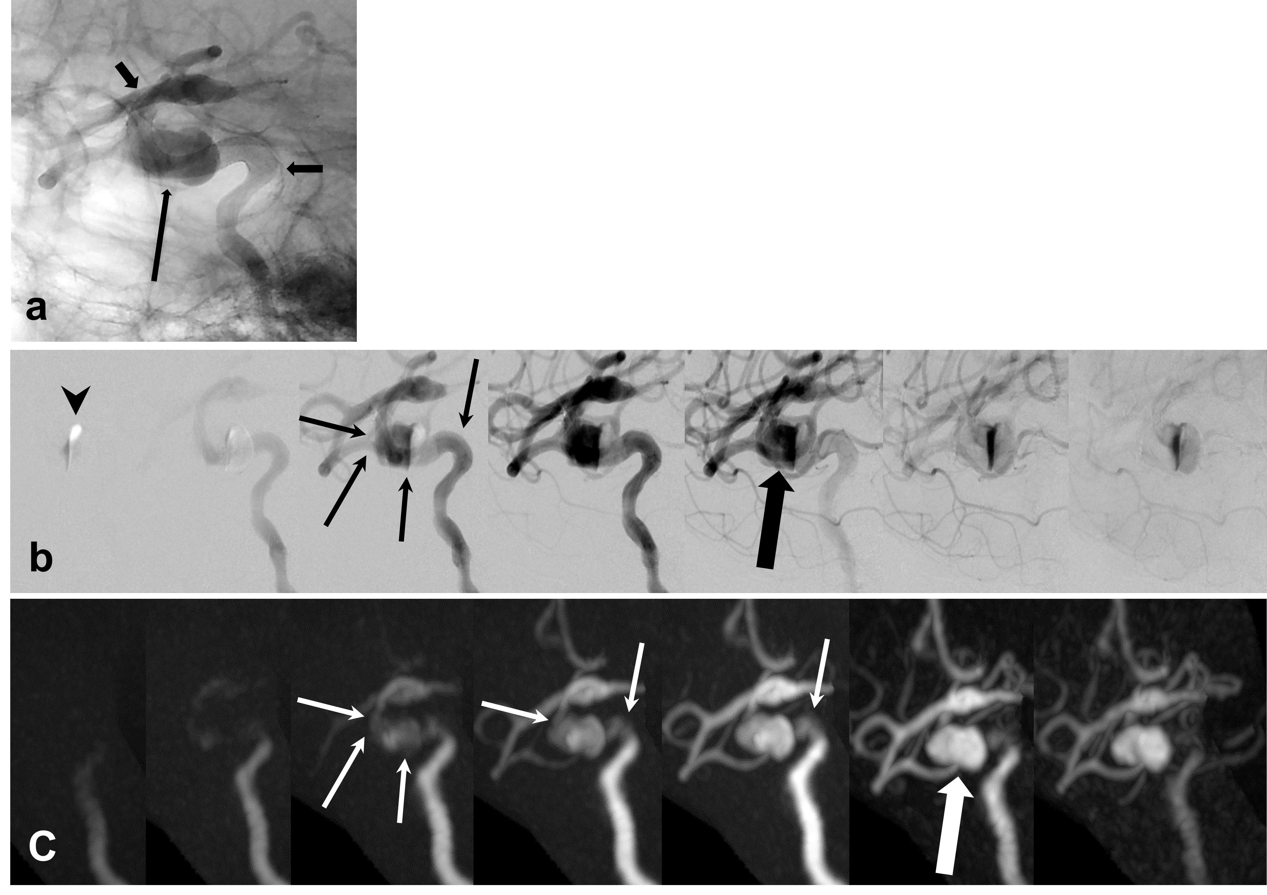
Fig.1
67 y.o Female (Left ICA : 4.0 × 25)
a.Digital angiography immediately after flow-diverter stent deployment is successful. Short arrows are stent edges. Long arrow is vascular flow in aneurysm.
b.DSA demonstrates flow in the stent (thin arrows) and vascular flow in aneurysm (thick arrow). First image shows the remains of contrast media (arrow head).
c.UTE 4D-MRA with 1day interval visualize flow in the stent at the acceptable image quality for diagnosis (thin arrows). Vascular flow in aneurysm appears gradually with time progress (thick arrow).
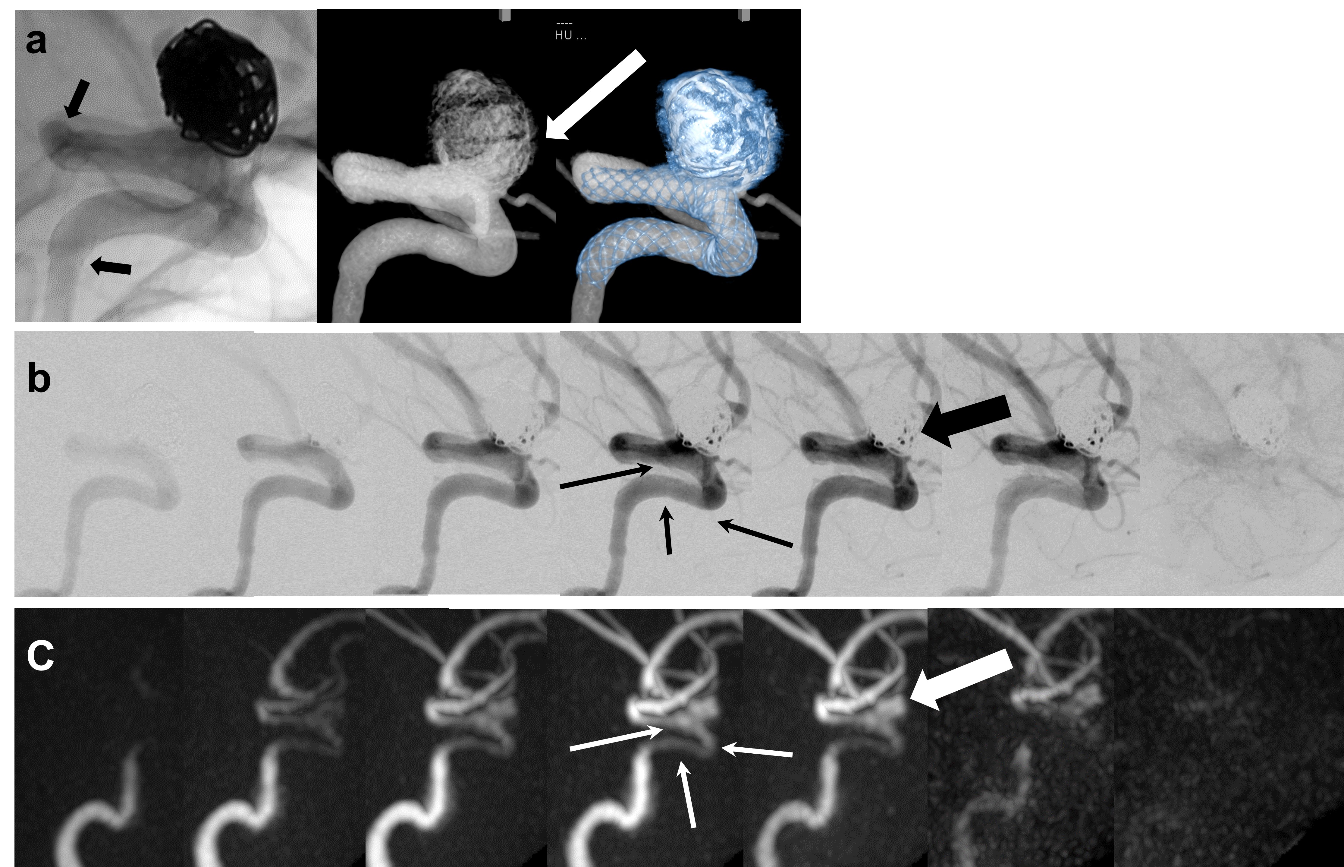
Fig.2
44 y.o Female (Left ICA : 4.75 × 25)
a.Digital angiography immediately after flow-diverter stent deployment is successful. Black arrows are stent edge. Cone-beam CT shows neck remnant (white arrow).
b.DSA demonstrates flow in the stent (thin arrows). Neck remnant is suspected (thick arrow).
c.UTE 4D-MRA with 1day interval visualize flow in the stent at the good image quality for diagnosis (thin arrows). Neck remnant is depicted clearly than DSA (thick arrow). UTE 4D-MRA demonstrates intraaneurysmal flow within the coil which was difficult to visualize even with X-ray DSA.
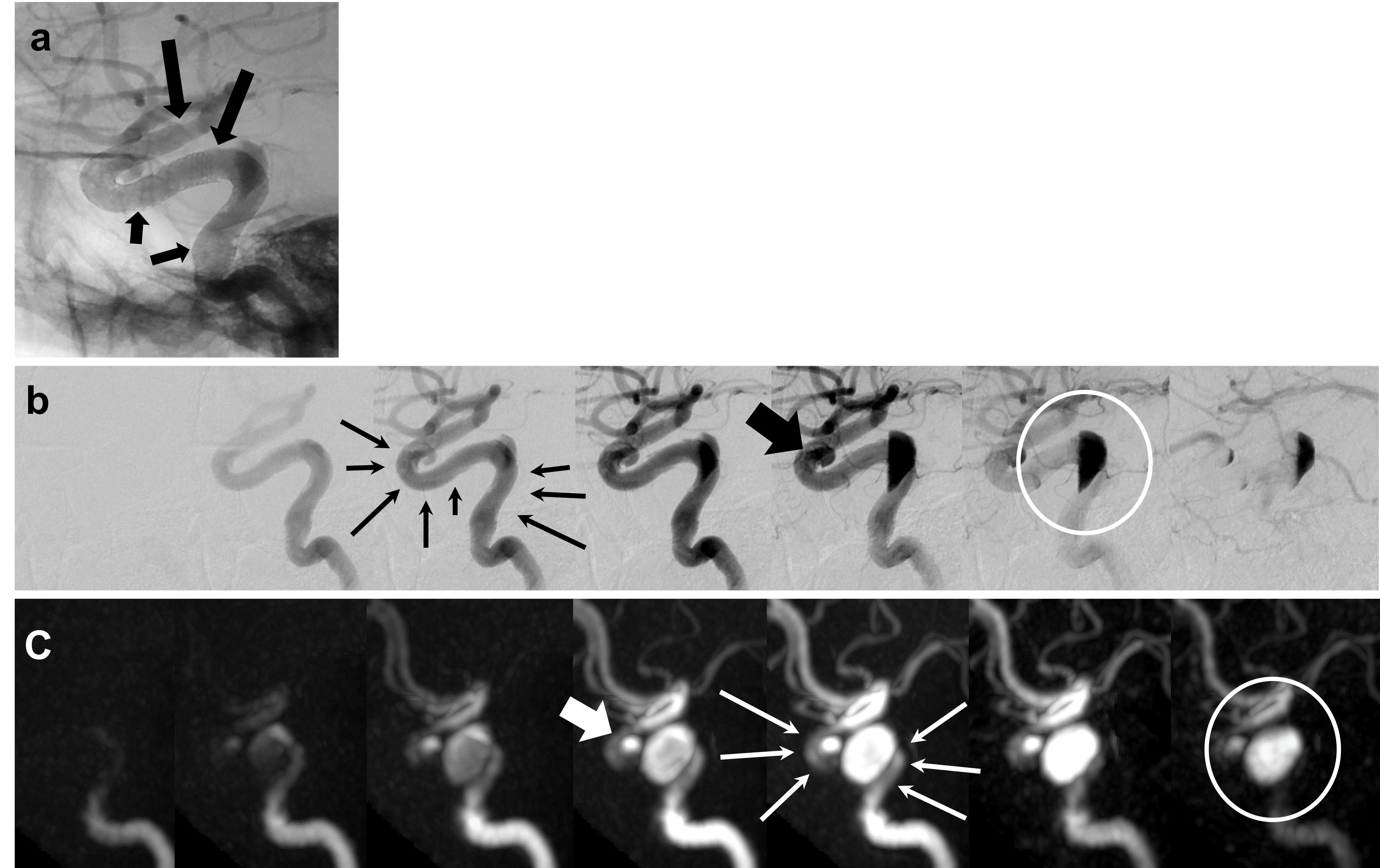
Fig.3
60 y.o Female (Left ICA : 4.25 × 25, 4.75 × 25)
a. Digital angiography immediately after treatment is successful. Black arrows are stent edge.
b. DSA shows flow in the stents and vascular flow in aneurysm (thin and thick arrows). In this case, blood flow is affected by gravity and injection speed of contrast media (white circle).
c. UTE 4D-MRA with 1day interval visualize flow in the stents (thin arrows). The appearance of vascular flow in aneurysm is comparable
to DSA (thick arrow). However, this case is different (white circle). UTE
4D-MRA reveals hemodynamic vascular flow than DSA.