1605
The feasibility of High resolution MRI in Length Measurement of Cerebral Arterial Thrombosis : a correlation study with Catheter Angiography1The First Affiliated Hospital of Shandong First Medical University, Jinan, China, 2GE Healthcare, MR Research China, Beijing, China, 3Qilu Children's Hospital of Shandong University, Jinan, China
Synopsis
This study aimed to investigate the feasibility of CUBE MRI for high resolution imaging in the length measurement of intraluminal thrombi for acute stroke patients. The T1 weighted CUBE images displayed the segments proximal and distal to the thrombus with sufficient quality for clinical diagnosis, and showed the length of it as high signal or iso-signal filling in the lumen. As a result, about 88.9% of the thrombus lengths assessed by CUBE T1 were consistent with the reference DSA. We therefore, demonstrated that the T1 weighted CUBE MRI can effectively evaluate the length of intraluminal thrombi.
INTRODUCTION
Detecting arterial occlusion and evaluating the length of intraluminal thrombi have prognostic and therapeutic implications for acute stroke patients[1]. Due to insufficient spatial resolution, conventional MR and CT examinations are limited in the diagnosis of thrombosis and reveal the location and morphological details for the thrombus[2].CUBE magnetic resonance imaging (MRI), as a three-dimensional (3D) high resolution imaging technique, has been widely applied in the diagnosis and evaluation of lesions on the intracranial arterial wall[3]. Due to the high spatial resolution, isotropic imaging and good suppression of blood flow signals reported previously, this technique was assumed to be able to identify and measure intracranial arterial thrombosis.
Therefore, this study aimed to investigate the feasibility of CUBE MRI for high resolution imaging in the length measurement of intraluminal thrombi in acute stroke patients. Digital subtraction angiography (DSA) examination during endovascular reperfusion therapy was also applied and served as a reference.
MATERIALS AND METHODS
PatientsEighteen patients (10 males and 8 females, mean age 57±9 years old, onset time from 8h to 14d) with cerebral artery thrombosis, diagnosed by DSA and having revascularization, were enrolled for T1-weighted CUBE imaging in this study. Written informed consent was obtained from each patient.
MRI experiment
All MR experiments were performed on a 3T clinical scanner (Discovery 750w, GE Healthcare, USA) equipped with a 32-channel coil. 3D T1 weighted CUBE sequence was applied with scan parameters of repetition time (TR) = 600ms, echo time (TE) = 14.4ms, slice thickness = 1mm, slice gap= 0.5mm, field of view (FOV) = 200mm * 200mm for whole brain coverage, matrix size = 288 * 288 and the echo chain length = 24. The total scan time was 4 minutes 16s.
Data analysis
DSA imaging during endovascular reperfusion therapy was used as the reference standard to image occlusion of the intracranial artery. Intraluminal thrombus on the T1-weighted CUBE images was defined as the presence of high signal or iso-signal filling in the lumen relative to low signal intensity of the normal vessel. Assessed by two senior radiologists with 10 years experience, the affected arteries were divided into segments either proximal or distal to the lesion. The image quality of acquired CUBE images was graded as identifiable, indistinct or unrecognizable. The thrombus length was determined by evaluating the position of proximal or distal to the lesion.
RESULTS
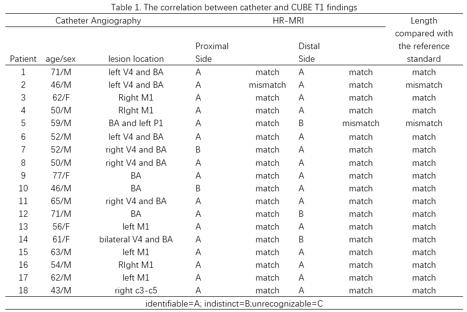
For in total 18 patients enrolled in the study, the acquired CUBE images have been rated with A (identifiable) or (B)indistinct level, indicating sufficient quality for clinical diagnosis (Table 1).Among these patients, on T1w CUBE images, 16 subjects (88.9%) showed the segments proximal to the lesion and 15 subjects (83.3%) clearly displayed the segments distal to the lesion.
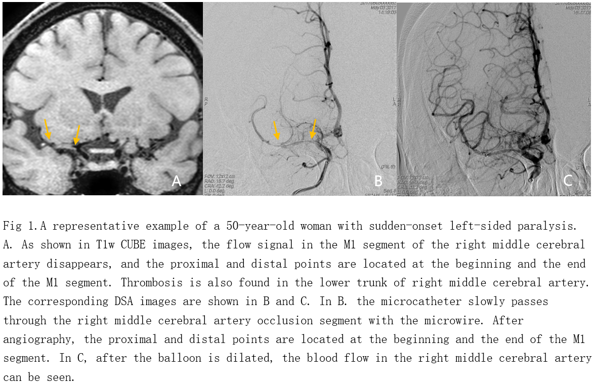
Compared with the reference standard DSA images, consistent proximal and end point positions were separately shown in 94% of the thrombus on T1w CUBE images. In addition, about 88.9% of the thrombus lengths assessed based on CUBE images were agreed with the reference DSA (Fig.1).
DISCUSSION
As a 3D high resolution MRI method, T1-weighted CUBE imaging has been used in this study to visualize the cerebral artery thrombosis and evaluate the length of intraluminal thrombi. As shown in CUBE images (Fig.1), all the segments proximal and distal to the lesion were displayed with sufficient quality for clinical diagnosis. Moreover, most of the thrombus lengths assessed by CUBE T1 were consistent with the results obtained from the reference DSA images.CONCLUSION
In conclusion, as comparable length measurement for intraluminal thrombi has been achieved relative to the reference standard of DSA images, T1-weighted CUBE MRI can be demonstrated for the feasibility in cranial cerebral thrombosis measurement.Acknowledgements
No acknowledgement found.References
[1] Derex L, Nighoghossian N, Hermier M, et al. Early detection of cerebral arterial occlusion on magnetic resonance angiography: predictive value of the baseline NIHSS score and impact on neurological outcome.[J]. Cerebrovascular Diseases, 2002, 13(4):225-229.
[2] Bash S ,Villablanca J P , Jahan R , et al. Intracranial Vascular Stenosis and Occlusive Disease: Evaluation with CT Angiography, MR Angiography, and Digital Subtraction Angiography[J]. Ajnr Am J Neuroradiol, 2005, 26(5):1012-1021.
[3]Li M L , Xu Y Y , Hou B , et al. High-resolution intracranial vessel wall imaging using 3D CUBE T1 weighted sequence[J]. European Journal of Radiology, 2016, 85(4):803-807.
Figures