1599
Association between type 2 diabetes mellitus and characteristics of intracranial atherosclerotic plaque: a high-resolution MRI study
Sheng Jiao1, Juan Huang2, Yan Song2, Yuhui Chen3, Chengwen Liu4, Jinxia Zhu4, Jintao Zhang2, and Min Chen2
1Radiology, Beijing hospital, Beijing, China, 2Radiology, Beijing Hospital, Beijing, China, 3Neurology, Beijing Hospital, Beijing, China, 4MR Collaboration, Siemens Healthcare Ltd., Beijing, China
1Radiology, Beijing hospital, Beijing, China, 2Radiology, Beijing Hospital, Beijing, China, 3Neurology, Beijing Hospital, Beijing, China, 4MR Collaboration, Siemens Healthcare Ltd., Beijing, China
Synopsis
In this study we used high-resolution MRI to image intracranial atherosclerotic plaque more precisely, and then characteristics of plaque were obtained to compare with the blood glycemic (BG) level in Type 2 Diabetes mellitus (T2DM). Results demonstrated that patients with poor BG control had heavier intracranial plaque burden and more vulnerability than the patients without DM. While compared with non-DM, patients with good BG control showed no significant different in plaque burden. It suggested that BG level is an independent risk factor for the vulnerability of the intracranial atherosclerotic plaque.
Purpose
Type 2 Diabetes mellitus (T2DM) is a chronic disease that always starts in middle- and late-adulthood, it will cause major health complications, particularly in the blood vessels. T2DM patients frequently have significant multiple atherosclerotic plaques, such as intracranial atherosclerotic plaque. However the relationship between blood glycemic (BG) level in T2DM and intracranial atherosclerotic plaque properties were unknown. In this study, by imaging plaque with high-resolution Magnetic Resonance Imaging (MRI), we investigated the association between T2DM and characteristics of intracranial atherosclerotic plaque, then plaque burden and vulnerability were investigated.Method
Three hundred and eleven patients who underwent high-resolution MRI examination for evaluating intracranial atherosclerosis were enrolled in this study. They were categorized into 3 groups according to health status and Hemoglobin A1C (HbA1c) level: patients without DM, n=172; DM patients with good BG control: HbA1c < 7%, n=55; DM patients with poor BG control: HbA1c ≥ 7%, n=84. All patients underwent MRI examinations on a MAGNETOM Prisma 3T MR scanner (Siemens Healthcare, Erlangen, Germany) with a 32-channel head coil. High-resolution (HR) intracranial artery wall images were acquired using a T1-weighted sampling perfection with application optimized contrasts (SPACE) sequence before and after contrast agent injection with following parameters: TR= 900 ms, TE= 15 ms, FOV= 170 × 170 mm2, voxel size= 0.53 × 0.53 × 0.53 mm3, and acquisition time = 8 min. Postcontrast T1WI was performed 5 min after the injection of single-dose (0.1mmol per kg of body weight) gadolinium-based contrast agent (Magnevist, Schering, Berlin, Germany). Characteristics of the intracranial plaques including plaque length, plaque thickness, lumen stenosis degree and strength of plaque enhancement were obtained after depicting the plaques region on HR intracranial artery vessel wall MR images. All these quantitative values were compared among 3 groups. The risk factors of the plaques for heavy burden and vulnerability were also analyzed. All continuous variables were expressed as means ± standard deviation and categorical variables were summarized as count. Characteristics of plaque between the 3 groups were compared using the analysis of variance for continuous variables and the chi-square test for categorical variables. Multivariate logistic regression was performed to determine independent indicators for heavy burden and strongly enhancement of plaque. All statistical analyses were performed by using SPSS 25.0. A P value < 0.05 indicated statistical significance.Result
Results showed that plaque length were significant different between the non-DM group and the DM with poor BG control group (non-DM: 5.95 ± 3.67 mm, DM with poor BG: 8.39 ± 5.14 mm, P < 0.001), plaque thickness (non-DM: 1.66 ± 0.83 mm, DM with poor BG: 1.98 ± 0.89 mm, P = 0.039) and lumen stenosis degree (non-DM: 45.05 ± 32.00%, DM with poor BG: 62.51 ± 29.73%, P < 0.001) reached significant different as well. The proportion of strongly enhanced plaques was significant different between the 3 groups (non-DM: 63.4%, DM with good BG: 72.7%, DM with poor BG: 92.9%, all P < 0.001). The results of multivariate logistic regression analysis shows that the proportion of strongly enhancement plaque was significantly correlated with BG level (OR = 2.21, 95% CI: 1.53 - 3.19, 𝑃 < 0.001).Discussion/Conclusion
In this study, we compared the BG level in patients and characteristics of intracranial atherosclerotic plaque using high-resolution MRI. The results indicated that patients with poor BGC had heavier intracranial plaque burden and more vulnerability than the patients without DM. When compared with non-DM, patients with good BGC showed no significant difference in plaque burden and plaque vulnerability. Our finding shows that the proportion of strongly enhancement plaque was significantly correlated with BG level, it suggests that BG level is an independent risk factor for the vulnerability of the intracranial atherosclerotic plaque.Acknowledgements
No acknowledgement found.References
- Selvin E, Marinopoulos S, Berkenblit G, et al. Meta-analysis: glycosylated hemoglobin and cardiovascular disease in diabetes mellitus. Ann Intern Med. 2004; 141:421-431.
- American Diabetes Association. Standards of Medical Care in Diabetes-2019 Abridged for Primary Care Providers. Clinical Diabetes 2019 Jan; 37(1): 11-34.
- Lee HN, Ryu CW, Yun SJ, et al. Vessel-Wall Magnetic Resonance Imaging of Intracranial Atherosclerotic Plaque and Ischemic Stroke: A Systematic Review and Meta-Analysis. Frontiers in Neurology. 2018 Dec 3; 9:1032.
Figures
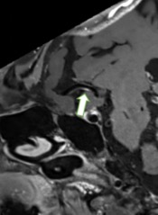
Post-contrast T1W image (axial acquisition) shows slightly enhancement atherosclerotic plaque (arrow) in the right middle cerebral artery (MCA) of the patient without DM.
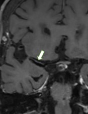
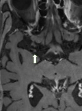
Post-contrast T1W image (longitudinal reconstruction) shows slightly enhancement atherosclerotic plaque (arrow) in the right middle cerebral artery (MCA) of the patient without DM.

Post-contrast T1W image (axial acquisition) shows strongly enhancement atherosclerotic plaque (arrow) in the right middle cerebral artery (MCA) of the patient with poor BG control.
Post-contrast T1W image (longitudinal reconstruction) shows strongly enhancement atherosclerotic plaque (arrow) in the right middle cerebral artery (MCA) of the patient with poor BG control.