1562
Detecting brain structural abnormality associated with breast cancer and chemotherapy using GQI1Department of Medical Imaging and Radiological Sciences, Chang Gung University, Taoyuan, Taiwan, 2School of Medicine, Chang Gung University, Taoyuan, Taiwan, 3Department of Psychiatry, Chang Gung Memorial Hospital, Chiayi, Taiwan, 4Department of Diagnostic Radiology, Chang Gung Memorial Hospital, Chiayi, Taiwan, 5Medical Imaging Research Center, Institute for Radiological Research, Chang Gung University and Chang Gung Memorial Hospital at Linkou, Taoyuan, Taiwan
Synopsis
Breast neoplasms are the most common cancer among women. Whether received adjuvant chemotherapy or not, lots of them presented cognitive impairment which decreased their quality of life. Therefore, we attempted to use generalized q-sampling imaging (GQI) to detect the white matter alteration between groups that may due to post-traumatic stress disorder (PTSD), post-traumatic growth (PTG) and chemotherapy. Our results showed the differences in the brain regions associated with cognition, emotion, and execution, such as angular gyrus, cingulate gyrus and so on. It indicted traumatic stressor or chemotoxicity can cause myelin injuries in these regions.
Introduction
The most common cancer in women worldwide is breast cancer (BC), and it also occupies the second leading cause of cancer-related mortality. BC is potentially a traumatic stressor that may lead to post-traumatic stress disorder (PTSD) and/or post-traumatic growth (PTG). In a review article, cognitive impairment bothered 11 to 35% of breast cancer patients before treatment, which was caused by PTSD 1,2. Adjuvant chemotherapy is an extensively used therapy in BC, which can induce emotional disturbance and cognitive impairment that has mostly been attributed to neurotoxic effects, so-called chemo-brain. Previous research showed that women treated with chemotherapy had decreased white matter integrity by diffusion tensor imaging (DTI) 3,4. DTI was widely used to investigating cerebral white matter, however, DTI cannot resolve the regions of complex fiber orientations. Therefore, we used generalized q-sampling imaging (GQI) 5 to investigate the white matter structure difference among women with a history of BC but did not receive any treatment (BB), women who had completed their chemotherapy less than 12 months (BA) and healthy control (BH).Methods
We divided participants into three groups, 45 of BB, 62 of BA and 55 of BH. All subjects were acquired by 3T MRI (Verio, Siemens). Echo-planar imaging (EPI) pulse sequence was performed to obtained images with TR / TE = 8943 / 115 ms, and voxel size = 1.9×1.9×4 mm3. Data were processed eddy current correction by FSL (FMRIB Software), and SPM (Statistical Parametric Mapping) was used for normalized all diffusion images of participants. Then, the GQI indices, including generalized fractional anisotropy (GFA) and normalized quantitative anisotropy (NQA), were reconstructed with model-free methods, also called q-space imaging methods to reduce the chance of calculating wrong fiber orientations. Finally, two voxel-based statistical analysis, Analysis of Variance (ANOVA) and post-hoc two-sample t-test were used to detect the change of GQI indices among these three groups. Age and education years were used as covariates. Participants were excluded if they were diagnosed with conditions that are known to affect cognitive function such as psychiatric, neurologic, or comorbid medical diseases.Results
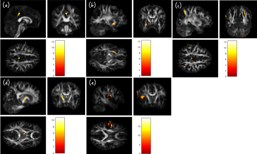
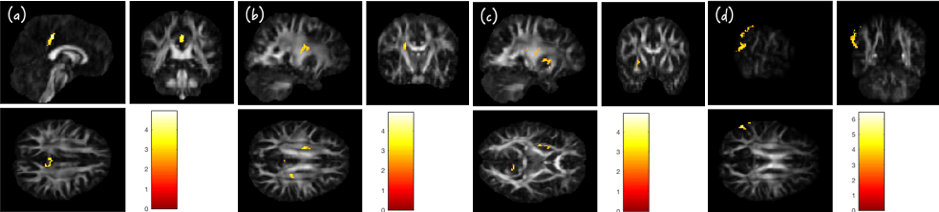
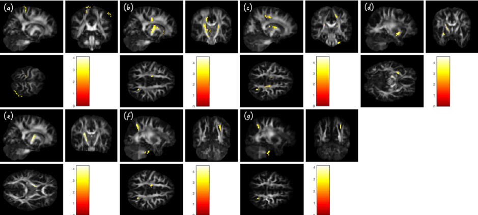
The value of GFA and NQA can represent the integrity of white matter. Lower GQI indices indicated lower myelination of white matter. The result of ANOVA showed that the GFA or NQA among these three groups had the difference in the region of the right posterior cingulate gyrus, left putamen, right angular gyrus, left posterior limb of the internal capsule and left postcentral gyrus (Fig. 1).In the result of the t-test, first, BB had higher GFA in the right posterior cingulate gyrus, bilateral corona radiata, left putamen and lower NQA in left angular gyrus compared with BA (Fig. 2). Second, BB had lower GFA in the left postcentral gyrus than BH (Fig.3). Third, BA had lower GFA in the left cingulate gyrus, right corona radiata, left putamen, left posterior limb of the internal capsule and both GFA and NQA in the right angular gyrus compared with BH (Fig.3). All of the above was with a p-value of less than 0.01.
Discussion
Our study found the smaller GFA of postcentral gyrus in BB compared with BH. Postcentral gyrus is the location of the primary somatosensory cortex, which is known for processing sensory information from various parts of the body. It also plays an important role in emotional processing, which may cause cognitive changes by PTSD. We also found that GFA and/or NQA of angular gyrus was BH > BA > BB. Angular gyrus involves visual information transfer to Wernicke’s area, language, spatial cognition, memory retrieval, and attention. It may result from PTSD and PTG that had related to myelin injury and recovery. From previous studies, they supported that postcentral gyrus and angular gyrus had an association with traumatic stressor 6,7. Additionally, we found significant differences between BA and BH in corona radiata, internal capsule, cingulate gyrus, and putamen. BA had lower GFA than BH indicated BA had higher demyelination may due to chemotherapy. Corona radiata and internal capsule play an important role in nerve conduction ascending and descending. Cingulate gyrus belongs to the limbic system, which is associated with emotion, learning, and memory. These functions have an impact on cingulate gyrus linking motivational outcomes to behavior. Putamen has many interconnect with other structures, its primary function is motor skills, including motor planning, learning, and execution. Our results were highly consistent with a systematic review of article 8.Conclusion
Myelin injury, whether through traumatic stressor or chemotoxicity, can have adversely affect cognition. This cross-sectional study demonstrates that GQI indices provide a good choice to detect white matter alterations of BC patients which may due to PTSD, PTG, and chemotherapy, and theses abnormalities could be potential imaging markers of cognitive change.Acknowledgements
This study was supported by the research grants MOST108-2813-C-182-032-B, MOST107-2221-E-182-054-MY3, MOST106-2221-E-182-079, and MOST104-2314-B-040-001 from the Ministry of Science and Technology, Taipei, Taiwan. This study was also supported by grants BMRPD1H0102 and BMRPD1G1322 from Chang Gung University, Taoyuan, Taiwan and CORPG6G0102 and CORPG6G0122 from Chang Gung Memorial Hospital, Chiayi, Taiwan.References
1. Parikh, D., et al. Post-traumatic stress disorder and post-traumatic growth in breast cancer patients--a systematic review. Asian Pac J Cancer Prev. 2015; 16(2): 641-646.
2. Pendergrass, J. C., et al. Cognitive Impairment Associated with Cancer: A Brief Review. Innov Clin Neurosci. 2018;15(1-2): 36-44.
3. Hermelink, K. Chemotherapy and Cognitive Function in Breast Cancer Patients: The So-Called Chemo Brain. J Natl Cancer Inst Monogr. 2015; 2015(51): 67-69.
4. Ganz, P. A. "Doctor, will the treatment you are recommending cause chemobrain?" J Clin Oncol. 2012; 30(3): 229-231.
5. Yeh, F. C., et al. Generalized q-sampling imaging. IEEE Trans Med Imaging. 2010; 29(9): 1626-1635.
6. Olson, E. A., et al. Disruption of white matter structural integrity and connectivity in posttraumatic stress disorder: A TBSS and tractography study. Depress Anxiety. 2017; 34(5):437-445.
7. O’Doherty, D. C. M., et al. White matter integrity alterations in post-traumatic stress disorder. Hum Brain Mapp. 2018; 39(3): 1327-1338.
8. Simo, M., et al. Chemobrain: a systematic review of structural and functional neuroimaging studies. Neurosci Biobehav Rev. 2013; 37(8): 1311-1321.
Figures