1503
Functional compensation on disrupted cingulate structural network in Parkinson’s disease1Zhejiang university, Hangzhou, China
Synopsis
Parkinson’s disease (PD) is the second most common neurodegenerative disease and its related brain changes appear not to be localized in isolate region but rather in the networks. We desire to exploring the large-scale structural and functional networks change which are of great importance for understanding the mechanism of disease. Moreover, it deserves to further answer the unsolved question that whether a functional compensation resulting from structural network disruption and whether such effect would be lasting or changed longitudinally.
Introduction
Parkinson’s disease (PD) related brain changes appear not to be localized in isolate region like substantia nigra but rather in the network of a multitude of brain regions1, 2, 3. Exploring the large-scale networks changes is of great importance for diagnosis or understanding the mechanism of PD.Methods
One hundred and four PD patients and 77 healthy controls were included in this study. Deformation-based morphometry and independent component analysis (ICA) were conducted to assess the disruption of large-scale structural networks in PD patients4, 5, 6. Functional ICA was performed in the same set of subjects. The components showed best spatial overlap with disrupted structural networks were selected as the corresponding functional networks and the changes of network connectivity in PD patients were assessed. Then, the association between structural network integrity and the strength of functional network connectivity were evaluated. Moreover, longitudinal analysis was conducted to further validate the functional connectivity alterations in 37 of the patients who had follow-up data.Results
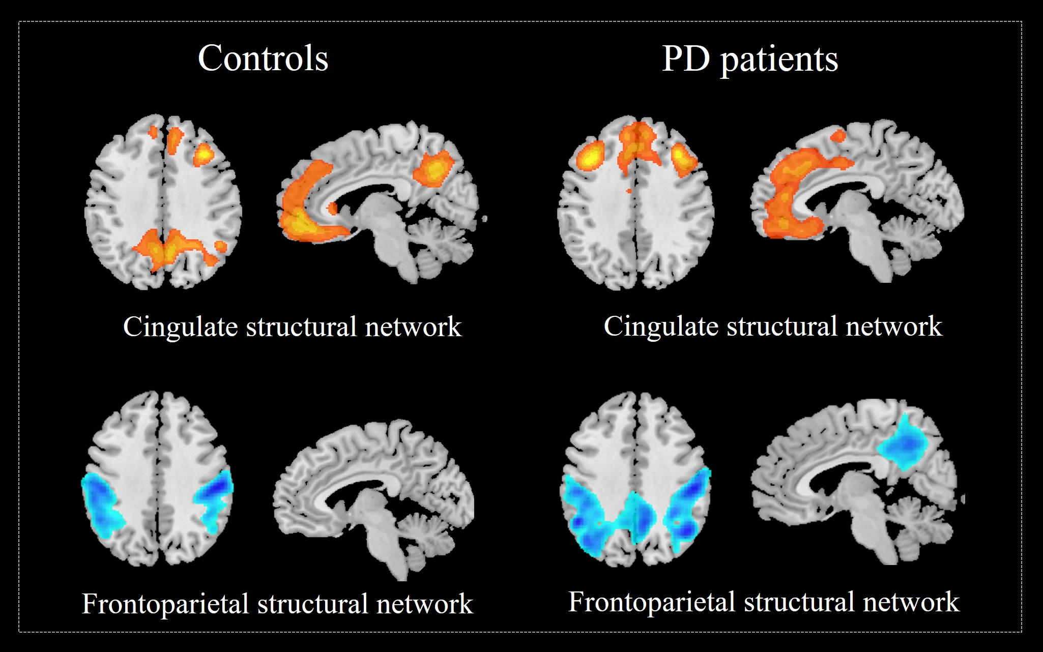
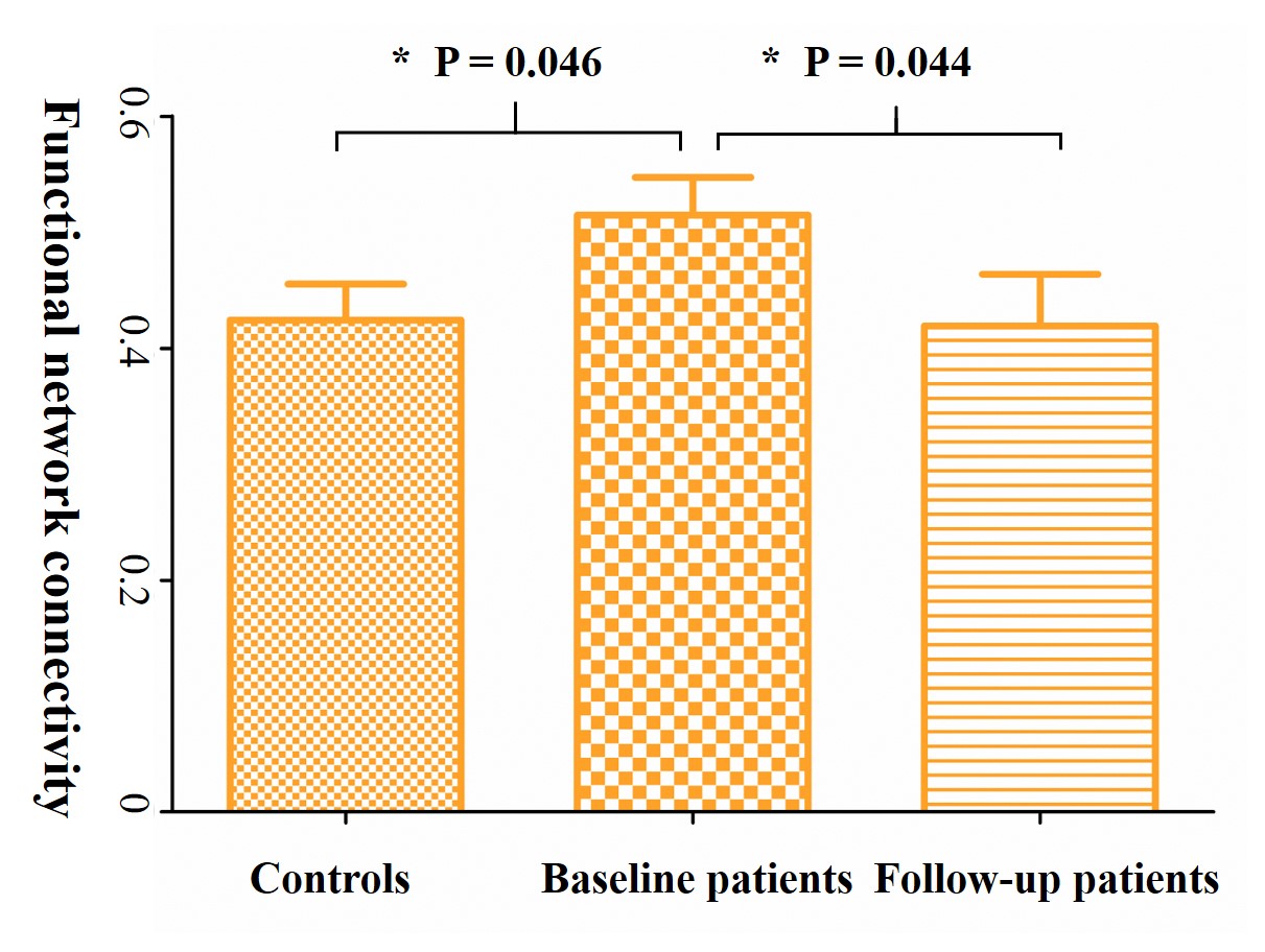
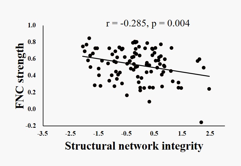
PD patients showed decreased structural covariance between anterior and posterior cingulate networks (Figure 1). PD patients showed increased connectivity between anterior and posterior cingulate functional networks at baseline and decreased connectivity at follow-up (Figure 2 and 3). Increased functional connectivity was negatively correlated with decreased integrity of the cingulate structural network (Figure 4).Conclusion
Our findings suggested that cingulate structural network which was similar to the default mode network display a high susceptibility in PD patients. Decreased covariance between anterior and posterior cingulate structural networks might indicate the structural basis of cognitive impairment, and increased functional connectivity between them could provide a temporal compensation on structural disruption.Acknowledgements
The authors thank the team at the department of Radiology, the Second Affiliated Hospital, Zhejiang University School of Medicine, Hangzhou 310009, China.References
1, Braak, H., Ghebremedhin, E., Rub, U., Bratzke, H., & Del Tredici, K. (2004). Stages in the development of Parkinson's disease-related pathology. Cell Tissue Res, 318(1), 121-134. doi:10.1007/s00441-004-0956-9
2, Jellinger, K. A. (2012). Neuropathology of sporadic Parkinson's disease: evaluation and changes of concepts. Mov Disord, 27(1), 8-30. doi:10.1002/mds.23795
3, de Schipper, L. J., van der Grond, J., Marinus, J., Henselmans, J. M. L., & van Hilten, J. J. (2017). Loss of integrity and atrophy in cingulate structural covariance networks in Parkinson's disease. NeuroImage: Clinical, 15, 587-593. doi:10.1016/j.nicl.2017.05.012
4, Beckmann, C. F., & Smith, S. M. (2004). Probabilistic independent component analysis for functional magnetic resonance imaging. IEEE Trans Med Imaging, 23(2), 137-152. doi:10.1109/tmi.2003.822821
5, Chung, M. K., Worsley, K. J., Robbins, S., Paus, T., Taylor, J., Giedd, J. N., . . . Evans, A. C. (2003). Deformation-based surface morphometry applied to gray matter deformation. NeuroImage, 18(2), 198-213.
6, Xu, L., Groth, K. M., Pearlson, G., Schretlen, D. J., & Calhoun, V. D. (2009). Source-based morphometry: The use of independent component analysis to identify gray matter differences with application to schizophrenia. Human Brain Mapping, 30(3), 711-724. doi:10.1002/hbm.20540
Figures