1502
Localization of the iron deposits along myelinated fibers within the substantia nigra of progressive supranuclear palsy on brain MRI1Department of Biomedical Engineering, Ulsan National Institute of Science and Technology, Ulsan, Korea, Republic of, 2Department of Anatomy, Pusan National University School of Medicine, Yangsan, Korea, Republic of, 3Department of Neurology, Pusan National University Hospital, Busan, Korea, Republic of, 4Department of Forensic Medicine, Pusan National University School of Medicine, Yangsan, Korea, Republic of, 5Department of Neurology, Research Institute for Convergence of Biomedical Science and Technology, Pusan National University Yangsan Hospital, Yangsan, Korea, Republic of
Synopsis
The purpose of this study was to determine the morphology change in the substantia nigra of progressive supranuclear palsy using MRI with histopathological validation. MR experiments for progressive supranuclear palsy brains were operated using 3T in vivo and 7T postmortem imaging systems. Perls’ Prussian blue staining, Luxol fast blue staining, and LA-ICP-MS for 2D iron mapping confirmed the large amount of iron deposits along the myelinated fibers within substantia nigra of PSP brain. The iron deposits along the myelinated fibers could be the potential source causing the blurred boundary between red nucleus and substantia nigra in in vivo MRI.
Purpose
Progressive supranuclear palsy (PSP) is a rare neurodegenerative disorder with predominant features of parkinsonism from abnormal tau protein aggregation and marked loss of dopaminergic neurons1, 2. The atrophy of midbrain including substantia nigra (SN) is one of the typical pathological characteristics of PSP identified in MRI studies2. In the SN, neuromelanin pigments and deposited iron molecules are dominant contributors in MRI contrast3. T1-weighted image with magnetization transfer (MT) effects for neuromelanin-sensitive MRI and transverse relaxometry-based imaging for iron-sensitive MRI have been investigated to non-invasively monitor the progression of Parkinsonism3. Additionally, the dopaminergic projection appears mainly from SN pars compacta and GABAergic neuron originates from SN pars reticulata. This study aimed to investigate the morphological alterations of microarchitecture within the SN with disease progression of PSP through direct comparison among MRI, histology, and mass spectrometer.Methods
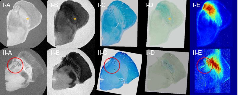
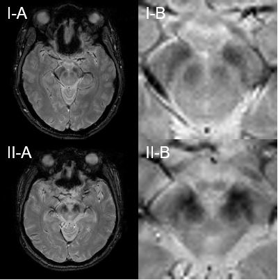
The postmortem midbrain samples were obtained from one normal subject and one patient with a diagnosis of PSP. High-resolution MRI of postmortem brains was conducted at 7T preclinical scanner (Bruker, Germany) for T1-weighted image with additional MT contrast preparation pulses and T2*-weighted image. After MR scans, the cryoprotected brain tissues were sectioned to 50 m thickness for Perls’ Prussian blue staining and Luxol fast blue staining. 2D iron mapping (56Fe/13C) was performed by Laser ablation inductively coupled plasma mass spectrometry (LA-ICP-MS). The postmortem MRI, stained slides, and 56Fe/13C iron map were co-registered for direct comparison. The normal controls and PSP patients underwent 3T in vivo MRI. T2*-weighted image with the oblique-axial plan was acquired parallel to the AC-PC line with the following parameters: TR = 406 ms, TE = 13.47 ms, and FA = 20 °.Results
For both normal and PSP brains, the hypointense area in the T1-weighted image with MT effects (Figure 1I-A and 1II-A) showed the myelinated fiber within anteriomedial SN identified from Luxol fast blue staining (Figure 1I-C and 1II-C). For the normal brain, the structure of nigrosome 1 was detected as a hyperintense area in the T2*-weighted image (orange asterisk in Figure 1I-B). This area corresponded to the hyperintensity in the T1-weighted image with lower iron content in co-registered Perls’ Prussian blue staining (Figure 1I-D). The positively stained region of Perl’s Prussian blue staining highly co-localized with the elevated signals in 56Fe/13C intensity from LA-ICP-MS imaging (Figure 1I-E). For the PSP brain, red circles in the T1-weighted image with MT effects (Figure 1II-A) and Luxol fast blue staining (Figure 1II-C) showed the neuronal projection of the 3rd cranial nerve and myelinated fiber within anteriomedial SN. In the high-resolution T2*-weighted image (Figure 1II-B) within SN of PSP brain, the hypointensity showed the linear patterns across the SN. However, their linearities were identified in Perls’ Prussian blue staining (Figure 1II-D) and were not shown in Luxol fast blue staining. The iron deposits along myelinated fibers in the red circles were identified in 56Fe/13C intensity of LA-ICP-MS imaging (Figure 1II-E), which was not detected in Perls’ Prussian blue staining. In the 3T in vivo T2*-weighted image, the boundary between SN and red nucleus was clearly visible in normal controls (Figure 2I). On the other hand, it disappeared in the PSP brain with the connection of hypointense signals (Figure 2II).Discussions
In this study, the evidence of iron deposition along myelinated fiber within anteriomedial SN was identified in PSP through the validation utilizing diverse approaches. Defining anatomy and delineating the subregions of the SN are still controversial even in the high-field MRI. The various sources within the SN of Parkinsonism complicate the interpretation of in vivo MRI. The iron deposits along myelinated fibers in the PSP brain reflect the iron deposits causing loss of myelinated nerve fibers with neurodegenerative diseases. However, the linear hypointense patterns in the T2*-weighted image of the postmortem PSP brain were not precisely determined in direct histopathological validation. The perivascular iron deposition could be one of the potential candidates bringing T2* shortening effect within SN. Further studies with more samples of PSP brains with additional histopathological experiments are needed for the improved interpretation of in vivo brain MRI of Parkinsonism.Acknowledgements
This work was supported by the Korea Health Industry Development Institute by the 2019 Research Fund (HI18C0713).References
1. Gerstenecker, A., Duff, K., Mast, B., Litvan, I., & ENGENE-PSP Study Group. (2013). Behavioral abnormalities in progressive supranuclear palsy. Psychiatry research, 210(3), 1205-1210.
2. Liscic, R. M., Srulijes, K., Gröger, A., Maetzler, W., & Berg, D. (2013). Differentiation of progressive supranuclear palsy: clinical, imaging and laboratory tools. Acta Neurologica Scandinavica, 127(5), 362-370. Sasaki, M., Shibata, E., Kudo, K., & Tohyama, K. (2008). Neuromelanin-sensitive MRI. Clinical Neuroradiology, 18(3), 147-153.
3. Lehéricy, S., Sharman, M. A., Santos, C. L. D., Paquin, R., & Gallea, C. (2012). Magnetic resonance imaging of the substantia nigra in Parkinson's disease. Movement disorders, 27(7), 822-830.
Figures