1484
Age and location related hemodynamic changes in the carotid artery of healthy adults assessed by 4D flow MRI
Guiling Zhang1 and Wenzhen Zhu1
1Tongji hospital, Wuhan, China
1Tongji hospital, Wuhan, China
Synopsis
Atherosclerosis is the predominant risk factor of ischemic stroke. Age and location are intrinsic factors in the development of carotid artery plaque. Therefore, the relationship between hemodynamic changes and location/age in healthy subjects can help understand how the hemodynamic states influence carotid incidents. Our study found velocity/WSS/PG decreased with age. And proximal ICA, the most common point arising atherosclerotic plaque, displayed the lowest velocity/WSS/PG. It may imply this state of hemodynamics are more likely to cause atherosclerotic plaques. The multi-parameter analysis of 4D flow MRI may offer suggestions for choosing age and location matched control cohorts in future study.
Introduction
Carotid diseases, especially atherosclerosis, are the predominant risk factor of ischemic stroke1. Patient age, hypertension, hyperlipidemia, diabetes, smoking, etc, are usually associated with the formation of atherosclerotic plaque2, in addition, carotid artery bifurcation is the predilection site for atherosclerotic plaque3. Among these risk factors, patient age and the anatomy of carotid bifurcation are immutable, while the status of hypertension, hyperlipidemia, diabetes can be interfered. Therefore, the relationship between hemodynamic changes and different territories of carotid artery, patient age in healthy subjects can help understand how the hemodynamic states influence carotid incidents.We use 4D flow MRI, a newly developed complementary method, to detect the changes of multiple hemodynamic parameters in different age and locations. The aim of this study is to demonstrate the hemodynamic distribution of normal carotid arteries and to explore the possible influence of these factors because age and location are intrinsic factors in the development of carotid artery disease. In addition, it may also provide age and location matched control cohorts for the assessment of carotid artery disease.
Method
Sixty-two normal volunteers aging from 20 to 75 were enrolled in our study, A rigorous medical history was taken from the volunteers to exclude those with cardiovascular risk factors (hypertension, hyperlipidemia, diabetes, smoking, drinking et al) or former cardiovascular events. 4D flow MRI examination was performed for each subject and analyzed using CVI42 platform to get hemodynamic parameters. Hemodynamic parameters were then compared in different ages and locations (pro-CCA, dis-CCA, pro-ICA and dis-CCA). The relationship between age and hemodynamic parameters was then qualified by Pearson's correlation coefficient.Result
Hemodynamics changes in different locationsHemodynamics changes in different locations of carotid artery were displayed in figure1. Pro-ICA had significant lower velocity, WSS and PG than other parts(P<0.05). Velocity and PG all first showed a downward trend and started rising when it reached the lowest point pro-ICA. Figure 2 visually displayed the MIP and streamline map of velocity/WSS in different age group.
Correlation between age and hemodynamics
Linear correlations between hemodynamic parameters in different locations and age were summarized in figure 3. The velocity, WSS and PG showed significantly inverse correlation with age (P<0.05). The velocity and PG better correlation with the age than the WSS. Moreover, the correlation between age and the velocity/PG at CCA were better than ICA, but for WSS, the correlation at ICA was better than CCA. Only distal ICA showed no correlation with age.
Discussion
The shape of carotid bifurcation has a large impact to the distribution of blood flow and velocity4. Previous study found low WSS was mainly distributed on the outer side wall of carotid bifurcation5. Pressure was the mechanical force of blood flow acting perpendicular to the luminal surface6 and PG reflected the magnitude of the pressure change. One study found that PGmean decreased after the percutaneous transluminal angioplasty and stenting (PTAS) when stenosis existed7.Our research showed that velocity, WSS and PG were all reduced with age, and the same phenomenon were also found in aorta for velocity and WSS8, 9.Vessel diameter can increase with age due to decreased elasticity10. So the decreased velocity may be driven by diameter expansion. How age affects hemodynamics remains unclear, it may be explained by the change of vascular elasticity and endothelial cell function with age11.
Why does this state of hemodynamic led to the formation of plaques? The decrease of velocity prolonged the blood duration in the vessel, resulting in lasting lipid retention and interaction with vessel wall. WSS is the frictional force on the endothelial cells induced by blood flow and is paralleled to the luminal surface of vessel6. Low WSS increased the uptake of oxidized low-density lipoprotein12, causing increasing lipid components in plaques. Moreover, low WSS altered vascular endothelium flow patterns in molecular and cellular levels. All these reactions promote the development of atherosclerosis13.
Conclusion
In conclusion, different locations of the carotid artery showed different hemodynamic changes. Among them, proximal ICA showed significantly lower volume, velocity and WSS. In addition, the hemodynamic parameters in different location were inversely correlated with age. It may imply that low volume, velocity and WSS are more likely to cause atherosclerotic plaques. The multi-parameter analysis of 4D flow MRI identified age and location changes of hemodynamic parameters in carotid artery of healthy adults, indicating that age and location impacted the blood flow. This study offered suggestion for choosing age and location matched control cohorts for the assessment of carotid artery disease in future study.Acknowledgements
No acknowledgement found.References
1. Lovett JK, Coull AJ, Rothwell PM. Early risk of recurrence by subtype of ischemic stroke in population-based incidence studies. Neurology 2004;62(4):569-573. 2. Moskau S, Golla A, Grothe C, Boes M, Pohl C, Klockgether T. Heritability of carotid artery atherosclerotic lesions: an ultrasound study in 154 families. Stroke 2005;36(1):5-8. 3. Ackerman RH, Candia MR. Identifying clinically relevant carotid disease. Stroke 1994;25(1):1-3. 4. Zhang Q, Steinman DA, Friedman MH. Use of Factor Analysis to Characterize Arterial Geometry and Predict Hemodynamic Risk: Application to the Human Carotid Bifurcation. J Biomech Eng-T Asme 2010;132(11). 5. Zhao SZ, Ariff B, Long Q, Hughes AD, Thom SA, Stanton AV, Xu XY. Inter-individual variations in wall shear stress and mechanical stress distributions at the carotid artery bifurcation of healthy humans. Journal of Biomechanics 2002;35(10):1367-1377. 6. Li C-H, Gao B-L, Wang J-W, Liu J-F, Li H, Yang S-T. Hemodynamic Factors Affecting Carotid Sinus Atherosclerotic Stenosis. World Neurosurgery 2019;121:e262-e276. 7. Han Y-F, Liu W-H, Chen X-L, Xiong Y-Y, Yin Q-, Xu G-L, Zhu W-S, Zhang R-L, Ma M-M, Li M-, Dai Q-L, Sun W-, Liu D-Z, Duan L-H, Liu X-F. Severity assessment of intracranial large artery stenosis by pressure gradient measurements: A feasibility study. Catheterization and Cardiovascular InterventionsCatheterization and Cardiovascular Interventions 2016;88(2):255-261. 8. van Ooij P, Garcia J, Potters WV, Malaisrie SC, Collins JD, Carr JC, Markl M, Barker AJ. Age-related changes in aortic 3D blood flow velocities and wall shear stress: Implications for the identification of altered hemodynamics in patients with aortic valve disease. Journal of Magnetic Resonance Imaging 2016;43(5):1239-1249. 9. Callaghan FM, Bannon P, Barin E, Celemajer D, Jeremy R, Figtree G, Grieve SM. Age-related changes of shape and flow dynamics in healthy adult aortas: A 4D flow MRI study. Journal of Magnetic Resonance Imaging 2019;49(1):90-100. 10. Wolak A, Gransar H, Thomson LEJ, Friedman JD, Hachamovitch R, Gutstein A, Shaw LJ, Polk D, Wong ND, Saouaf R, Hayes SW, Rozanski A, Slomka PJ, Germano G, Berman DS. Aortic Size Assessment by Noncontrast Cardiac Computed Tomography: Normal Limits by Age, Gender, and Body Surface Area. Jacc-Cardiovasc Imag 2008;1(2):200-209. 11. Redheuil A, Yu WC, Mousseaux E, Harouni AA, Kachenoura N, Wu CO, Bluemke D, Lima JAC. Age-Related Changes in Aortic Arch Geometry Relationship With Proximal Aortic Function and Left Ventricular Mass and Remodeling. J Am Coll Cardiol 2011;58(12):1262-1270. 12. Roustaei M, Nikmaneshi MR, Firoozabadi B. Simulation of Low Density Lipoprotein (LDL) permeation into multilayer coronary arterial wall: Interactive effects of wall shear stress and fluid-structure interaction in hypertension. Journal of Biomechanics 2018;67:114-122. 13. Cunningham KS, Gotlieb AI. The role of shear stress in the pathogenesis of atherosclerosis (vol 85, pg 9, 2005). Lab Invest 2005;85(7):942-942.Figures
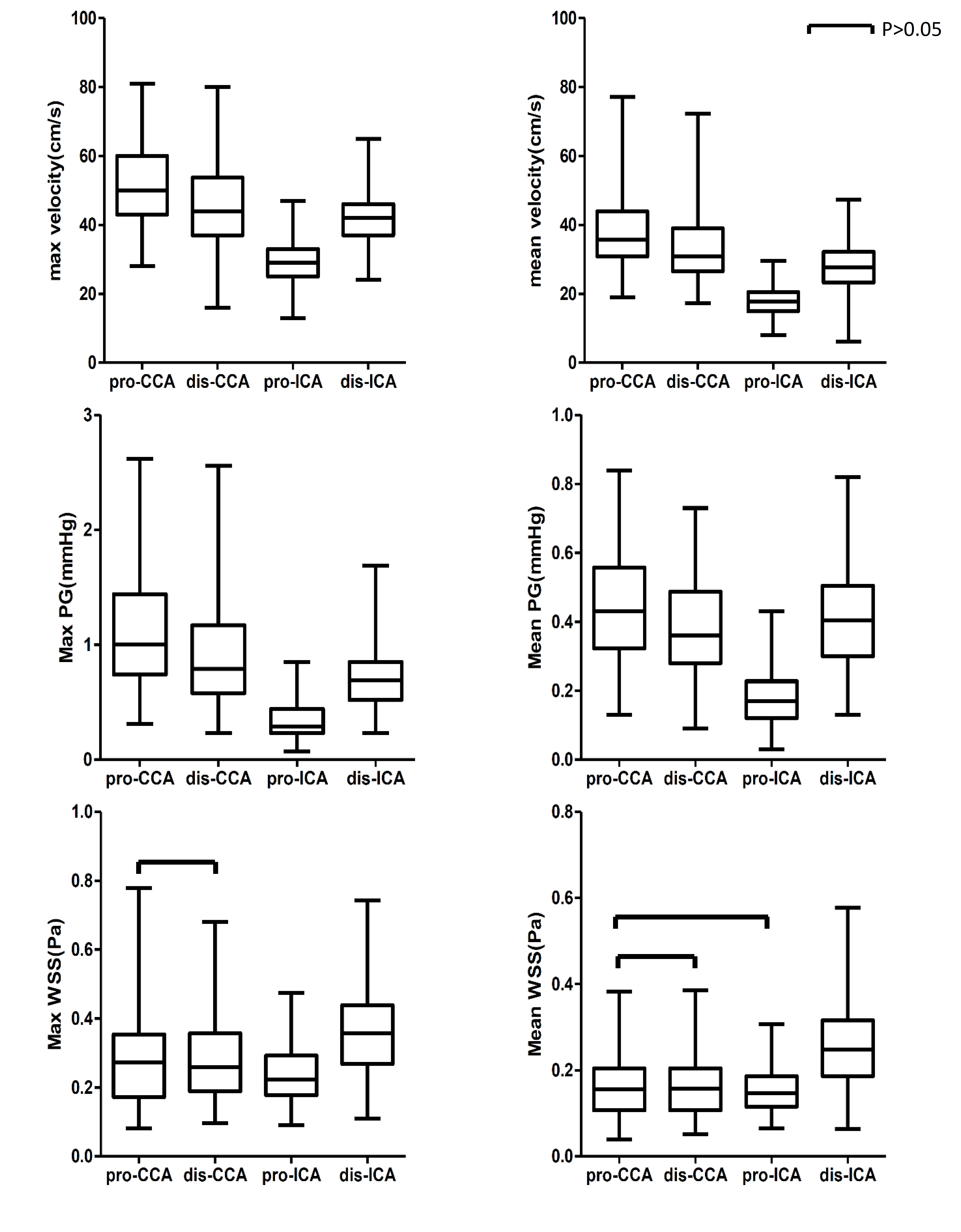
Figure1:
Velocity, WSS and PG changes at different locations(pro-CCA, dis-CCA, pro-ICA,
dis-ICA). WSS: wall shear stress; PG: pressure gradient
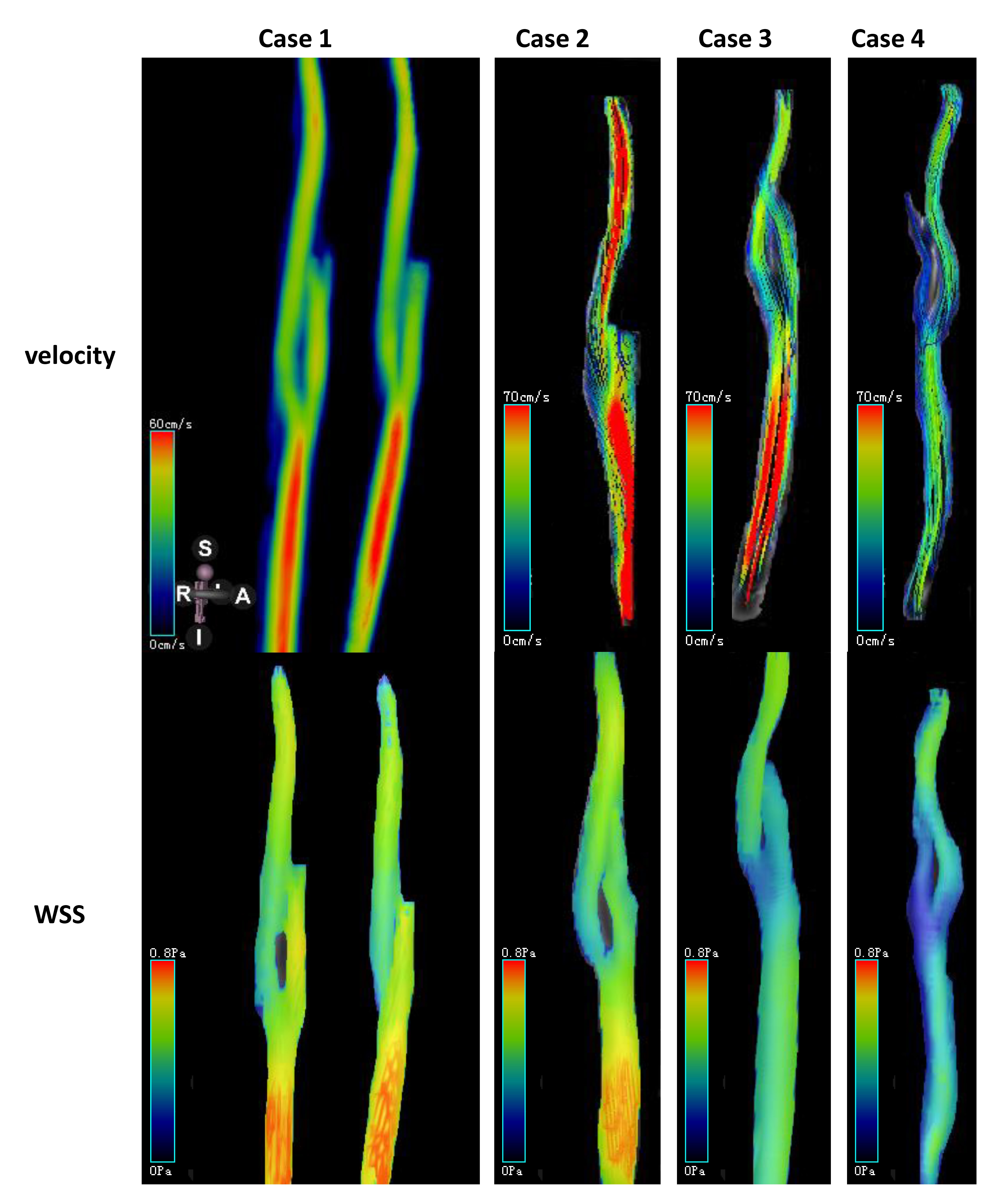
Figure2: Examples of hemodynamic parameters visibly showed by
4D flow.
Case 1: mip map, man, 23 years old,
velocity and WSS showed no difference in bilateral artery, proximal ICA
displayed lowest velocity and WSS.
Case 2,3,4: Streamline and mip map, man,
24y(case2), 40y(case3), 62y(case4).
These cases showed that hemodynamic
parameters decreased with age visibly , proximal ICA still displayed lowest
velocity and WSS.

Figure 3: Velocity, WSS(except at distal
ICA), PG decreased with aging. (p<0.05)