1476
Source Based Brain Structural Features extraction and Classification on Alzheimer’s Disease1Nanjing Drum Tower Hospital, Nanjing, China, 2Clinical science, Philips Health Care, Shanghai, Shanghai, China
Synopsis
We used independent components analysis to analysis the inter-subject variation of the voxel-based morphometry results of 446 subjects of Alzheimer's spectrum. 20 components which reflect the inter subject volume variation were extracted and used as features in SVM. We observed better performance in SVM classification by these features than those using atlas-based method. These findings might be helpful for classification of AD, MCI and NC populations, which provide assistance to the early diagnosis and intervention of Alzheimer’s disease.
Introduction
Auto classification of typical neurology and psychiatry diseases by MRI have been a hot issue in recent year. However, original MRI data often have extreme large dimensions which may lead to over-fitting, while feature extraction is often based on specific assumption. A data-driven method would be a potential solution and improve the performance of classification. The current study utilized a source-based morphometry (SBM) method on the images from ADNI database, and aims to extract brain structural features which can reflect the variation among AD spectrum populations. We assume using these features in classification would improve classification performances.Methods
T1 weighted structural MRI images of AD (Alzheimer’s disease, 151), MCI (mild cognitive impairment, 124) and NC (normal control, 171) populations was downloaded from ADNI database. SBM analysis was performed on these data, which extract the components reflecting the variation of the voxel-based morphometry (VBM) results across subjects through independent component analysis (ICA). Brain atlas was also used to extract volume features. We used Anatomical Automatic Labeling (AAL, divided cerebrum grey matters into 90 regions) and a further divided AAL (into 1024 regions, AAL1024). Support vector machine (SVM) was used to classify each pair among 3 populations, using SBM components, volume in each region of AAL90 or AAL1024 as features, respectively. Age, gender and education years was controlled as covariances. We employed leave-one-out method for classification test.Results
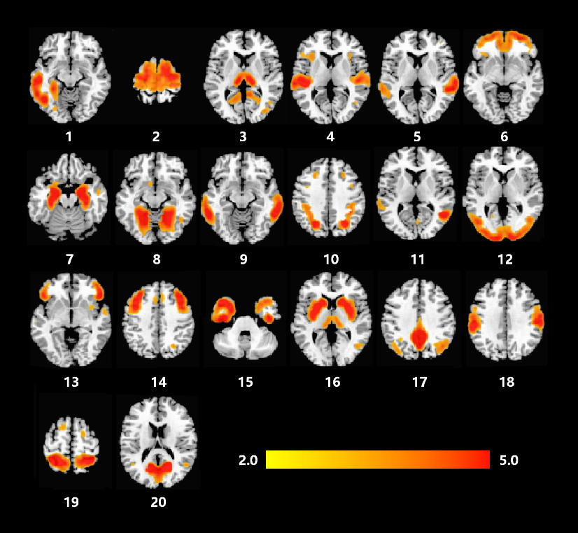
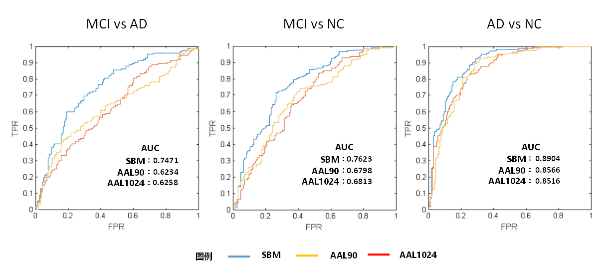
The across subject variation of brain volume as decomposed into 20 components (Figure 1). Using these 20 SBM components as features, SVM classifier achieves the accuracy 81.68% between AD and NC, while the accuracy is 78.57%, 79.19% respectively for AAL90 and AAL1024. The area under curve (AUC) value of the receiver operating characteristic (ROC) curve using SBM features was also significantly larger than those using AAL90 and AAL1024 features (Figure 2). Similarly, SVM classifier based on SBM also have higher accuracy than AAL90 and AAL1024 when classify MCI-NC (SBM: 69.82%, AAL90: 58.18%, AAL1024: 60.19%) and MCI-AD (SBM: 71.53%, AAL90: 64.07%, AAL1024: 67.80%). The AUC of ROC curve is also significantly larger when SBM features utilized (Figure 2).Discussion&Conclusion
Although our results did not achieve very high performance, SBM based classification indeed showed significant better performance than those based on previous defined atlas but using even less features. These findings might be helpful for classification of AD, MCI and NC populations, which provide assistance to the early diagnosis and intervention of Alzheimer’s disease.Acknowledgements
No acknowledgement found.References
Arbabshirani MR, Plis S, Sui J, Calhoun VD. 2017. Single subject prediction of brain disorders in neuroimaging: Promises and pitfalls. Neuroimage. 145:137-165.
Filippi M, Agosta F, Frisoni GB, De Stefano N, Bizzi A, Bozzali M, Falini A, Rocca MA, Sorbi S, Caltagirone C, Tedeschi G. 2012. Magnetic resonance imaging in Alzheimer's disease: from diagnosis to monitoring treatment effect. Curr Alzheimer Res. 9:1198-1209.
Ten Kate M, Dicks E, Visser PJ, van der Flier WM, Teunissen CE, Barkhof F, Scheltens P, Tijms BM, Alzheimer's Disease Neuroimaging I. 2018. Atrophy subtypes in prodromal Alzheimer's disease are associated with cognitive decline. Brain. 141:3443-3456.
Tzourio-Mazoyer N, Landeau B, Papathanassiou D, Crivello F, Etard O, Delcroix N, Mazoyer B, Joliot M. 2002. Automated anatomical labeling of activations in SPM using a macroscopic anatomical parcellation of the MNI MRI single-subject brain. Neuroimage. 15:273-289.
Xu L, Groth KM, Pearlson G, Schretlen DJ, Calhoun VD. 2009. Source-based morphometry: the use of independent component analysis to identify gray matter differences with application to schizophrenia. Hum Brain Mapp. 30:711-724.
Figures