1453
Blood-brain barrier integrity and cognitive function in a healthy aging population
SAMUEL BARNES1, SHILPY CHOWDHURY2, JENNIFER GARCIA-CANO3, NICOLE M GATTO4, and GRACE J LEE3
1RADIOLOGY, LOMA LINDA UNIVERSITY HEALTH, Loma Linda, CA, United States, 2LOMA LINDA UNIVERSITY, LOMA LINDA, CA, United States, 3PSYCHOLOGY, LOMA LINDA UNIVERSITY SCHOOL OF BEHAVIORAL HEALTH, LOMA LINDA, CA, United States, 4School of Community and Global Health, Claremont Graduate University, CLAREMONT, CA, United States
1RADIOLOGY, LOMA LINDA UNIVERSITY HEALTH, Loma Linda, CA, United States, 2LOMA LINDA UNIVERSITY, LOMA LINDA, CA, United States, 3PSYCHOLOGY, LOMA LINDA UNIVERSITY SCHOOL OF BEHAVIORAL HEALTH, LOMA LINDA, CA, United States, 4School of Community and Global Health, Claremont Graduate University, CLAREMONT, CA, United States
Synopsis
Dynamic-contrast enhanced (DCE) MRI can detect changes in the blood brain barrier integrity which can be associated with cognitive changes in aging populations. 40 participants from a large cohort underwent contrast MRI and battery of neuropsychological tests. DCE Ktrans values showed a negative correlation with verbal learning and memory testing (RAVLTIR and RAVLTSD). Blood brain barrier disruption, defined by higher Ktrans values, is associated with worse performance on verbal learning, memory and category fluency tests.
INTRODUCTION
Recently, dynamic contrast enhanced MR techniques have been able to detect very small changes in blood-brain barrier (BBB) integrity.4 This has allowed cognitive dysfunction to be associated with BBB disruption in elderly populations.1 Advancements in MR imaging and improvement in post-processing techniques have provided a non-invasive approach to examine associations between cognition and BBB integrity in humans. Disruption of BBB integrity may be a key mechanism early in cognitive decline.METHODS
The Adventist Health Study-2 (AHS-2) is a longstanding prospective cohort study of over 96,000 members of the Seventh-Day Adventist Church in the US and Canada. The AHS-2 CAN sub-study examined the relationship between dietary patterns and cognitive function. Here we present data from a sample of 40 AHS-2 CAN participants. Participants were 60-70 years old, had baseline neuropsychological assessment approximately one year prior, did not have dementia, or any other neurological disorder. All participants underwent repeat neurocognitive testing with the same battery of neuropsychological tests used to detect age-related changes in elderly populations. Tests included measures of global cognitive functioning (Mini-Mental State Examination), language (Boston Naming Test, Letter Fluency/FAS and Category Fluency/Animals), and verbal learning and memory (Rey Auditory Verbal Learning Test, 2nd Edition (RAVLT)2, Rey Auditory Verbal Learning Test- Total recall trials (RAVLTIR), Rey Auditory Verbal Learning Test- Short-delay recall (RAVLTSD) and Rey Auditory Verbal Learning Test- Long delay recall (RAVLTLD)). Participants had a brain MRI with and without contrast on a 3T (Siemens Medical Systems, Erlangen, Germany) scanner using a 32-channel array head coil. Sequences included 3D T1-weighted MPRAGE, Dynamic Contrast Enhanced (DCE), 30-direction Diffusion Tensor Imaging (DTI) and SWI. DCE was used to evaluate BBB integrity. Data was processed with the software package ROCKETSHIP3 using the Patlak model to calculate Ktrans. Ktrans values were evaluated in the hippocampus, frontal white matter, corpus callosum, thalamus, caudate nucleus and internal capsule. Statistical analyses were performed using SPSS. Pearson correlation coefficients were calculated for Ktrans values in different brain regions and scores on individual cognitive tests.RESULTS
The mean age of the participants was 76.25 years (SD=8.28, min age=63 years, max=90 years). Participants were well-educated and their estimated VIQ indicated high level of premorbid intelligence. Ktrans values were negatively correlated with scores on the RAVLTIR and RAVLSD for all brain regions, but only RAVLTIR reached statistical significance for the thalamus. Ktrans values were negatively correlated with category fluency (animals) for all brain regions, however; associations were statistically significant for frontal white matter, thalamus and caudate nucleus.DISCUSSION
The hippocampus along with the anterior and medial nuclei of the thalamus, the medial and basal parts of the striatum and hypothalamus form the limbic system, which helps coordinate declarative (explicit) memory for recent events. In this study, disruption of BBB integrity estimated by DCE-MRI was associated with worse performance on tests for verbal learning and memory and category fluency.CONCLUSION
This pilot study supports the need for additional research in which participants are followed with annual neuropsychological assessment and neuroimaging to further understand relationship between cognitive functioning and neuro-biomarkers such as measures of BBB.Acknowledgements
This study was supported by the GRASP award by Loma Linda University.References
- B.V. Zlokovic. Neurovascular pathways to neurodegeneration in Alzheimer’s disease and other disorders. Nat Rev Neurosci. 2011; 12 (12): 723-738.
- Delis D.C., J.H. Kramer, et al. California Verbal Learning Test. San Antonio: Psychological Corporation. 2000; second edition.
- S.R. Barnes, T.S. Ng, et al. ROCKETSHIP: a flexible and modular software tool for the planning, processing and analysis of dynamic MRI studies. BMC Med Imaging. 2015; 15(1): p.19.
- S.R. Barnes, T.S. Ng, A. Montagne, M. Law, et al. Optimal acquisition and modeling parameters for accurate assessment of low ktrans blood-brain barrier permeability using dynamic contrast-enhanced MRI. Magn Reson Med. 2016; 75(5): 1967-1977.
Figures
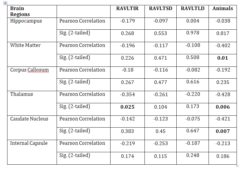
Correlation between brain regions Ktrans and
RAVLTIR, RAVLTSD, RAVLTLD, and Category fluency (animals). Correlation is significant with
p<0.05, in bold (2-tailed).
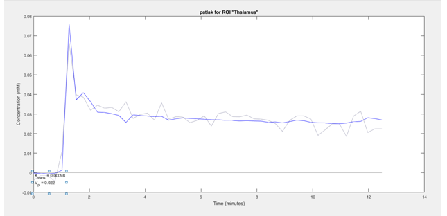
Thalamus with
high Ktrans=0.00098