1451
INVESTIGATING THE LINK BETWEEN CEREBROVASCULAR AND NEURO HEALTH IN COGNITIVE DECLINE1University of the Sunshine Coast, Sunshine Coast, Australia
Synopsis
Current markers of early neurodegeneration include β-amyloid plaque accumulation and Tau-mediated neuronal injury; both of which are thought to emerge prior to significant cognitive deficits and measurable brain atrophy. Unfortunately, the role of these biomarkers within the cascade of pathophysiological processes of AD remains poorly understood. Impairment in the glymphatic clearance system has garnered attention and is thought to represent a pathway to decline. Here we demonstrate that quantification of glymphatic clearance measures are feasible in healthy controls. Combining these measures with cognitive performance scores and diagnosis will provide insight into the role of the glymphatics system with decline.
BACKGROUND
It is becoming increasingly recognized that cardio and cerebro-vascular (CV) health is a contributing factor in the pathogenesis of AD, with most current clinical trials now employing some form of exercise intervention1-3. Results are promising, indicating that exercise can decrease AD specific atrophy and slow cognitive decline. Yet the association between CV and neuro health and neurodegeneration is still poorly understood. The glymphatic system, a recently discovered transport system, mediated by cerebral spinal fluid (CFS), that clears metabolic and cellular waste products in the brain provides a theoretical link between CV function, neuronal dysfunction and subsequent cognitive decline4. While there is growing recognition of the critical role this waste clearance system plays in maintaining normal brain health, there are very few non-invasive human imaging methods that characterize the glymphatic transport5.The combined neuro and cerebrovascular MR imaging (NCI) protocol presented here builds on the work of Taoka et al. 20175 to assess perivascular diffusivity and quantify the transport system as well as the mediators of clearance function, CFS flow and CV dynamics to evaluate the system as a whole. This imaging project will be implemented within the Healthy Brain Ageing (HBA) program at the Sunshine Coast Mind and Neuroscience Thompson Institute (SCMN-TI) to investigate the links between neuro- and cardio- health and cognitive decline. Here we present preliminary data in a healthy cohort to demonstrate feasibility.
METHODS
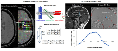
The NCI protocol was acquired in a pilot cohort of 8 healthy controls (4 female) aged between 21 and 59 on a 3-Tesla Siemens Skyra MRI (Germany, Erlangen) with a 64-channel head and neck receive coil recruited at SCMN-TI. In brief the protocol consists of MPRAGE: to assess differences in cortical and subcortical thickness and volume (hippocampal and prefrontal regions implicated in memory and executive function and shown to be atrophied in MCI and AD)6-8 to determine association with CV function, glymphatic clearance and cognitive decline. SWI: axial SWI to visualise and locate the parenchymal vessels and perivascular region of interest. DTI: diffusion weighted imaging to evaluate perivascular diffusivity of the glymphatic clearance system2. SWI scans was used to inform DTI ROI placement. DTI data is used to quantify the principle components of the diffusion tensor on a voxel-wise basis. Three ROIs are placed in the projection area (Proj - blue tract), the association area (Asso - red tract) and subcortical area (Subc - red tract) (see Figure 1). All three principle diffusivity components (Dxx, Dyy, Dzz) are then quantified for each ROI. Perivascular diffusivity is then quantified using the previously published APLS index5. Higher scores indicated impaired glymphatic. rsfMRI: resting-state functional MRI to quantify functional network signatures that distinguish diagnosis and subsequent correlation to cognitive function. CSF-PC: phase contrast imaging optimised for CSF flow rates to quantify CSF inflow through locations such as the cerebral duct (CD). 4DPC: 4D phase contrast imaging to quantify cerebral flow and haemodynamics such as pulse wave velocity (PWV)9 and turbulence within and around the circle of Willis. Association between haemodynamics, glymphatic clearance system and cognitive decline will then be investigated.Pilot data: Association between perivascular diffusivity and CSF-CD flow was evaluated to investigate the relationship between glymphatic clearance system measures and age.
RESULTS
Bivariate correlation analysis indicates that lower CD flow is associated with impaired glymphatic clearance (R2 = 0.30) and glymphatic clearance declines with age (R2 = 0.23). However, CD CSF flow is not directly associated with age indicating that CD CSF flow is a larger mediator of glymphatic function than just age (see Figure 2). Correlation between PWV, glymphatic clearance and CD CSF flow will provide further insight into the relationship between Glymphatic system function, CV haemodynamics and brain structure and function.CONCLUSION
Quantification of glymphatic clearance measures are feasible in a healthy cohort and show an association with age. Translating this combined neuro and CV MRI protocol, will, for the first time investigate the link between the two fluid dynamic systems (CV and glymphatics) and association with cognitive function in healthy ageing and MCI and impact of exercise intervention. Understanding the neuronal underpinnings of exercise-based gains will aid in targeted treatment approaches.Acknowledgements
No acknowledgement found.References
1. Gates, N.J., et al. Study of Mental Activity and Regular Training (SMART) in at risk individuals: a randomised double blind, sham controlled, longitudinal trial. BMC Geriatr 11, 19 (2011).
2. Ngandu, T., et al. A 2 year multidomain intervention of diet, exercise, cognitive training, and vascular risk monitoring versus control to prevent cognitive decline in at-risk elderly people (FINGER): a randomised controlled trial. Lancet 385, 2255-2263 (2015).
3. Northey, J.M., Cherbuin, N., Pumpa, K.L., Smee, D.J. & Rattray, B. Exercise interventions for cognitive function in adults older than 50: a systematic review with meta-analysis. Br J Sports Med (2017)
4. Jessen, N.A., Munk, A.S., Lundgaard, I. & Nedergaard, M. The Glymphatic System: A Beginner's Guide. Neurochem Res 40, 2583-2599 (2015).
5. Taoka, T., et al. Evaluation of glymphatic system activity with the diffusion MR technique: diffusion tensor image analysis along the perivascular space (DTI-ALPS) in Alzheimer's disease cases. Jpn J Radiol 35, 172-178 (2017).
6. Apostolova, L.G., et al. Conversion of mild cognitive impairment to Alzheimer disease predicted by hippocampal atrophy maps. Arch Neurol 63, 693-699 (2006).
7. Apostolova, L.G., et al. Subregional hippocampal atrophy predicts Alzheimer's dementia in the cognitively normal. Neurobiol Aging 31, 1077-1088 (2010).
8. Costafreda, S.G., et al. Automated hippocampal shape analysis predicts the onset of dementia in mild cognitive impairment. Neuroimage 56, 212-219 (2011).
9. Broadhouse, K. Quantification of aortic pulse wave velocity in preterm infants using 4D phase contrast MRI. Journal of Cardiovascular Magnetic Resonance 15(2013).
Figures


Glymphatic system-CSF relationship with healthy ageing. Eight healthy controls (4 female) 21-59 years were scanned. Bivariate correlation analysis indicates that lower CD flow is associated with impaired glymphatic clearance (left) and glymphatic clearance declines with age (middle). However, CD CSF flow is not directly associated with age indicating that CD CSF flow is a larger mediator of glymphatic function than just age.