1440
Development and evaluation of a high spatial resolution diffusion tensor template of the older adult human brain1Biomedical Engineering, Illinois Institute of Technology, Chicago, IL, United States, 2Rush Alzheimer's Disease Center, Rush University Medical Center, Chicago, IL, United States
Synopsis
A high-resolution older adult brain template containing full diffusion tensor (DT) information has not been constructed. Available DT templates of low spatial resolution lack fine details, and most are not representative of the older adult brain. Both factors limit spatial normalization accuracy in studies of aging. This work a) introduced a novel approach for constructing a DT template with high spatial resolution based on principles of super-resolution, and using this technique, b) developed and quantitatively evaluated a DT template of the older adult brain with high spatial resolution using high-quality data from 202 non-demented older adults.
Introduction
Although high spatial resolution diffusion tensor imaging (DTI) studies of the aging human brain are on the rise, a high-resolution older adult brain template containing full diffusion tensor information has not been constructed. Available templates of low spatial resolution lack fine details due to partial volume effects, and most are not representative of the older adult brain. Both factors limit spatial normalization accuracy in studies of aging. The purpose of this work was to a) introduce a novel approach for constructing a DTI template with high spatial resolution based on principles of super-resolution, and using this technique, to b) develop a DTI template of the older adult brain with high spatial resolution using high-quality data from 202 non-demented older adults. The new template was quantitatively compared to available templates in terms of image quality and spatial normalization accuracy achieved when used as reference for alignment of DTI data from older adults.Methods
Data:DTI data of 2mm isotropic resolution collected on 202 non-demented older adults (65-95 age-range, 50% male) participating in the Rush Memory and Aging Project1 were used in this work. The data were processed using TORTOISE2 and were used in the following method to develop a super-resolved DTI template of the older adult brain.
Template construction approach:
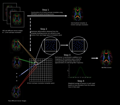
The proposed method for constructing a high spatial resolution DTI template consisted of the following steps (Fig.1):
Step 1: Raw DTI data of 2mm isotropic resolution from all participants were spatially normalized in a 0.5mm isotropic resolution space using DRTAMAS3
Step 2: The resulting non-linear deformations were utilized to map the signals from the raw space to exact physical locations in the 0.5mm template space. This process eliminated interpolations incurred in conventional template building methods.
Step 3: In addition to the spatial signal relocation, each tensor in template space was reoriented based on the rotation information of the corresponding deformation field.
Step 4: The final tensor in a 0.5mm isotropic voxel in template space was calculated as the weighted average of all tensors included in that voxel. The weights were derived using a Gaussian kernel with a standard deviation equal to the standard deviation of the diffusion tensor trace values included in the template space voxel, and centered at the median trace value included in that voxel. This approach is less sensitive to the effects of residual misregistration.
Evaluation:
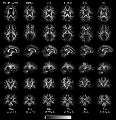
The resulting high-resolution DTI template is part of the 0.5mm version of the MIITRA atlas4 (www.nitrc.org/projects/miitra) and is referred to here as the MIITRA 0.5mm DTI template. The new template was compared to 5 publicly available DTI templates: ICBM815, NTU6, IIT v5.07, EVE8 of 1mm isotropic resolution and IXI9 of resolution 1.75mm X 1.75mm X 2.25mm (Fig.2). First, fractional anisotropy (FA) maps of all templates were compared by visual inspection and also in terms of image sharpness demonstrated by the normalized power spectral density in all axes. Next, the accuracy of inter-subject DTI spatial normalization was compared using each template as reference for normalization of DTI data from 90 non-demented ADNI10 participants. Normalization accuracy was assessed by means of the standard deviation of FA and trace, coherence of primary eigenvectors (COH), average Euclidean distance of diffusion tensors (DTED) and total variance of the diffusion tensors (TVDT) in white matter.
Results
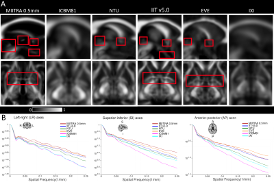
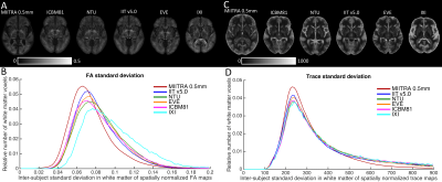
Fine white matter structures such as the a) anterior commissure, b) posterior commissure, c) inter-thalamic adhesion and d) optic chiasm were clearly resolved in the MIITRA 0.5mm DTI template (Fig.3A). In contrast, these features were either less visible in some of the 1mm templates, or not present in others (Fig.3A). Image sharpness assessed by means of the high spatial frequency content in the normalized power spectra was higher in the FA maps of MIITRA 0.5mm template, IIT v5.0 and EVE compared to other templates (Fig.3B). The MIITRA 0.5mm template was free of image artifacts. Artifacts in ICBM81, NTU and EVE have been pointed out in the literature7. Finally, MIITRA 0.5mm allowed higher inter-subject spatial normalization accuracy for DTI data from older adults compared to other templates (Figs.4,5). This was demonstrated by the higher relative number of white matter voxels with low FA and trace standard deviation (Fig.4A,B,C,D), high COH (Fig.5A,B), and low DTED and TVDT (Fig.5C,D,E,F) for registration to MIITRA 0.5mm compared to other templates.Discussion
The new MIITRA 0.5mm DTI template is the first high-resolution population-based template of the older adult brain. This work demonstrated that the MIITRA 0.5mm template exhibits high image quality, high sharpness, is free of artifacts, and resolves fine white matter structures. Furthermore, the MIITRA 0.5mm template provided higher spatial normalization accuracy of older adult DTI data compared to other available templates. This was due to the higher image quality and the fact that it was more representative of the older adult brain than other templates (NTU, IIT v5.0, EVE are all young adult templates).Conclusion
The findings of this work have important implications in regard to template selection in DTI studies on older adults. The MIITRA 0.5mm template is a high-quality, high-resolution diffusion tensor template of the older adult brain that provides higher DTI spatial normalization accuracy than other available templates even for normalization of lower resolution older adult data.Acknowledgements
This study was supported by National Institutes of Health grant R01AG052200, P30AG010161, UH2NS100599, UH3NS100599, R01AG064233, R01AG17917References
1. Bennett DA, et al. Curr Alzheimer Res. 2012;9:646-663.
2. Pierpaoli C, et al. TORTOISE: an integrated software package for processing of diffusion MRI data. Proc. Intl. Soc. Mag. Reson. Med. 2010;18:1597.
3. Irfanoglu MO, et al. DR-TAMAS: diffeomorphic registration for tensor accurate alignment of anatomical structures. Neuroimage 2016;132:439–454.
4. Ridwan AR, et al Evaluation of standardized as well as study-specific and age-specific structural T1-weighted brain templates for use in studies on older adults. Proc. Int. Soc. for Magn. Reson. In Med. 2019
5. Mori S, et al. Stereotaxic white matter atlas based on diffusion tensor imaging in an ICBM template. Neuroimage 2008;40(2):570-582
6. Hsu YC, et al. NTU-DSI-122: A diffusion spectrum imaging template with high anatomical matching to the ICBM-152 space. Hum Brain Mapp. 2015; 36(9):3528-41.
7. Zhang S, Arfanakis K, Evaluation of standardized and study-specific diffusiontensor imaging templates of the adult human brain: template characteristics, spatialnormalization accuracy, and detection of small inter-group fa differences.Neuroimage 2018; 172:40–50.
8. Oishi K, et al. Atlas-based whole brain white matter analysis using large deformation diffeomorphic metric mapping: application to normal elderly and Alzheimer’s disease participants. Neuroimage. 2009;46:486–99.
9. Zhang, H., et al. A computational white matter atlas for aging with surface-based representation of fasciculi. Biomedical Image Registration. WBIR 2010. Lecture Notes in Computer Science, vol 6204.
10. http://adni.loni.usc.edu/
Figures

Figure 1: Schematic of the approach for constructing a high-resolution DTI template.

Figure 2: Axial, sagittal and coronal slices of the FA maps of MIITRA 0.5mm DTI template, ICBM81, NTU, IIT v5.0, EVE and IXI.

Figure 3: A) Top row: Magnified sagittal FA maps at the mid sagittal plane. The anterior commissure, posterior commissure and optic chiasm are visible in the MIITRA 0.5mm DTI template and some of the other templates. In addition, MIITRA 0.5mm was able to resolve the inter-thalamic adhesion. Bottom row: Magnified axial view of the anterior commissure.
B) Normalized power spectra for the left-right (LR), superior-inferior (SI) and anterior-posterior (AP) axes.

Figure 4: Maps and histograms of the standard deviation in (A,B) FA and (C,D) trace across spatially normalized older adult DTI data after registration to the different templates.

Figure 5: Maps and histograms of the (A,B) COH, (C,D) DTED, and (E,F) TVDT across spatially normalized older adult DTI data after registration to the different templates.