1439
Multi-component relaxation in relapsing remitting multiple sclerosis:Variability of myelin water fraction estimates of individual lesions
1Institut und Poliklinik für Diagnostische und Interventionelle Neuroradiologie, University Hospital Carl Gustav Carus, Dresden, Germany, 2Neurology, University Hospital Carl Gustav Carus, Dresden, Germany
Synopsis
Multiple Sclerosis lesions are pathologically heterogeneous. The Multi-component relaxation method (mcDESPOT) which allows the determination of relative myelination by estimating the myelin water fraction (MWF) could be valuable to capture the high variability of disease development. In a pilot study n=112 RRMS patients got MRI examination and MWF values were determined of white matter (WM) and individual lesions. The MWF of WM was 0.230 for healthy controls and 0.224 for RRMS patients. 2787 WM FLAIR lesions were examined. Patients with a higher degree of disability of EDSS ≥ 4 significantly have lower MWF values than patients with EDSS < 2.5.
INTRODUCTION
Myelin Water Imaging (MWI) bears great potential as a biomarker of Multiple Sclerosis (MS) inflammatory myelin loss and allows detecting the in vivo relative myelin content of single MS lesions. However, such MS lesions are pathologically heterogeneous and the net effect of demyelination and/or remyelination of single lesions can be very different. Further the lesion demyelination variations within MS cohorts are unknown. However, this may be a prerequisite to interpret single patient lesion demyelination as their individual subclinical disease activity. The aim of this study was to assess the intra-individual and inter-individual variability of myelin water fraction (MWF) in relapsing-remitting MS (RRMS) white matter (WM) lesions.METHODS
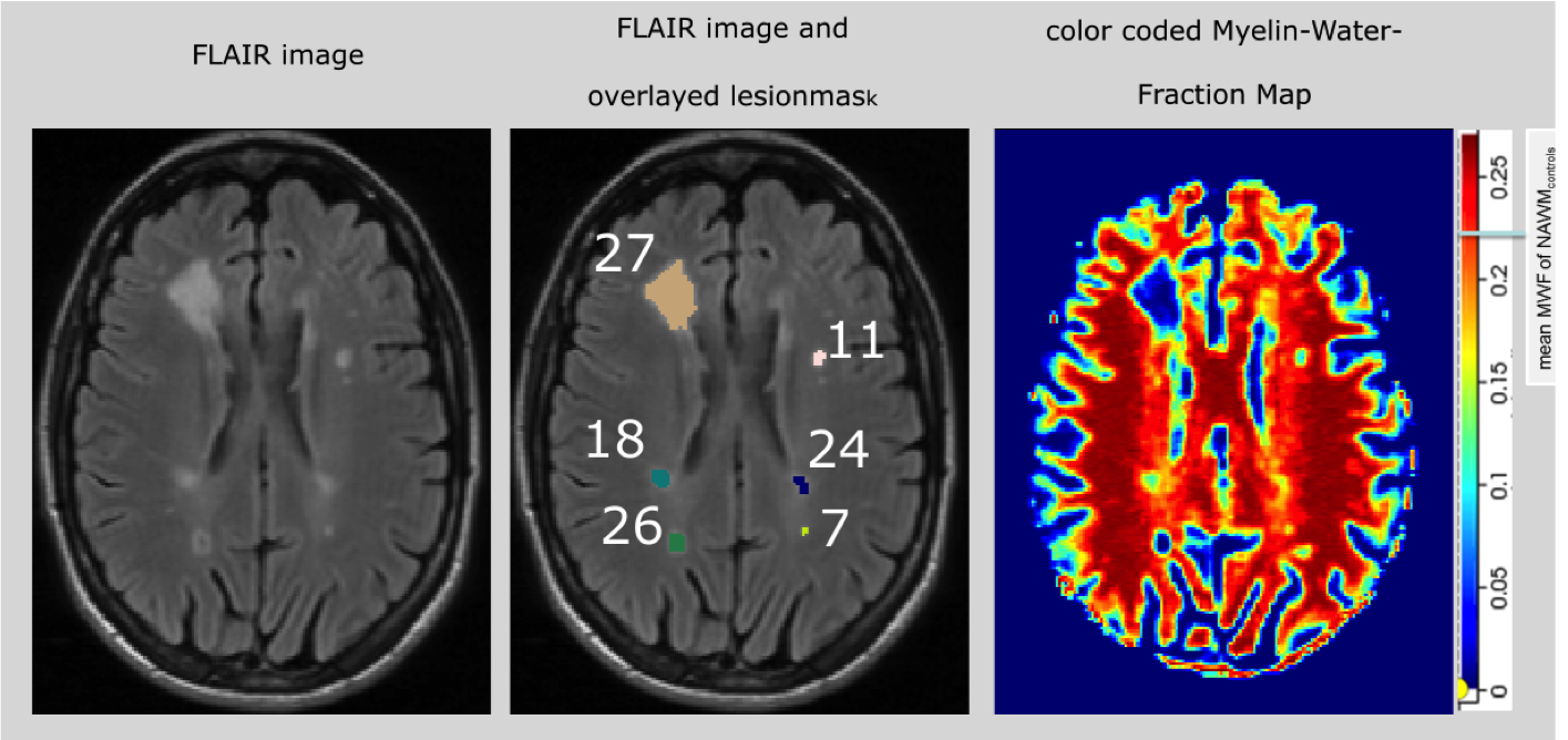
In a pilot study 112 RRMS patients (mean age = 38, mean EDSS= 2.7) and 63 age-matched healthy controls got conventional MRI and quantitative multi-component relaxation imaging derived from mcDESPOT. DATA ACQUISITION: A 1.5T MR scanner (Siemens Magnetom Sonata equipped with a 8-channel radio-frequency coil was used and sets of 2D-T1 spin echo (T1w), 3D Fluid-Attenuated Inversion Recovery (FLAIR), Spoiled Gradient Recalled (SPGR) and Balanced Steady- State Free-Precession (bSSFP) images were obtained. According to the mcDESPOT protocol SPGR and bSSFP scans were acquired over a range of flip angles at constant echo time (TE) and repetition time (TR) using the following specifications: field of view (FOV) = 22 cm, slice thickness = 1.7 mm; SPGR: TE/TR = 2.0/ 5.7 ms, flip angle (α) = [5, 6, 7, 8, 9, 11, 13, 18]°; bSSFP: TE/ TR = 1.71/3.42 ms, α = [9, 14, 19, 24, 28, 34, 41, 51, 60]° Data post-processing was executed by sets of python scripts to automate the FMRIB Software Library (FSL). MWF maps were calculated from the SPGR and bSSFP data using the established mcDESPOT theory and processing method [1]. A semi-automatic method was used to generate binary masks of WM, normal appearing white matter (NAWM) and MS lesions. In order to obtain individual lesion parameters, the binary lesion mask was automatically labelled and these individual labels served as region of interest to quantify their MWF (Fig. 3).RESULTS
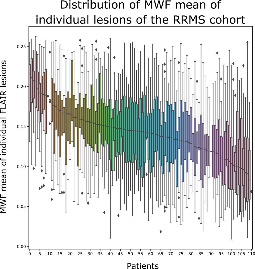
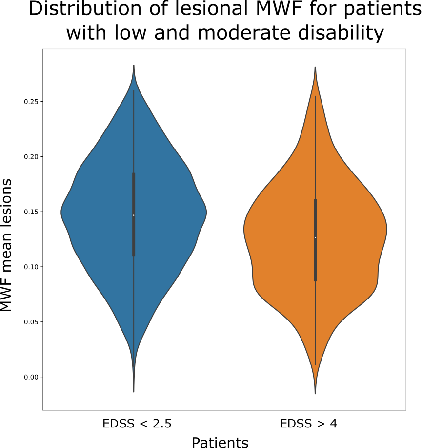
The median MWF of white matter (WM) was 0.23 for healthy controls and 0.224 for RRMS patients. 2787 WM FLAIR lesions were examined. The median MWF loss within the lesions varied from 6% to 70% for different patients compared to MWF white matter levels of the controls (Fig. 1). In a group comparison it could be shown that patients with a higher degree of disability of EDSS ≥ 4 significantly (p<0.05) have lower MWF values than patients with EDSS < 2.5 (Fig. 2).DISCUSSION
The analysis of the intra-subject variability of MWF in lesions showed that almost all patients had both lesions with a major, intermediate and minor MWF loss. For different patients, however, the severity and amount of lesions with MWF loss varied. While MWF was preserved to a greater extent in a smaller subset of patients, the majority of patients had a significant MWF loss within their T2L.CONCLUSION
A significant reduction and heterogeneity of MWF in MS lesions was detected within patients and between patients, reflecting varying degrees of demyelination.Acknowledgements
References
[1] Deoni SC, Rutt BK, Arun T, Pierpaoli C, et al., Gleaning multicomponent T1 and T2 information from steady-state imaging data. Magn Reson Med. 2008; 6:1372-87
[2] Laule C, Vavasour I. Kolind SH, et al., Magnetic resonance imaging of myelin. Neurotherapeutics. 2007 Jul;4(3):460-84
Figures