1424
Network alterations at rest in multiple sclerosis patients with tremor1Neuroscience, Monash University, Melbourne, Australia, 2University of Melbourne, Melbourne, Australia, 3Royal Melbourne Hospital, Melbourne, Australia, 4Bionics Institute, Melbourne, Australia, 5Monash University, Melbourne, Australia, 6University of Tübingen, Tübingen, Germany
Synopsis
Almost half of patients with multiple sclerosis (MS) experience tremor, which significantly worsens disability. Pathophysiology studies of MS tremor have highlighted the importance of the cerebello-thalamo tract. This study aimed to use resting-state fMRI to identify brain networks that are dysfunctional in MS tremor. We found significantly higher connectivity within the motor network in tremor patients compared to controls, and the mean activation within the motor network was negatively correlated to tremor. Resting-state fMRI could provide useful markers for studying the pathophysiology of tremor in MS.
Introduction
Tremor is defined as an ‘involuntary, rhythmic, oscillatory movement of a body part’, 1 and it is reported in approximately 45% of people with MS. 2 The presence of tremor significantly impacts patients’ quality of life, with more than half of patients with mild tremor being unemployed. 2, 3 Recent studies using both structural MRI and task-based fMRI, showed the importance of atrophy along the cerebello-thalamic tract and sensorimotor neuroplasticity. 4, 5 However, no studies have examined functional connectivity in the context of MS tremor. This study aimed to examine potential connectivity differences in the sensorimotor network between patients with tremor compared to health and MS control subjects.Methods
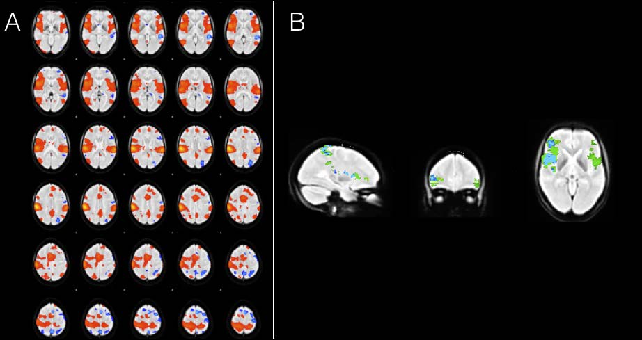
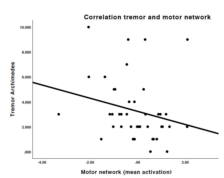
Fifteen healthy controls, 27 MS tremor patients and 42 MS control patients without tremor were included. Tremor was quantified using the Bain score (0–10) for overall severity, handwriting and Archimedes spiral drawing. A 5 min resting-state sequence was performed on a 3T MRI system (Trio, Siemens, Erlangen; voxel size: 2.5x2.5x2.5mm and 1.5s repetition time). Pre-processing of the data was performed using FSL FIX and MATLAB to remove noise and motion parameters from the signal, and a high-pass temporal filter with a cut-off point of 100 s was applied. We applied group-ICA analysis (FSL, MELODIC) to identify the sensorimotor network across all subjects. Subsequently, dual regression was performed to identify differences in functional connectivity within the sensorimotor network between groups. Significant voxels were identified based on threshold-free cluster enhanced p < 0.05. Mean connectivity (normalised beta weight from regression against IC timecourse) within the network was correlated to tremor severity using Spearman correlation.Results
Compared to MS controls, the sensorimotor network in tremor patients was larger with a small number of significant voxels in left inferior frontal lobe. No significant differences were detected between healthy controls and either MS controls or MS tremor patients. Interestingly, within the MS tremor patients the mean activation correlated with the tremor Archimedes spiral drawing (rho=-0.352, p=0.022; Fig. 3). No correlation was found with the other two tremor measures.Discussion
In this study we found increased connectivity within the sensorimotor network in MS tremor patients compared to MS controls that was inversely correlated with clinical tremor severity.Conclusion
This study demonstrated the use of resting-state fMRI in characterizing the pathophysiology of tremor in MS.Acknowledgements
No acknowledgementsReferences
1. Deuschl G, Bain P, Brin M. Consensus statement of the Movement Disorder Society on Tremor. Ad Hoc Scientific Committee. Movement disorders : official journal of the Movement Disorder Society 1998;13:2-23.
2. Rinker JR, Salter AR, Walker H, Amara A, Meador W, Cutter GR. Prevalence and characteristics of tremor in the NARCOMS multiple sclerosis registry: a cross-sectional survey. BMJ Open 2015;5.
3. Koziarska D, Król J, Nocoń D, Kubaszewski P, Rzepa T, Nowacki P. Prevalence and factors leading to unemployment in MS (multiple sclerosis) patients undergoing immunomodulatory treatment in Poland. PloS one 2018;13:e0194117.
4. Boonstra FMC, Noffs G, Perera T, et al. Functional neuroplasticity in response to cerebello-thalamic injury underpins the clinical presentation of tremor in multiple sclerosis. Manuscript sumbitted for publication 2018.
5. Boonstra F, Florescu G, Evans A, et al. Tremor in multiple sclerosis is associated with cerebello-thalamic pathology. Journal of neural transmission (Vienna, Austria : 1996) 2017;124:1509-1514.
Figures