1421
Simultaneous T2 and T2* Mapping of Multiple Sclerosis Lesions with a Radially Sampled RARE-EPI Hybrid1Berlin Ultrahigh Field Facility (B.U.F.F.) Max Delbrück Center For Molecular Medicine in the Helmoltz Association, Berlin, Germany, 2Experimental und Clinical Research Center (ECRC), Charité-Campus Berlin-Buch, Berlin, Germany, 3NeuroCure Clinical Research Center, Charité – Universitätsmedizin, Berlin, Germany, 4Department of Neurology, Charité – Universitätsmedizin, Berlin, Germany
Synopsis
MRI examinations commonly involve a series of multiple imaging contrasts and MR-metrics, resulting in long examination times and propensity to motion and slice misregistration. Dual or multi-contrast techniques offer substantial scan time reduction. Recognizing this opportunity this work presents a dual contrast RARE-EPI hybrid, that provides T2 (RARE module) and T2* (EPI module) contrast and facilitates simultaneous T2 and T2* mapping in a single radially (under)sampled scan (2-in-1 RARE-EPI). The applicability of 2-in-1 RARE-EPI is demonstrated in an in vivo feasibility study involving patients suffering from multiple sclerosis and healthy controls and benchmarked versus conventional T2 and T2* weighted/mapping techniques.
Introduction
MRI protocols tailored for the diagnosis and longitudinal monitoring of multiple sclerosis (MS) comprise several imaging contrasts including T1, DWI, T2, and T2* [1, 2]. T2-weighted imaging (T2-WI) is frequently used in clinical practice for identification of MS lesions, which are characterized by hyperintense appearance [2]. T2*-weighted imaging revealed a central vein sign, which is specific for MS lesions [1,3]. Furthermore T2* is sensitive to iron deposition, which may relate to disease duration and activity [1,4]. Recognizing these features of MS lesions it is conceptually appealing to pursue MRI mapping techniques that are sensitive to multiple contrast mechanisms and support simultaneous generation of quantitative maps of multiple contrasts [5] with the goal to enhance imaging speed, to reduce slice misregistration, to lower the propensity to bulk motion and to boost clinical utility. To facilitate simultaneous T2 and T2* mapping we proposed a radially (under)sampled RARE-EPI hybrid (2-in-1 RARE-EPI) [6].Here, we carefully examine the applicability of 2-in-1 RARE-EPI in an in vivo study involving healthy controls and patients with MS. For validation of T2 and T2* mapping, 2-in-1 RARE-EPI is benchmarked versus conventional T2 and T2* mapping techniques.
Methods
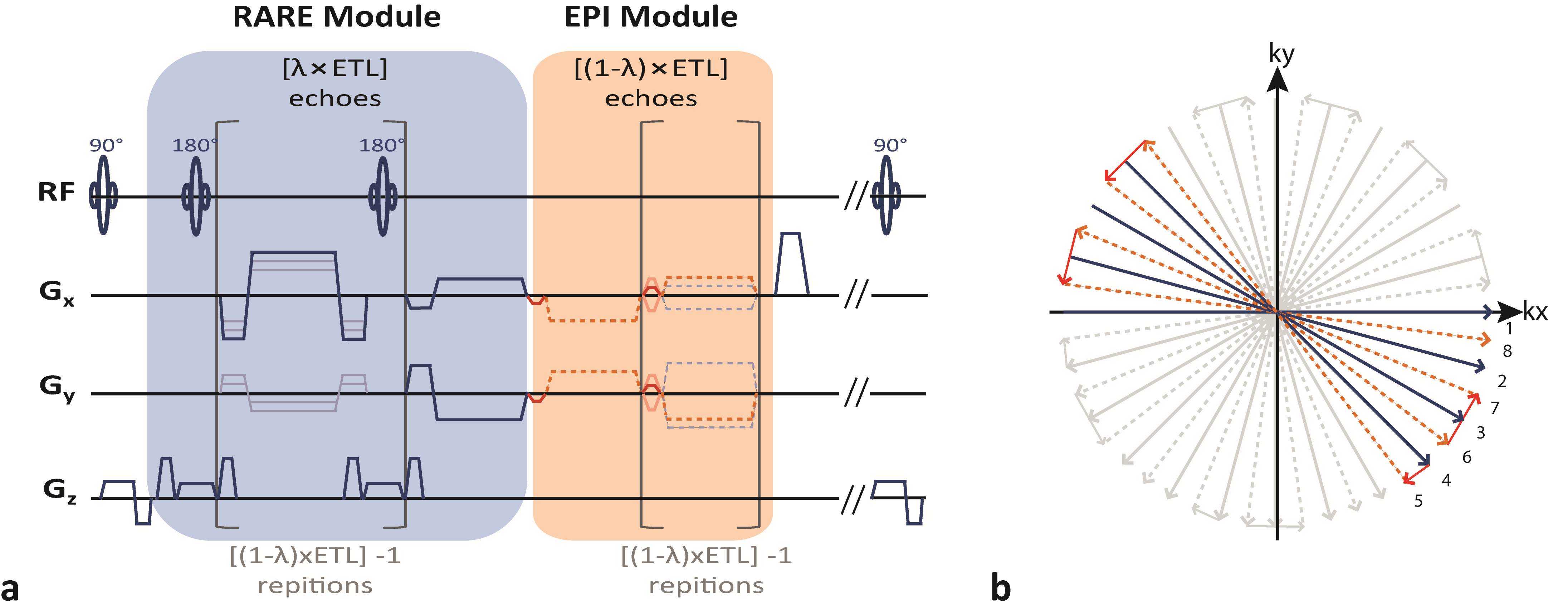
2-in-1 RARE-EPI is based on a RARE-EPI hybrid [7]. The first echoes in the echo train are acquired with a RARE module, later echoes are acquired with an EPI module (Figure 1, left), with the combined-acquisition-technique (CAT) factor λ governing the fraction of RARE echoes in the echo train. Here we used an echo train length of 16 with 8 RARE and EPI echoes (λ=0.5), acquired in an interleaved manner with respect to the angular position in radially sampled k-space (Figure 1b). To reduce motion propensity of individual TE images (used for mapping) the spokes of each TE are acquired to cover the whole π radians of k-space over a cycle of four TRs. Gradient delay artifacts were corrected using the method proposed by Block et al. [8]. For image reconstruction regridding with linear density compensation [9] and a magnitude sum-of-squares channel combination were employed.Reference T2 and T2* maps were acquired with conventional multi-spin-echo (MSE) and multi-gradient-echo (MGRE) techniques. T2 and T2* maps were calculated with a linear least-square-fit of the logarithmic signal magnitude. For T2* mapping a spatial-adaptive-non-local-means filter was applied to the images prior to the fitting to improve the fit quality [10].
Images were acquired at 3.0 T (Siemens Magnetom SkyraFit, Erlangen, Germany) using a 32-channel head coil (Siemens, Erlangen, Germany) for signal reception and the body coil for transmission.
For the in vivo study we scanned MS patients (sex: male, mean age: 42 years) and healthy controls (sex: male, mean age: 42 years). MS specific lesions were selected to be imaged with the 2-in-1 RARE-EPI, based on a whole brain coverage scan with a pd-weighted RARE sequence.
Imaging parameters: FoV = 256 mm, base resolution = 256, TR = 500 ms, TA(2-in-1 RARE-EPI/MSE/MGRE) = 1:58/2:12/2:10 min, ESP(RARE-module/MSE) = 9.94 ms, ESP(EPI-module/MGRE) = 2.27 ms, ETL(2-in-1 RARE-EPI/MSE/MGRE) = 16/12/12, No. of Spokes(2-in-1 RARE-EPI) = 3200 (twofold undersampling for individual TE images).
Results
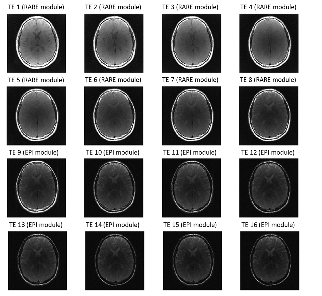
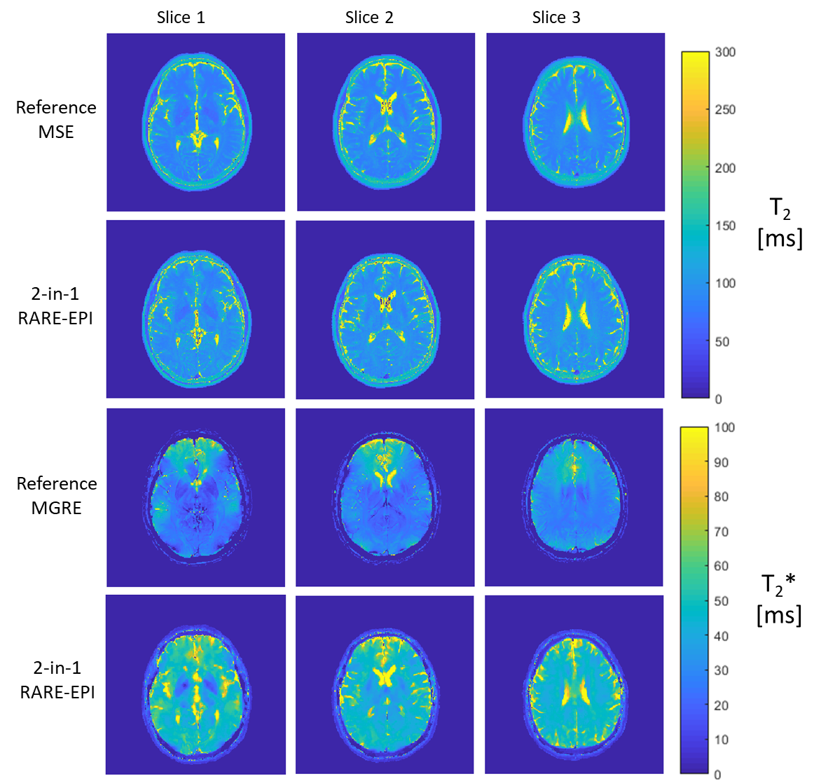
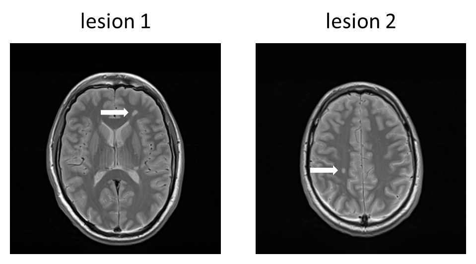
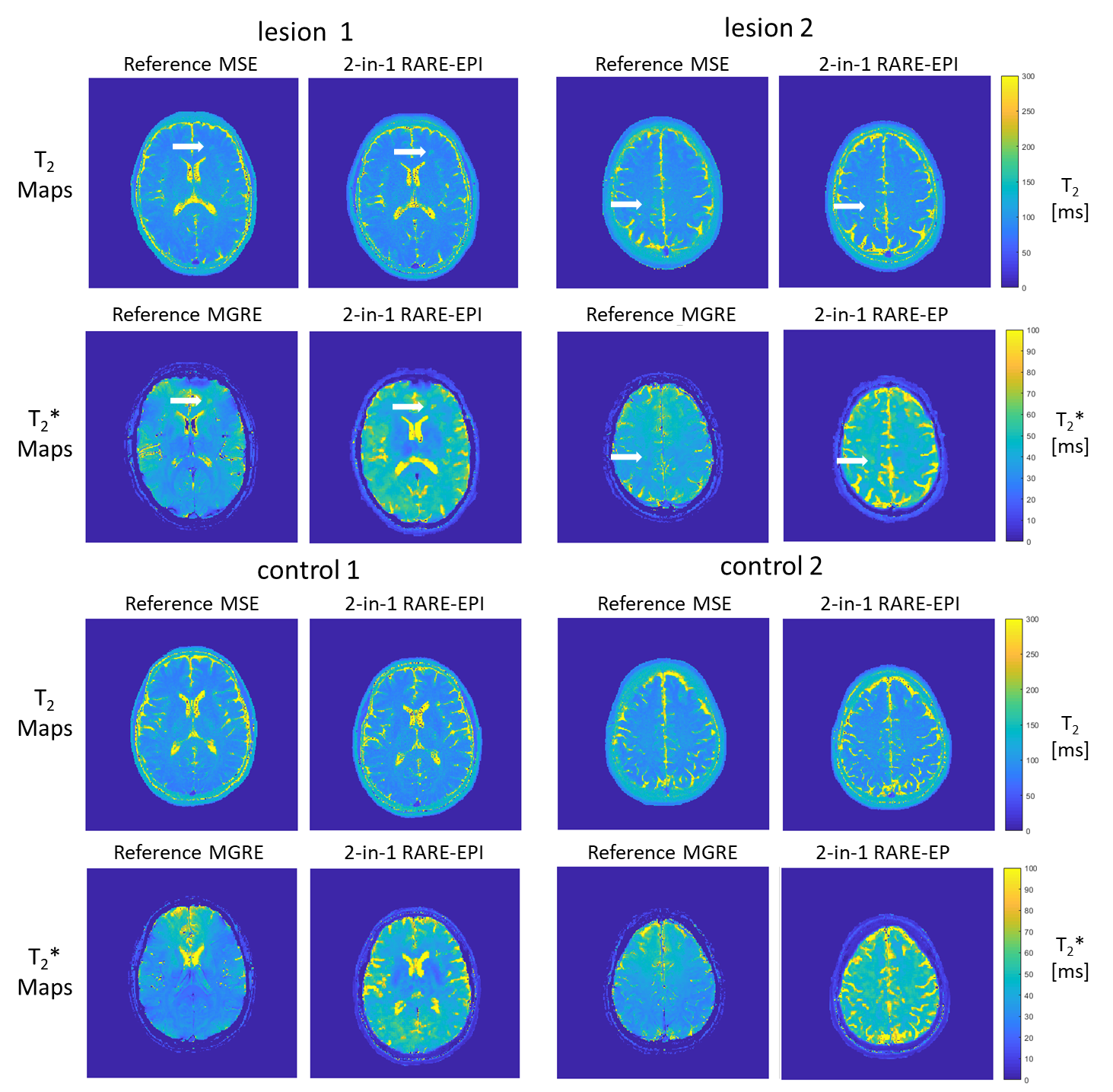
To demonstrate the feasibility of 2-in-1-RARE-EPI in vivo, three brain slices covering the corpus callosum (bottom edge, upper edge and center) were acquired. Figure 2 shows a series of 16 individual TE images covering the center of the corpus callosum of a healthy subject. Each individual-TE image was reconstructed from echoes acquired at the same TE. T2 and T2* maps obtained with 2-in-1 RARE-EPI are depicted in Figure 3 along with the reference maps obtained with MSE and MGRE for all three slices covering the corpus callosum. The T2 and T2* maps obtained with the 2-in-1-RARE-EPI are in accordance with the reference maps.Figure 4 shows proton density weighted images of two MS lesions selected to be imaged with 2-in-1 RARE-EPI. The T2 and T2* maps highlighting the two exemplary MS lesions are depicted in Figure 5 along with the T2 and T2* reference maps. Both lesions can be detected and delineated in the T2 and T2* maps obtained from the 2-in-1 RARE-EPI which is confirmed by the reference maps derived from MSE/MGRE.
Discussion and Conclusion
This work demonstrates the feasibility of radially sampled 2-in-1 RARE-EPI for dual contrast weighted (T2/T2*) imaging and for simultaneous T2 and T2* mapping for the detection of MS lesions. MS lesions detected in the T2 and T2* maps obtained with 2-in-1-RARE-EPI were confirmed by the reference T2 and T2* maps deduced from MSE/MGRE. Our undersampled 2-in-1 RARE-EPI hybrid approach substantially decreases acquisition time for T2 and T2*mapping by a factor of two versus conventional mapping methods and hence permits whole brain coverage in a reasonable scan time.Simultaneous T2 and T2* mapping with 2-in-1 RARE-EPI promises to support the differential diagnosis of MS and other (orphan) neurodegenerative diseases. However, our 2-in-1 RARE-EPI hybrid is not limited to MS lesions or brain imaging, as shown in this work. 2-in-1 RARE-EPI offers the capacity for simultaneous T2 and T2* mapping in applications dealing with bulk or physiological motion such as cardiac imaging including T2 imaging/mapping of myocardial edema and myocardial T2* mapping in HCM patients [11], as well as MRI of the eye, abdomen and liver.
Acknowledgements
No acknowledgement found.References
1. Hemond C et al., Magnetic resonance imaging in multiple sclerosis. Cold Spring Harbor perspectives in medicine, 2018. 8(5): p. a028969.
2. Zivadinov R. et al., Role of MRI in multiple sclerosis I: inflammation and lesions. Front Biosci, 2004. 9(665): p. C28.
3. Tallantyre E.C., et al., A comparison of 3T and 7T in the detection of small parenchymal veins within MS lesions. Investigative Radiology, 2009. 44(9): p. 491-494.
4. Bozin I., et al., Magnetic resonance phase alterations in multiple sclerosis patients with short and long disease duration. Public Library of Science one, 2015. 10(7): p. e0128386.
5. Fuchs K., et al., Simultaneous dual contrast weighting using double echo rapid acquisition with relaxation enhancement (RARE) imaging. Magnetic Resonance in Medicine, 2014. 72(6): p. 1590-1598.
6. Herrmann C., et al. Dual Contrast Weighting and Simultaneous T2 and T2* Mapping with Radially Sampled RARE-EPI. in Proc Int Soc Magn Reson Med Sci Meet Exhib 27. 2019.
7. Paul K., et al., Multiband diffusion‐weighted MRI of the eye and orbit free of geometric distortions using a RARE‐EPI hybrid. NMR in Biomedicine, 2018. 31(3): p. e3872.
8. Block K.T. and M. Uecker. Simple Method for Adaptive Gradient-Delay Compensation in Radial MRI. in Proc Int Soc Magn Reson Med Sci Meet Exhib 19. 2011.
9. Fessler, J., B. Sutton, and Y. Zhang. Michigan Image Reconstruction Toolbox. Available from: https://web.eecs.umich.edu/~fessler/code/
10. Manjón, J.V., et al., Adaptive non‐local means denoising of MR images with spatially varying noise levels. Journal of Magnetic Resonance Imaging, 2010. 31(1): p. 192-203.
11. Huelnhagen T., et al., Myocardial T2* mapping with ultrahigh field magnetic resonance: Physics and frontier applications. Frontiers in Physics, 2017. 5: p. 22.
Figures