1415
Combined 3D FLAIR and T2-weighted (FLAIR3) improves the detection of infratentorial lesions in multiple sclerosis1Diagnostic and Interventional Imaging, University of Texas Health Science Center at Houston, Houston, TX, United States, 2Neurology, University of Texas Health Science Center at Houston, Houston, TX, United States
Synopsis
Infratentorial lesions play an important role in the diagnosis of multiple sclerosis (MS), but are challenging to detect on conventional FLAIR protocols. Synthetic FLAIR3 images were generated by combining FLAIR and T2-weighted images to improve lesion contrast while maintaining CSF suppression. We evaluated FLAIR3 for detecting infratentorial lesions in 10 patients with MS. Our results show 11-fold increase in the lesion contrast-to-noise ratio and 163% increase in the number of detected lesions using FLAIR3 as compared to FLAIR.
Introduction
Multiple sclerosis (MS) is the most common non-traumatic demyelinating disease in young adults, affecting about 2.5 million persons worldwide.1 Diagnosis of MS requires demonstration of the dissemination of lesions in time and space.2 The dissemination in space criterion is established by detection of one or more T2-hyperintense lesions characteristic of MS in two or more of four areas of the CNS: periventricular, juxtacortical, and infratentorial brain regions, and spinal cord.2 Fluid-attenuated inversion recovery (FLAIR) is the most common imaging sequence used for identifying brain T2-hyperintense lesions. However, the contrast of infratentorial lesions is suboptimal on FLAIR images due to the partial T1 weighting and the different relaxation properties between supratentorial and infratentorial regions.Advanced methods to improve detection of infratentorial lesions include optimizing 3D FLAIR scan parameters3, and combining T2 and proton density weighted images.4 A previous study has shown substantial improvement in lesion contrast by combing 3D FLAIR and 3D T2-weighted images5, but performance for infratentorial lesions was not specifically addressed. In this work, we investigated whether the detection of infratentorial lesions can be improved with FLAIR3.
Methods
This is a prospective study that included 10 patients with MS. The MRI protocol included 3D sagittal turbo spin echo (TSE) FLAIR (FOV = 256×256×180 mm3; voxel size = 1×1×1 mm3; TR/TI/TE=4800/1650/300 ms; 125-ms T2-preparation; scan time 5:31 min) and 3D TSE T2w (TR/TE = 2500/252 ms; scan time 4:33 min) with matching coverage and resolution. After acquisition, the FLAIR and T2w images were co-registered using a rigid-body transformation and combined to produce the FLAIR3 image as follows5: FLAIR3 = FLAIR1.55 × T2w1.45. To improve the dynamic range of the reconstructed FLAIR3 image, intensity uniformity correction was used.6Two neuroimaging experts in neuroradiology and MS neurology examined the images, in a random order, and identified all infratentorial and upper cervical lesions seen on FLAIR and FLAIR3. Regions of interest were placed in the identified lesions and in adjacent normal-appearing white matter, and the contrast-to-noise ratio (CNR) was calculated.
Results
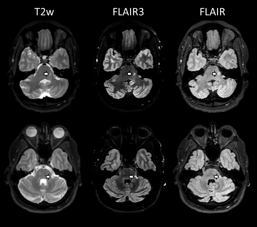
Figure 1 shows slices from FLAIR, T2w, and reconstructed FLAIR3 images from two MS patients, demonstrating the improved lesion contrast in the infratentorial region on FLAIR3. The first/second expert identified 18/20 lesions on FLAIR, and 43/57 lesions on FLAIR3. Higher lesion CNR for the infratentorial lesions was observed on FLAIR3 compared to FLAIR (rater 1: 73±114 vs. 5.4±2.4, P<0.001, Rater 2: 88±125 vs. 7.7±3.8, P<0.0001). Both neuroimaging experts qualitatively found an improvement in the infratentorial lesion visualization on FLAIR3.Discussion
Sensitive detection of infratentorial lesions can increase the diagnostic confidence in MS. Synthetic images combining two or more conventional images provide images with higher lesion conspicuity compared to the source images.4,5,7,8 In this study, FLAIR3 detected a significantly larger number of infratentorial lesions compared to FLAIR, and lesions on FLAIR3 had significantly higher CNR. These promising results suggest that FLAIR3 may be a good option for assessing brain lesions, including infratentorial lesions, in MS.Similar to other image-combining methods, FLAIR3 could be susceptible to image registration errors or artifacts. To minimize these errors, it is recommended that the FLAIR and T2w sequences be executed in succession with minimal delay. Interleaving the sequences may eliminate the need for co-registration9, greatly simplifying FLAIR3 reconstruction. Further qualitative and quantitative assessment in a larger cohort will be conducted to assess the performance of FLAIR3.
Conclusion
FLAIR3 significantly improves the visualization and detection of infratentorial MS lesions.Acknowledgements
We thank Vipulkumar Patel for help with the MRI experiments.References
1. Tullman MJ. Overview of the epidemiology, diagnosis, and disease progression associated with multiple sclerosis. Am J Manag Care. 2013;19:S15-20.
2. Thompson AJ, Banwell BL, Barkhof F, Carroll WM, Coetzee T, Comi G, Correale J, Fazekas F, Filippi M, Freedman MS, others. Diagnosis of multiple sclerosis: 2017 revisions of the McDonald criteria. Lancet Neurol. 2018;17:162–173.
3. Lecler A, El Sanharawi I, El Methni J, Gout O, Koskas P, Savatovsky J. Improving Detection of Multiple Sclerosis Lesions in the Posterior Fossa Using an Optimized 3D-FLAIR Sequence at 3T. Am J Neuroradiol. 2019;
4. Gaitán MI, Yañes P, Sati P, Romero C, Reich DS, Correale J. Optimal detection of infratentorial lesions with a combined dual-echo MRI sequence: “pT2.” Mult Scler. 2016;
5. Gabr RE, Hasan KM, Haque ME, Nelson FM, Wolinsky JS, Narayana PA. Optimal combination of FLAIR and T2-weighted MRI for improved lesion contrast in multiple sclerosis. J Magn Reson Imaging. 2016;44:1293–1300.
6. Li C, Gore JC, Davatzikos C. Multiplicative intrinsic component optimization (MICO) for MRI bias field estimation and tissue segmentation. Magn Reson Imaging. 2014;32:913–23.
7. Fujiwara Y, Inoue Y, Kanamoto M, Ishida S, Adachi T, Kimura H. The use of combined T2-weighted and FLAIR synthetic magnetic resonance images to improve white matter region contrast: a feasibility study. Radiol Phys Technol. 2019;12:118–125.
8. Wiggermann V, Hernandez-Torres E, Traboulsee A, Li DKB, Rauscher A. FLAIR2: A Combination of FLAIR and T2 for Improved MS Lesion Detection. Am J Neuroradiol [Internet]. 2015;1–7. Available from: http://www.ajnr.org/cgi/doi/10.3174/ajnr.A4514
9. Gabr RE, Pednekar AS, Kamali A, Lincoln JA, Nelson FM, Wolinsky JS, Narayana PA. Interleaved susceptibility-weighted and FLAIR MRI for imaging lesion-penetrating veins in multiple sclerosis. Magn Reson Med. 2018;80:1132–1137.
Figures