1413
Cerebral vasoreactivity reveals white matter tracts alteration in multiple sclerosis patients with motor symptoms1Neuroradiology, Hospital Gui de Chauliac, MONTPELLIER, France, 2Center for Mind/Brain Science, Rovereto, Italy, 3Hospital Pierre Wertheimer, Lyon, France, 4Hospital Gui de Chauliac, MONTPELLIER, France
Synopsis
One hypothesis in physiopathological mechanisms of multiple sclerosis (MS) involves astrocyte metabolism disruption as a consequence of axonal dysfunction, leading to cerebral vasoreactivity alterations (CVR). The aim was to explore a possible link between axonal tracts alterations and CVR disruption. 35 MS patients underwent MR. We selected motor clinical symptoms, and compared cerebral vasoreactivity and DTI maps. Both poCVR and DTI maps showed vasoreactivity alterations in white matter regions. Maps outlined the Inferior Fronto-Occipital Fasciculus in patients with “difficulties using upper limbs”. Vasoreactivity could be a new biomarker for assessing the evolution of white matter tracts’ alterations in MS follow-up.
Introduction
Multiple sclerosis (MS) is a common neurologic disease and a major source of handicap, especially in young people. Yet, its physiopathological mechanisms are still poorly understood. Studies have shown that microvascular alterations, especially cerebral vasoreactivity (CVR) alterations, played a role in the pathophysiological mechanisms of MS. One supported hypothesis is that axonal dysfunction can induce a disruption in astrocyte metabolism1, leading to vasoreactivity alteration. The VASOSEP study was created to evaluate CVR variations in patients with MS. A first article from this study highlighted CVR as a viable biomarker for cognitive impairment in MS patients2. However, to our knowledge, no other clinical study has investigated a link between altered vasoreactivity and white matter tracts’ alterations. Focusing on blood-steal phenomenon, the aim of this research was to compare CVR alterations in patients with and without specific motor symptoms. CVR alterations are expected to appear in white matter regions.Methods
Thirty-five patients with MS from the VASOSEP study were included. They underwent MRI with a hypercapnic challenge, a questionnaire and a clinical examination. For anatomical reasons, the relevant clinical parameters were selected to feature only symptoms that could be a consequence of supratentorial lesions. The selected symptoms were : 25-Foot-Walk Test, Nine-Hole Peg Test (NHPT) two hands, falls, spasticity, muscular weakness, shaking, clumsiness, amyotrophy, difficulties (including motor deficit, ataxia, clumsiness, abnormal movements) at using upper limbs, difficulties at using lower limbs, fatigue and vesico-sphincteric disorders.Blood steal phenomenon is characterized by a decreasing BOLD signal during hypercapnic phase recovering afterwards, consequence of heterogeneities in vasodilation ability after CO2 inhalation. This process was investigated by shifting the mean end-tidal carbon dioxide regressor by one phase to obtain phase opposed CVR (poCVR). A two-sample T-test (Threshold : pPeak <0.005; pcluster FWE <0.05) was used to compare poCVRs for each motor symptom. 25-foot-walk-test was tested using general linear model (Threshold : pPeak <0.005; pcluster FWE <0.05). Diffusion Tensor Imaging (DTI) was also performed, and tested for each symptom with a significant cluster observed in the poCVR analysis, in order to realize a visual comparison between poCVR maps and DTI maps.
Results
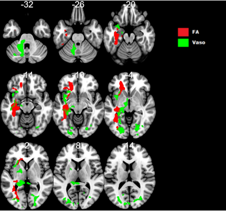
Patients with symptom “difficulties when using upper limbs” experienced higher CVR disruption in the poCVR analysis. As shown in Figure 1, two clusters were identified. A first cluster (2762 voxels; pcluster FWE < 0.001) located in white matter of left occipital and frontal lobes, left fusiform gyrus and left external capsule. Second cluster is (917 voxels; pFWE cluster = 0.001), located in cerebellum white matter.Other clusters of CVR disruption located in supratentorial white matter areas were identified for patients with “muscular weakness” (563 voxels; pcluster FWE = 0.026), “falls” (1961 voxels; pcluster FWE < 0.001), “spasticity” ( 707 voxels, pcluster FWE = 0.009) and “25-foot-walk-test” (616 voxels; pcluster FWE = 0.008).No significant cluster (pPeak <0.005; pcluster FWE <0.05) was observed for variables “NHPT”, “amyotrophy”, “shaking”, fatigue”, “difficulties at using lower limbs” and “vesico-sphincteric disorders”.In the DTI analysis, one cluster of reduced FA (pPeak <0.005; pcluster FWE <0.05) was identified for patients with symptom “difficulties when using upper limbs” (2183 voxels; pcluster FWE < 0.001). It is located in white matter in left occipital and frontal lobes, left fusiform gyrus and left external capsule, and left hemisphere of the cerebellum.Other clusters of FA alterations located in supratentorial white matter areas were identified for patients with symptoms “muscular weakness” (1058 voxels; pcluster FWE = 0.005), “falls” (788 voxels; pcluster FWE < 0.026), and “25-foot-walk-test” (1323 voxels; pcluster FWE = 0.005).No significant cluster (pPeak <0.005; pcluster FWE <0.05) was observed for variable “spasticity”.Discussion
These results are coherent with the current hypotheses about astrocytes metabolism’s abnormalities in white matter and their link with the known vascular alterations in MS patients. Both poCVR and DTI maps for variable “difficulties when using upper limbs” outline the left Inferior Fronto-Occipital Fasciculus, which is mainly known for its functions in language, but also in visual-motor integration and writing3. These roles are coherent with the studied clinical symptom. The other results are concordant and also reveal CVR alterations in white matter region. Vascular alterations are already studied in MS4 but still mostly unknowned. This study suggests a link between axonal tracts and vasoreactivity alterations.However, this study suffered from some limitations. First, a low number of patients limits its statistical power. A larger study would be necessary to further explore the link suggested by these results. Second, the variable “difficulties when using upper limbs” isn’t specific enough. A dedicated study with more specific clinical parameters could be useful.
Conclusion
This study showed CVR alteration in white matter regions, with similar results on DTI analyses. Therefore, it hints to a link between CVR and white matter tracts alterations. Vasoreactivity could be a new biomarker for assessing the evolution of white matter tracts’ alterations in MS follow-up. A new study with more patients could directly compare poCVR and DTI maps to explore this link.Acknowledgements
Thanks to my friend Jérémy Deverdun, for everything he did to make this research possible, and helping me in my first real research work.
Many thanks too for Céline Charroud, who was always there to answer my questions and correct my mistakes.
And I would like to thank the I2FH lab in Hospital Gui de Chauliac, Nicolas Menjot de Champfleur, Emmanuelle Le Bars and everybody else for there formidable support.
References
- D’haeseleer M, Cambron M, Vanopdenbosch L, De Keyser J. Vascular aspects of multiple sclerosis. Lancet Neurol. 2011;10:657–666.
- Metzger A, Bars EL, Deverdun J, et al. “Is impaired cerebral vasoreactivity an early marker of cognitive decline in multiple sclerosis patients?” Eur Radiol. 2018;28:1204–1214.
- Vassa F, Boutet C, Sontheimer A, Lemaire JJ. (2017). “Terminaisons corticales du faisceau fronto-occipital inférieur : une étude en tractographie.”; Neurochirurgie.63.48.10.1016/j.neuchi.2016.11.048.
- Marshall O, Lu H, Brisset J-C, et al. “Impaired cerebrovascular reactivity inmultiple sclerosis.”; JAMA Neurol. 2014;71:1275–1281.
Figures