1409
Reduced fibre-specific measures in multiple sclerosis patients with cerebellar dysfunction1The University of Melbourne, Melbourne, Australia, 2Monash University, Melbourne, Australia, 3Royal Melbourne Hospital, Melbourne, Australia, 4The Bionics Institute, Melbourne, Australia, 5University of Tübingen, Tübingen, Germany
Synopsis
Cerebellar dysfunction is a common feature of multiple sclerosis (MS), leading to disabling symptoms such as tremor. In MS, brain atrophy is the most accepted correlate of neurodegeneration; however cerebellar atrophy does not seem to correlate with the degree of cerebellar dysfunction. Here we explore axonal degeneration, a key driver of disability, with a fibre-specific marker based on diffusion-weighted MRI – fibre density and fibre bundle cross-section. We found that loss of cerebellar fibre density and fibre cross-section was associated with increased clinical cerebellar dysfunction. Fibre-specific measures could provide a useful marker of cerebellar dysfunction in MS.
Introduction
Multiple sclerosis (MS) is an autoimmune disease of the central nervous system. The cerebellum is commonly affected in MS, leading to symptoms such as tremor, ataxia and dysarthria. Understanding the underlying structural changes of cerebellar dysfunction is essential for targeted treatment development. Conventional measures of MRI, including atrophy, do not correlate with cerebellar dysfunction 1. A MRI based quantitation of axonal pathology could provide a better imaging correlate of cerebellar dysfunction.Diffusion-weighted MRI can non-invasively detect microstructural changes in white matter. Fixel-based analysis is a method that can potentially provide improved markers of axonal degeneration. Unlike standard voxel-based diffusion approaches, the fixel-based approach can detect changes for each fibre element (“fixel”) within a voxel.
This study aimed to understand the correlation between fixel-based MRI measures and cerebellar dysfunction in a cohort of MS patients with and without upper limb tremor and healthy controls.
Methods
Multi-shell diffusion weighted scans were acquired for 29 MS tremor, 26 matched MS patients without tremor, and 14 healthy controls (voxel size=2.4x2.4x2.4mm3, 16 non-diffusion, 31 b=1000s/mm2, 51 b=2000s/mm2, 64 b=2800s/mm2). A quantitative clinical score of cerebellar dysfunction (SARA) was obtained for all patients.To estimate fixel-specific measures, fibre orientation distributions (FODs) are estimated for each voxel with constrained spherical deconvolution techniques 2. Firstly, a population-average FOD template was created from subject specific FOD images generated from multi-shell 3-tissue constrained spherical deconvolution 3. Each subject’s FOD image was then registered to the population template to allow for whole-brain fixel-based comparison between controls and patient groups. A fixel-specific measure of fibre density and fibre bundle cross-section (FDC) was generated 4. Connectivity-based fixel enhancement allowed for fixel-specific analyses, corrected for age and sex (5000 permutations) 5. All results were family-wise error corrected for multiple comparisons. All pre-processing, statistical analyses and visualisation were conducted with MRtrix3 (www.mrtrix.org).
Results
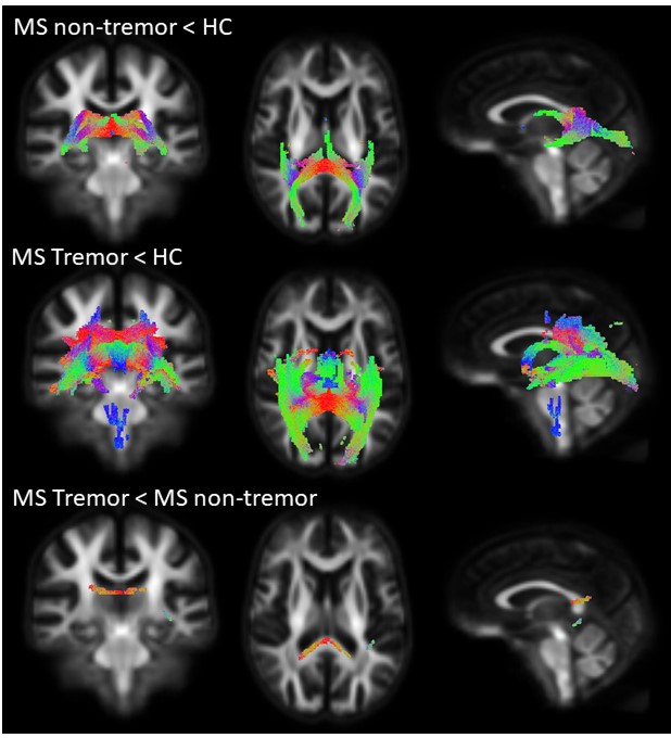
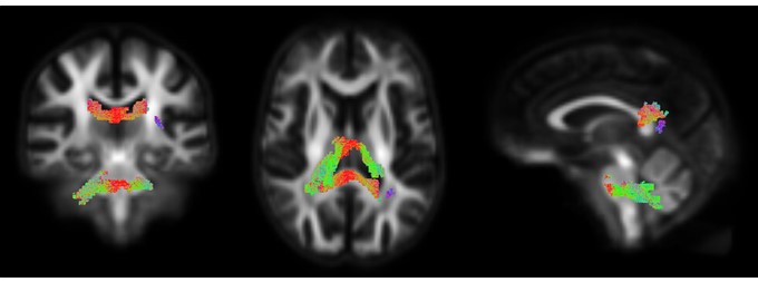
Compared to healthy controls, both MS non-tremor and MS tremor patients exhibited a significant loss of FDC. Furthermore, MS tremor patients exhibited a significant loss of FDC in the splenium of the corpus callosum compared to MS non-tremor patients (FWER corrected p<0.05) (Fig. 1). A loss of FDC in the splenium and cerebellum correlated with clinical scores of cerebellar dysfunction in patients (FWER corrected p<0.05) (Fig. 2).Discussion
In this study we have used a fixel-based approach to understand axonal MRI measures associated with cerebellar dysfunction in MS patients. Significant reduction in FDC were observed in MS non-tremor and MS tremor patients compared to healthy controls, with a greater degree of axonal degeneration in MS tremor patients. Furthermore, a significant reduction in FDC was observed in MS tremor patients compared to MS non-tremor patients. In addition, increased cerebellar dysfunction in MS patients was associated with reduced FDC within the splenium and cerebellum.Conclusion
This study demonstrated the advantage of the fixel-based approach in detecting the correlation between fibre-specific measures and cerebellar dysfunction in patients with MS.Acknowledgements
The authors would like to acknowledge the Murdoch Children’s Research Institute for providing access to the MRI suite and invaluable technical support.References
1. Boonstra F, Florescu G, Evans A, et al. Tremor in multiple sclerosis is associated with cerebello-thalamic pathology. Journal of Neural Transmission 2017;124:1509-1514.
2. Tournier JD, Yeh C-H, Calamante F, Cho K-H, Connelly A, Lin C-P. Resolving crossing fibres using constrained spherical deconvolution: Validation using diffusion-weighted imaging phantom data. NeuroImage 2008;42:617-625.
3. Jeurissen B, Tournier J-D, Dhollander T, Connelly A, Sijbers J. Multi-tissue constrained spherical deconvolution for improved analysis of multi-shell diffusion MRI data. Neuroimage 2014;103:411-426.
4. Raffelt D, Tournier JD, Smith RE, et al. Investigating white matter fibre density and morphology using fixel-based analysis. Neuroimage 2017;144, Part A:58-73.
5. Raffelt D, Smith RE, Ridgway GR, et al. Connectivity-based fixel enhancement: Whole-brain statistical analysis of diffusion MRI measures in the presence of crossing fibres. Neuroimage 2015;117:40-55.
Figures