1377
Thalamic volume relates to future memory performance in Multiple Sclerosis1Imaging Sciences, The Cleveland Clinic, Cleveland, OH, United States, 2Neurological Institute, The Cleveland Clinic, Cleveland, OH, United States, 3Schey Center for Cognitive Neuroimaging, The Cleveland Clinic, Cleveland, OH, United States
Synopsis
Cognitive dysfunction is a common symptom of Multiple Sclerosis (MS), and patients would benefit from a measure that estimates their risk of future decline. Previous work suggests that thalamic volume may have value as a predictive measure. Using 7 tesla MRI, we measured thalamic volume in 79 participants with MS. We found a strong cross-sectional relationship between volume and verbal episodic memory. The longitudinal relationship was significant, but was strongly modulated by disease duration. The current report highlights the need for careful modeling of potential confounding variables.
Introduction
Although cognitive dysfunction impacts nearly half of all patients with Multiple Sclerosis (MS), there is currently no measure that can predict which patients are at risk.1 Atrophy of the thalamus has been widely reported in MS, and measures of thalamic volume have been linked to episodic memory performance and overall cognitive decline.2,3 This relationship suggests that thalamic volume may have value as a predictor of future cognitive decline. Here we present preliminary results from a large longitudinal study of neuroimaging in MS, focusing on the relationship of thalamic volume to cognitive performance.Methods
In an IRB-approved protocol, 79 participants with MS (age: 51.53 ± 8.2 years, 19 males, EDSS: 3.9 ± 1.6) were scanned on a Siemens 7T Magnetom with SC72 gradient (Siemens Medical Solutions, Erlangen) using a 32-channel head coil (Nova Medical). All participants completed comprehensive cognitive testing, including measures of verbal and spatial episodic memory and speed of processing. Fifty-two participants returned for a follow-up scan and cognitive testing session after 1 year. A whole-brain anatomical MP2RAGE (voxel size = 0.75mm3) was used to segment the thalamus using the longitudinal analysis pipeline of Freesurfer v 6.0.0 (http://surfer.nmr.mgh.harvard.edu/). Individual segmentations were checked for accuracy. A volumetric scaling factor was used to correct for intracranial volume. Left and right volumes were averaged. Thalamic volume at baseline was related to cognitive measures at baseline and follow-up.Results
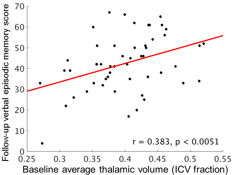
At baseline, average thalamic volume was not related to age or education, but was related to disease duration (r = -0.320, p < 0.0041). Verbal episodic memory was the only cognitive measure to show a significant relationship (r = 0.345, p < 0.0018; Figure 1), remaining significant after correction for disease duration (r = 0.318, p < 0.0046).In the 52 participants with follow-up data, baseline thalamic volume was related to verbal (r = 0.383, p < 0.0051; Figure 2) and spatial (r = 0.320, p < 0.0207) episodic memory, and to speed of processing (r = 0.274, p < 0.0489). However, after correction for disease duration, only verbal episodic memory remained significant (r = 0.289, p < 0.0398).
Discussion
In agreement with previous reports, we find that thalamic volume is related to episodic memory in MS. Volume was also related to later memory function, but the contribution of disease duration serves as a reminder that efforts to develop imaging predictors must account for demographic and disease-related variables. Longitudinal data collection is ongoing. In addition to ongoing assessment of overall thalamic volume, the resolution and contrast achievable at 7 tesla allows for future work to assess the relationship of individual thalamic nuclei to cognitive decline.Acknowledgements
This work was supported by the Department of Defense (MS150097). We thank Siemens Healthineer Tobias Kober for use of WIP944.References
1. Achiron A, Chapman J, Magalashvili D, Dolev M, Lavie M, Bercovich E, et al. Modeling of Cognitive Impairment by Disease Duration in Multiple Sclerosis: A Cross-Sectional Study. PLoS One. 2013;8(8):e71058
2. Benedict RH, Hulst HE, Bergsland N, Schoonheim MM, Dwyer MG, Weinstock-Guttman B, Geurts JJ, Zivadinov R. Clinical significance of atrophy and white matter mean diffusivity within the thalamus of multiple sclerosis patients. Multiple Sclerosis. 2013;19(11):1478-84.
3. Schoonheim MM, Hulst HE, Brandt RB, Strik M, Wink AM, Uitdehaag BM, Barkhof F, Geurts JJ. Thalamus structure and function determine severity of cognitive impairment in multiple sclerosis. Neurology. 2015;84(8):776-83.
Figures