1313
High resolution spiral simultaneous multi‐slice first‐pass perfusion imaging with whole-heart coverage at 1.5 T and 3 T1Biomedical Engineering, University of Virginia, Charlottesville, VA, United States, 2Biomedical Engineering and Imaging Institute and Department of Radiology, Icahn School of Medicine at Mount Sinai, New York, NY, United States, 3Medicine, University of Virginia, Charlottesville, VA, United States, 4Electrical and Computer Engineering, University of Iowa, Iowa City, IA, United States, 5Electrical and Computer Engineering, University of Virginia, Charlottesville, VA, United States, 6Radiology, University of Virginia, Charlottesville, VA, United States
Synopsis
First-pass contrast-enhanced myocardial perfusion imaging is a useful noninvasive tool to evaluate patients with known or suspected coronary artery disease, but current techniques are still limited in spatial-temporal resolution and ventricular coverage. We designed a spiral pulse sequence with simultaneous multi-slice (SMS) acquisition and utilized the SMS-L1-SPIRiT reconstruction technique to achieve ultra-high resolution (1.5 mm at 1.5 T and 1.25 mm at 3 T) perfusion imaging with whole-heart coverage. The proposed spiral SMS perfusion acquisition strategy was tested on heathy volunteers and clinical patients. High image quality was demonstrated with an SMS factor of 3 at both 1.5T and 3T.
Introduction
First-pass contrast-enhanced myocardial perfusion imaging is a valuable tool to evaluate patients with coronary artery disease (CAD)1. However, current techniques are still limited in spatial-temporal resolution and ventricular coverage, which reduces the sensitivity to detect perfusion differences between the endocardium and epicardium and quantify ischemic burden. Recently, we developed a simultaneous multi-slice (SMS) spiral perfusion pulse sequence and SMS-L1-SPIRiT reconstruction technique capable of whole-heart high resolution perfusion imaging2. We have demonstrated high-resolution SMS perfusion imaging with an SMS factor of 2 at 3 T, but image quality was not superior to our single-band technique3. The goal of this study is to further optimize the spiral pulse sequence and SMS-L1-SPIRiT reconstruction technique to achieve ultra-high resolution (1.5 mm at 1.5 T and 1.25 mm at 3 T) perfusion imaging with an SMS factor of 3 at comparable image quality to the single-band technique.Methods
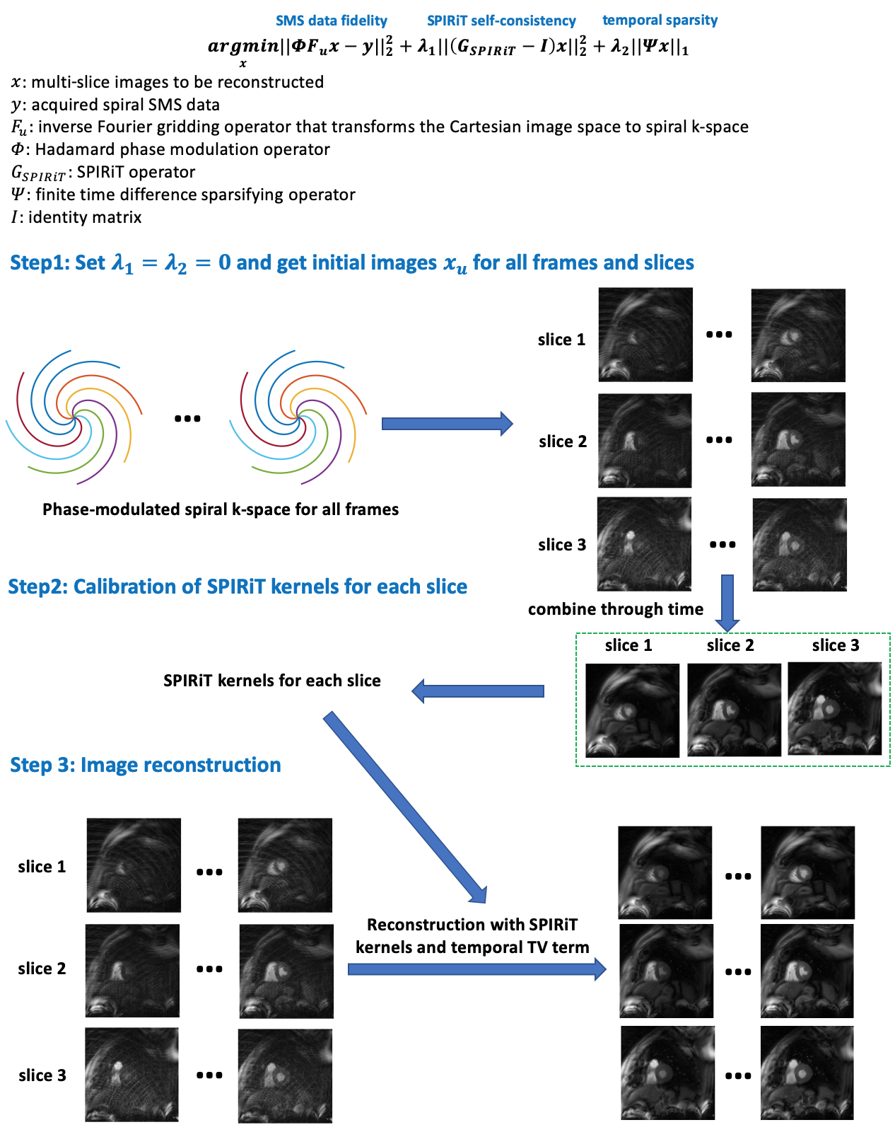
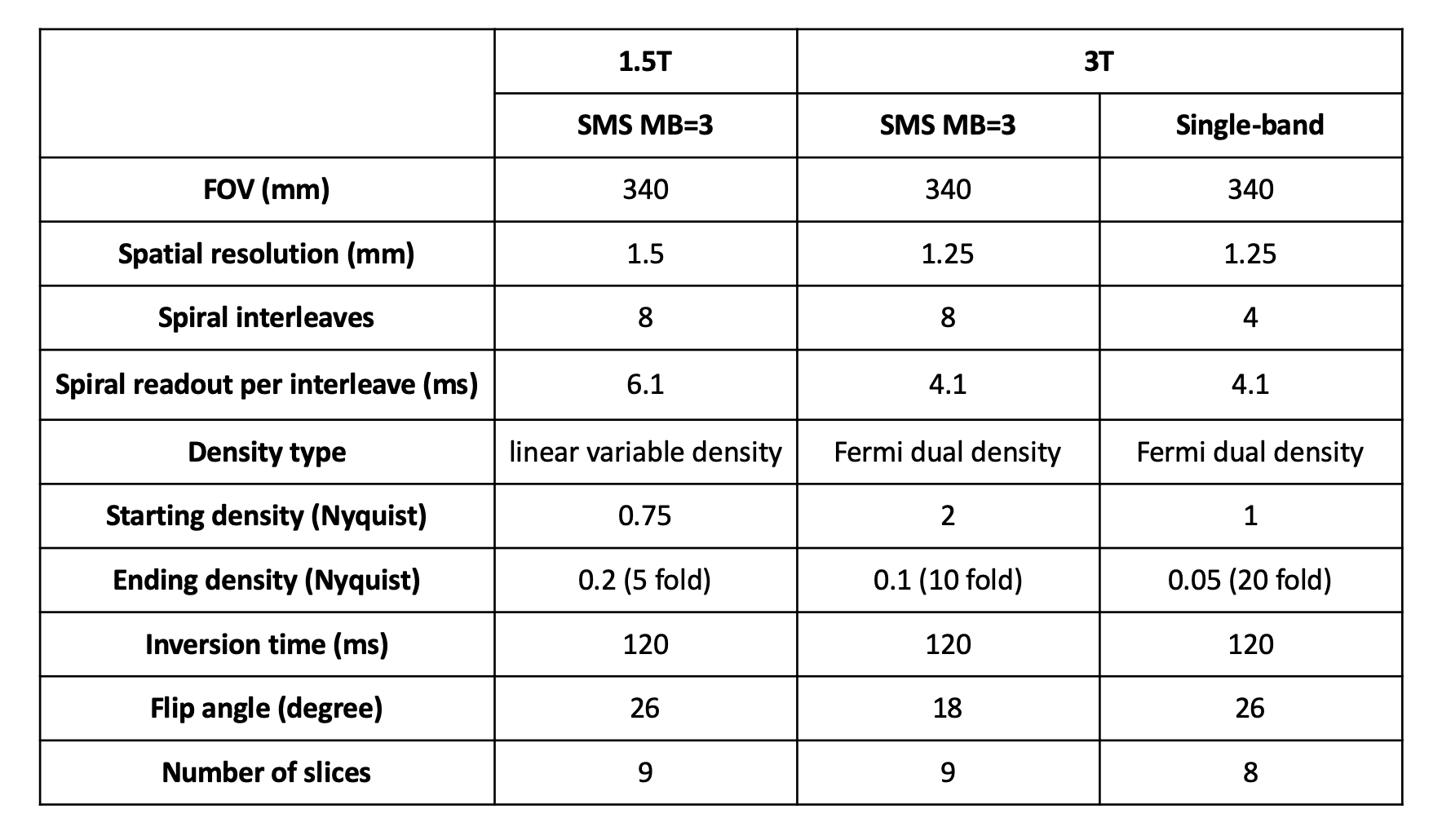
A pulse sequence with uniform distributed spirals in each heart beat with a Hadamard phase modulation4, which is rotated by the golden angle (137°) between time frames was used. To validate the SMS acquisition strategy, at 3 T scanners, we acquired data with both single-band interleaved acquisition5 and SMS acquisition on the same healthy subjects. We also utilized the same SMS strategy to perform high-resolution perfusion imaging at 1.5 T. Pulse sequence parameters for 1.5 T and 3 T are shown in Table 1.The SMS-L1-SPIRIT reconstruction method can be formulated as shown in the equation in Fig 1. The first term enforces SMS data fidelity, the second term (SPIRiT) enforces self-consistency6, the third term enforces temporal sparsity. The objective function was solved using non-linear conjugate gradient algorithm7. The SPIRiT operator was calibrated using a novel auto-calibration process whereby initial images at each slice position are summed through all temporal frames to achieve un-aliased calibration data (Fig 1). $$$\lambda_1$$$=1 and $$$\lambda_2$$$=0.005 were determined based on the results of a retrospective SMS experiment, and used in all reconstructions. Single-band data acquired at 3 T was reconstructed using L1-SPIRiT5. Coil selection to retain coils with low artifact power was performed before image reconstruction8.
Prospective SMS MB=3 spiral perfusion imaging was performed on 3 healthy volunteers undergoing clinically ordered CMR studies during injection of Dotarem contrast at 4 cc/s on a 3 T scanner (Siemens Skyra/Prisma). Single-band and multi-band perfusion images were acquired in subsequent injections on the healthy volunteers to compare the techniques. Imaging was also performed at 1.5 T (Siemens Aera) on 8 patients undergoing clinical CMR studies. The image quality was graded on a 5-point scale (5 excellent, 1 poor) by an experienced cardiologist.
Results
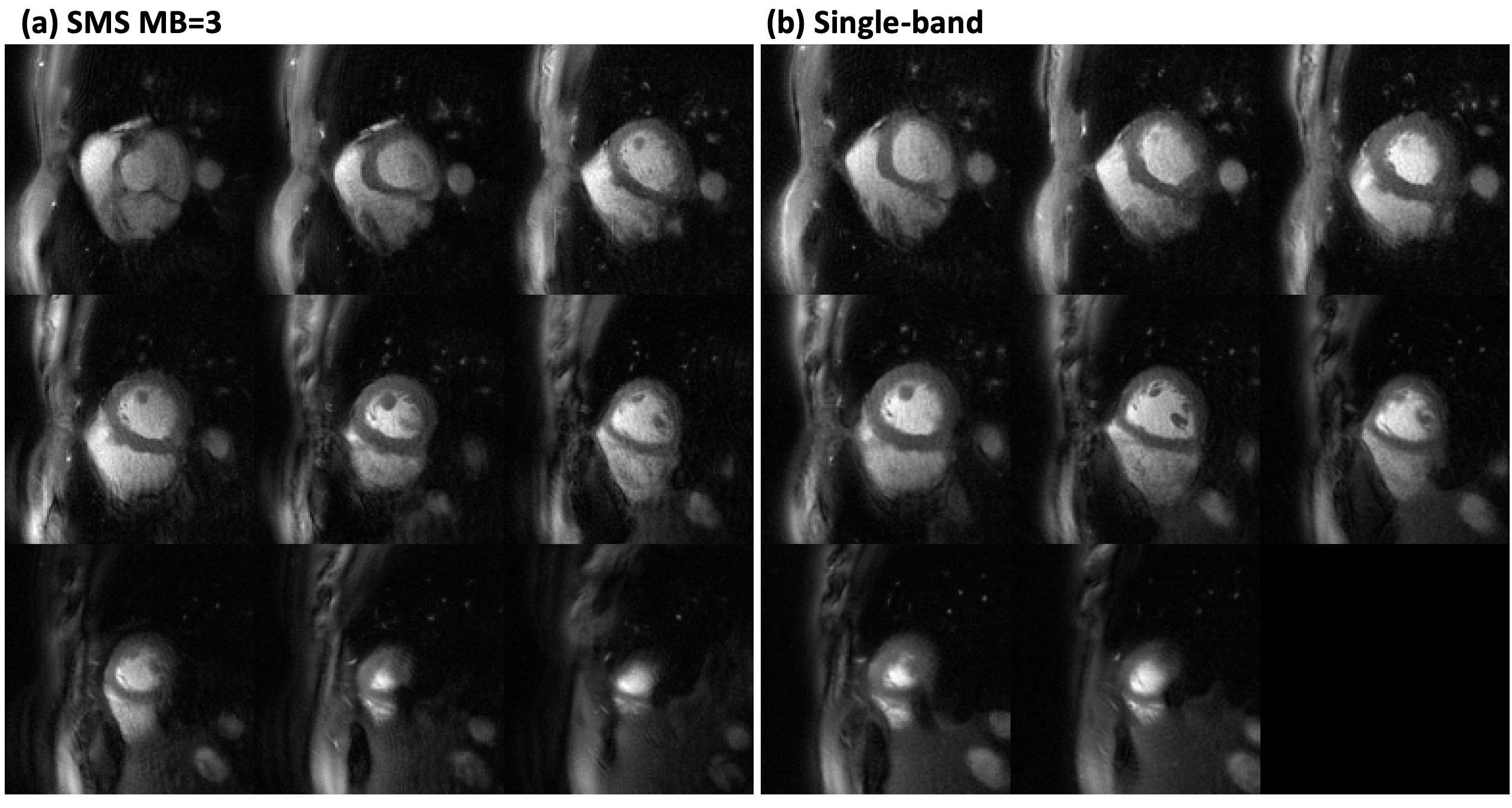
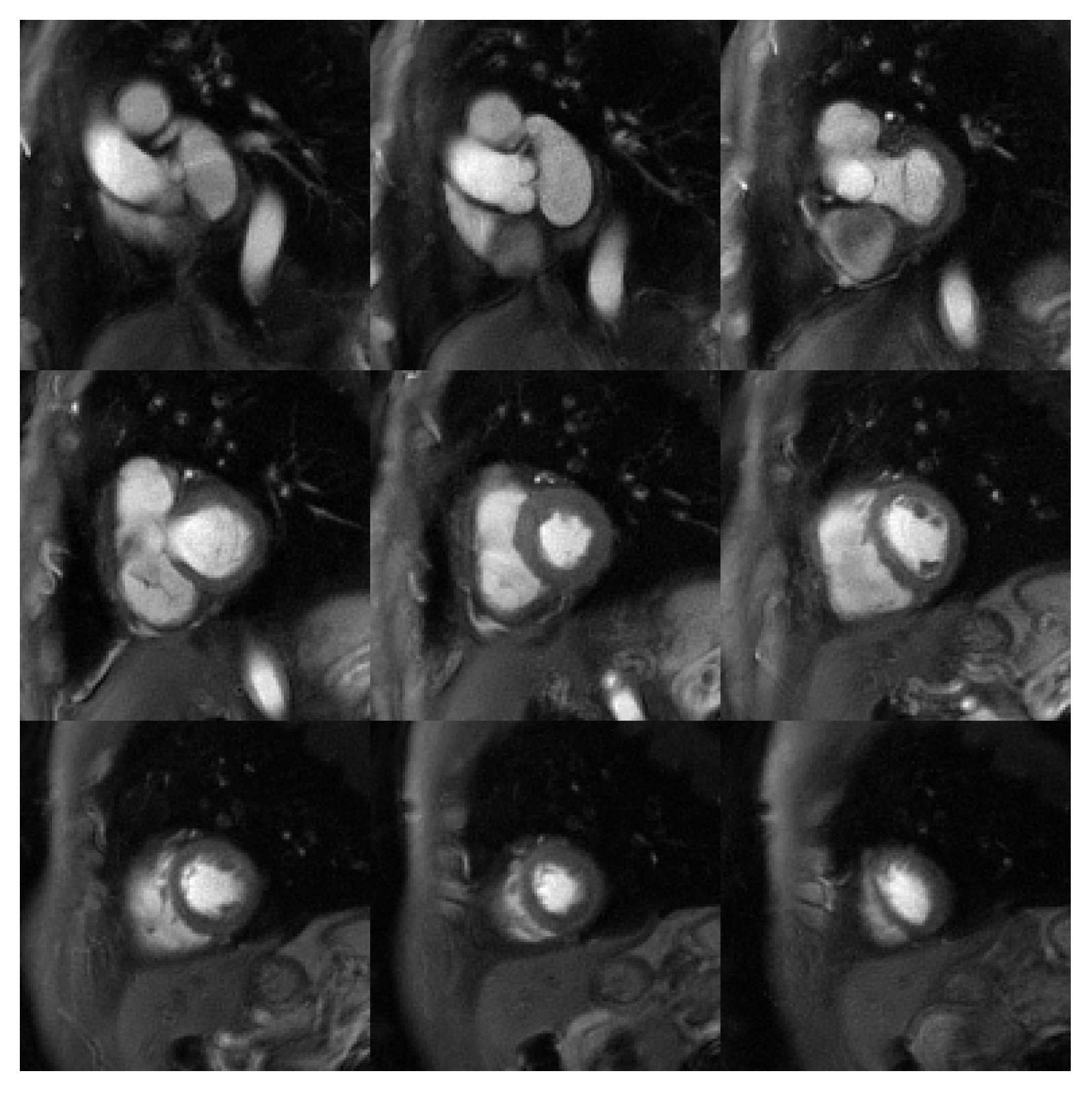
Fig 2 shows a direct comparison of whole-heart perfusion imaging at middle time frame using single-band acquisition and SMS acquisition with an SMS factor of 3 from the same healthy subject at 3 T. For the 3 volunteers image scores were 4, 3.5, 3.5 for the single-band technique and 3.5, 3.5, 3.5 respectively for the multi-band technique.Fig 3 shows an example of 9 slices of 1.5 mm resolution with an SMS factor of 3 at 1.5 T. The overall image score for SMS MB=3 at 1.5 T was 4.06±0.42. Fig 4 shows the corresponding first-pass perfusion movie of the same subject shown in Fig 3.
Good image quality was achieved with 1.5 mm spatial resolution at 1.5 T and 1.25 mm resolution at 3 T (Fig 2, 3 and 4).
Discussion and Conclusion
We demonstrated the successful application of ultra-high resolution spiral perfusion imaging using SMS acquisition and SMS-L1-SPIRiT reconstruction techniques with an SMS factor of 3 and whole-heart coverage at both 1.5 T and 3 T. High-resolution whole-heart perfusion will potentially serve as a tool to quantify regional differences in perfusion of the sub-endo and sub-epi myocardium. Further validation will be required in patients undergoing adenosine stress studies.Acknowledgements
This work was supported by NIH R01 HL131919.References
1. Nagel E, Klein C, Paetsch I, Hettwer S, Schnackenburg B, Wegscheider K, Fleck E. Magnetic resonance perfusion measurements for the noninvasive detection of coronary artery disease. Circulation. 2003 Jul 29;108(4):432-7.
2. Yang Y, Meyer CH, Epstein FH, Kramer CM, Salerno M. Whole‐heart spiral simultaneous multi‐slice first‐pass myocardial perfusion imaging. Magnetic resonance in medicine. 2019 Feb;81(2):852-62.
3. Yang Y, Kramer CM, Salerno M. Whole heart First-pass spiral perfusion imaging with 1.25mm resolution at 3T. Proc Intl Soc Mag Reson Med 2018;26:3321.
4. Souza SP, Szumowski J, Dumoulin CL, Plewes DP, Glover G. SIMA: simultaneous multislice acquisition of MR images by Hadamard-encoded excitation. J Comput Assist Tomogr. 1988 Nov 1;12(6):1026-30.
5. Yang Y, Kramer CM, Shaw PW, Meyer CH, Salerno M. First‐pass myocardial perfusion imaging with whole‐heart coverage using L1‐SPIRiT accelerated variable density spiral trajectories. Magnetic resonance in medicine. 2016 Nov;76(5):1375-87.
6. Lustig M, Pauly JM. SPIRiT: iterative self‐consistent parallel imaging reconstruction from arbitrary k‐space. Magnetic resonance in medicine. 2010 Aug;64(2):457-71.
7. Weller DS, Ramani S, Fessler JA. Augmented Lagrangian with Variable Splitting for Faster Non-Cartesian L1-SPIRiT MR Image Reconstruction. IEEE Transactions on Medical Imaging 2014; 33:351–361.
8. Zhou R, Yang Y, Mathew RC, Mugler III JP, Weller DS, Kramer CM, Ahmed AH, Jacob M, Salerno M. Free‐breathing cine imaging with motion‐corrected reconstruction at 3T using SPiral Acquisition with Respiratory correction and Cardiac Self‐gating (SPARCS). Magnetic resonance in medicine. 2019 Aug;82(2):706-20.
Figures