1312
Fully Self-gated Free-breathing 3D Cartesian Cardiac CINE with Isotropic Whole-heart Coverage in Less Than 2 Minutes1Biomedical Engineering Department, School of Biomedical Engineering and Imaging Sciences, King's College London, London, United Kingdom, 2MR Research Collaborations, Siemens Healthcare Limited, Frimley, United Kingdom
Synopsis
Free-breathing continuous acquisitions, so called free-running, enable 3D whole-heart coverage for motion-resolved functional cardiac MRI. In prior work approaches based on 3D radial imaging were proposed with scan times of ~10-15min which also require computationally demanding reconstructions. In this work, we propose a 3D Cartesian free-running water-selective sequence that provides isotropic 3D whole-heart CINE imaging in <2min. Data is acquired with a variable-density spiral-like 3D Cartesian out-inward sampling and sequence-adaptive tiny-golden and golden angle increment. Respiratory motion-corrected and cardiac motion-resolved CINE images are obtained from a multi-bin-PROST reconstruction which exploits spatial-temporal redundancies. High agreement to conventional 2D CINE was observed.
Introduction
Cardiac CINE imaging enables the assessment of morphology and function to diagnose cardiovascular diseases. 3D free-breathing continuous acquisitions1-5, so called free-running, have been proposed to reconstruct 3D whole-heart CINE images with respiratory motion correction or motion-resolved. These approaches retrospectively assign the data into different cardiac and respiratory phases based on ECG or self-gated cardiac and respiratory signals. Cardiac and respiratory resolved images are then reconstructed exploiting temporal redundancy in both cardiac and respiratory directions6. These approaches have shown promising image quality in reasonable scan times of ~10-15min (for high spatial resolution), however require long reconstruction times due to non-Cartesian data sampling and some rely on administration of contrast agent for sufficient image contrast.Here, we propose a 3D Cartesian free-running acquisition which enables translational beat-to-beat respiratory motion correction and cardiac motion-resolved images of the whole-heart without contrast administration in a clinically feasible scan time (<2 minutes) and reconstruction times (~10 minutes). A free-running CINE sequence with variable-density CASPR (VD-CASPR) 3D Cartesian sampling7 and embedded self-navigation enables incoherent and non-redundant sampling within and between respiratory and cardiac phases. A water-selective balanced steady-state free-precession (bSSFP) sequence is employed reducing the impact of fat-related motion aliasing. An efficient multi-bin PROST (MB-PROST)7 reconstruction exploits patch-based spatial-temporal redundancies to provide high quality images.
Methods
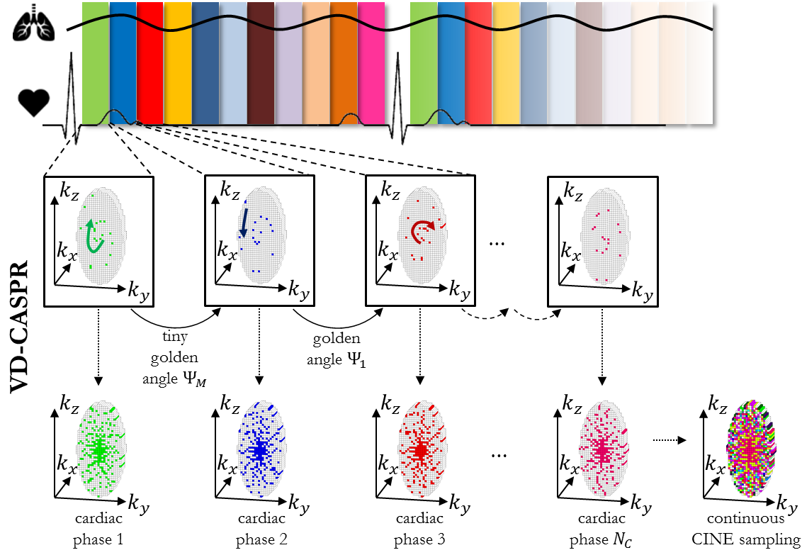
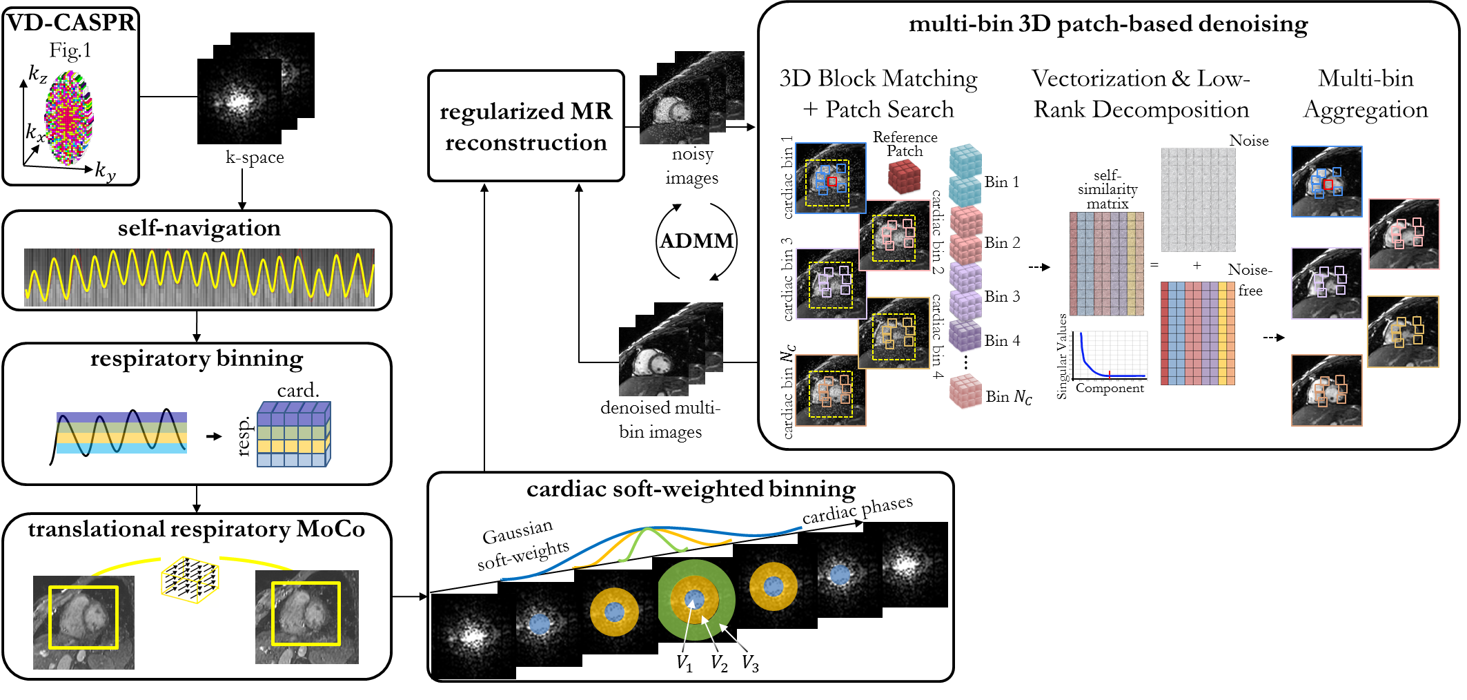
Acquisition: VD-CASPR sampling8,9 with alternating tiny-golden and golden angle increment between spirals continuously undersamples the Cartesian ky/kz plane with an acceleration factor L (Fig.1). An acquisition-dependent tiny-golden angle is calculated based on the total desired scan time TA and cardiac phases to ensure incoherent and non-redundant sample-to-bin allocation for the continuous free-breathing CINE acquisition. An out-inward trajectory is used to minimize the effect of eddy currents. The center line of k-space is periodically sampled to enable 1D cardiac and respiratory self-navigation.Reconstruction: As shown in Fig.2, data is first retrospectively binned into respiratory phases based on the respiratory self-navigation signal which is extracted via PCA6. An auxiliary respiratory motion-resolved image is reconstructed with MB-PROST for estimation of translational respiratory motion. Respiratory translational motion-corrected data is then binned into cardiac phases based on the cardiac self-navigation signal and considering a spatially varying Gaussian soft-weighting amongst neighboring cardiac phases (see Fig.2 V1 to V3). Subsequently, a cardiac motion-resolved 3D CINE image is reconstructed using MB-PROST. MB-PROST alternates between two optimization problems: 1) an L2-norm regularized reconstruction using the denoised data from step 2 as a prior, and 2) an efficient low-rank patch-based denoising. MB-PROST exploits redundant information on a local (within a patch), non-local (similar patches within a spatial neighborhood) and temporal (amongst all motion phases) scale with an implicit motion-alignment amongst patches.
The proposed free-running 3D Cartesian CINE was acquired with a water-selective (1-2-1 binomial pulse) bSSFP sequence in sagittal orientation in ten healthy subjects with 1.5T MRI (MAGNETOM Aera, Siemens Healthcare, Erlangen, Germany) and TE=2.33ms, TR=4.66ms, flip angle α=39°, acceleration L=8, 1.9mm3 isotropic resolution within TA=1:50min:s for whole-heart coverage. Motion-resolved images are reconstructed to 8 respiratory and 16 cardiac phases (40-50ms temporal resolution). An additional conventional multi breath-hold 2D CINE (short axis, non-fat suppressed, TE = 1.06ms, TR = 2.12ms, flip angle α=52°, 1.9x1.9x8mm resolution, LV coverage, TA=4:20min:s) was acquired for comparison of LV functional parameters: end-systolic volume (ESV), end-diastolic volume (EDV) and ejection fraction (EF), which were derived from manually segmented LV endo- and epicardium.
Results and Discussion
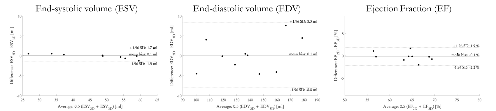
Animated Fig. 3 shows cardiac motion-resolved images for two subjects of the proposed free-running 3D Cartesian CINE with reformats into short axis, 2-chamber, 4-chamber and coronal views. Good agreement between free-running 3D Cartesian CINE and conventional 2D CINE can be observed in the short axis view. The isotropic resolution of the proposed approach enables high-resolution reformats into arbitrary orientations. Binning with the cardiac self-navigator signal shows good agreement with an additionally acquired ECG signal. MB-PROST could be used to simultaneously exploit redundancies in the respiratory and cardiac direction with the consequent tradeoff in reconstruction time. A respiratory-corrected approach was chosen to provide a fast reconstruction in around ~10min. Spatial coverage of the proposed free-running 3D CINE in comparison to conventional 2D CINE is shown in Fig. 4. Fig. 5 shows Bland-Altman plots for ESV, EDV EF between the proposed free-running 3D CINE and conventional multi breath-hold 2D CINE sequence indicating no bias and tight confidence intervals of ESV=±1.6ml, EDV=±8.2ml, EF=±2.1%.Conclusion
The proposed 3D Cartesian free-running CINE provides respiratory-corrected and cardiac motion-resolved images with high spatial and temporal resolution acquired under free-breathing in <2min scan time and ~10min reconstruction time.Acknowledgements
This work was supported by EPSRC (EPSRC EP/P032311/1, EP/P001009/1 and EP/P007619/1) and Wellcome EPSRC Centre for Medical Engineering (NS/A000049/1).References
1. Coppo S, Piccini D, Bonanno G, Chaptinel J, Vincenti G, Feliciano H, Van Heeswijk RB, Schwitter J, Stuber M. Free‐running 4D whole‐heart self‐navigated golden angle MRI: initial results. Magnetic resonance in medicine 2015;74(5):1306-1316.2. Liu J, Nguyen TD, Zhu Y, Spincemaille P, Prince MR, Weinsaft JW, Saloner D, Wang Y. Self-gated free-breathing 3D coronary CINE imaging with simultaneous water and fat visualization. PloS one 2014;9(2):e89315.
3. Pang J, Sharif B, Fan Z, Bi X, Arsanjani R, Berman DS, Li D. ECG and navigator‐free four‐dimensional whole‐heart coronary MRA for simultaneous visualization of cardiac anatomy and function. Magnetic resonance in medicine 2014;72(5):1208-1217.
4. Wu HH, Gurney PT, Hu BS, Nishimura DG, McConnell MV. Free‐breathing multiphase whole‐heart coronary MR angiography using image‐based navigators and three‐dimensional cones imaging. Magnetic resonance in medicine 2013;69(4):1083-1093.
5. Qi H, Jaubert O, Bustin A, Cruz G, Chen H, Botnar R, Prieto C. Free-running 3D whole heart myocardial T1 mapping with isotropic spatial resolution. Magnetic Resonance in Medicine 2019;82(4):1331-1342.
6. Feng L, Coppo S, Piccini D, Yerly J, Lim RP, Masci PG, Stuber M, Sodickson DK, Otazo R. 5D whole-heart sparse MRI. Magnetic Resonance in Medicine 2018;79(2):826-838.
7. Küstner T, Bustin A, Neji R, Botnar R, Prieto C. 3D Cartesian Whole-heart CINE MRI Exploiting Patch-based Spatial and Temporal Redundancies. Proceedings of the European Society for Magnetic Resonance in Medicine (ESMRMB); 2019.
8. Prieto C, Doneva M, Usman M, Henningsson M, Greil G, Schaeffter T, Botnar RM. Highly efficient respiratory motion compensated free-breathing coronary MRA using golden-step Cartesian acquisition. Journal of magnetic resonance imaging : JMRI 2015;41(3):738-746.
9. Bustin A, Ginami G, Cruz G, Correia T, Ismail TF, Rashid I, Neji R, Botnar RM, Prieto C. Five-minute whole-heart coronary MRA with sub-millimeter isotropic resolution, 100% respiratory scan efficiency, and 3D-PROST reconstruction. Magnetic Resonance in Medicine 2018;81(1):102-115.
Figures