1282
Integrated RF/Wireless Coil and Ultrasound-Based Sensors to Enable Wireless Physiological Motion Monitoring in MRI1Brain Imaging and Analysis Center, Duke University, Durham, NC, United States, 2Medical Physics Graduate Program, Duke University, Durham, NC, United States, 3Department of Radiology, Brigham and Women's Hospital, Harvard Medical School, Boston, MA, United States
Synopsis
An integrated RF/wireless coil, used to wirelessly transmit data and acquire MR images, and small ultrasound-based sensors, used to monitor physiological motion and correct MRI artifacts, were combined to enable wireless transmission of 1-MHz ultrasound data acquired from the sensors. Experiments were performed to validate that the coil could wirelessly transmit ultrasound data 1) while taking MR images and 2) from inside and outside the bore to a computer. This integrated system improves the mobility of the OCM sensors, thereby enabling them to accompany patients throughout the hospital, and demonstrates the coil’s ability to transmit high-fidelity analog data while imaging.
Introduction
Physiological motion can cause artifacts during medical imaging and/or degrade therapeutic treatment accuracy. Most MR-compatible methods that are available to monitor physiological motion, such as navigator echoes and optical tracking systems, are essentially fixed with respect to a room’s floor/walls and as such cannot accompany a patient through procedures performed in different rooms or buildings. In contrast, small MR-compatible ultrasound-based devices called organ-configuration motion (OCM) sensors1 attach to the skin and may accompany a given patient through serial diagnostic and/or therapeutic procedures, to potentially help combine data from different modalities in ways that take breathing motion into account. However, electronics and wired connections associated with these sensors can very much limit mobility.A novel integrated RF/wireless coil design2 has been proposed to perform simultaneous, but completely independent, RF signal reception and wireless data transfer by allowing RF currents at the Larmor frequency and in a wireless communication band to flow concurrently on the same coil (Fig. 1a). Previously, this design was combined with the integrated parallel reception, excitation, and shimming (iPRES) coil design to acquire MR images and wirelessly transmit a small data set to control DC currents for localized B0 shimming with the same coil3,4.
Here, we further develop and combine these two innovative technologies, OCM sensors and integrated RF/wireless coils. More specifically, we use an integrated RF/wireless coil to wirelessly transmit OCM ultrasound data acquired and digitized within an MRI scanner bore, or outside the MRI suite, to a computer in the console room, thereby demonstrating 1) the ability to simultaneously acquire MR images and wirelessly transmit high-fidelity analog data with an integrated RF/wireless coil and 2) improved OCM sensor portability.
Methods
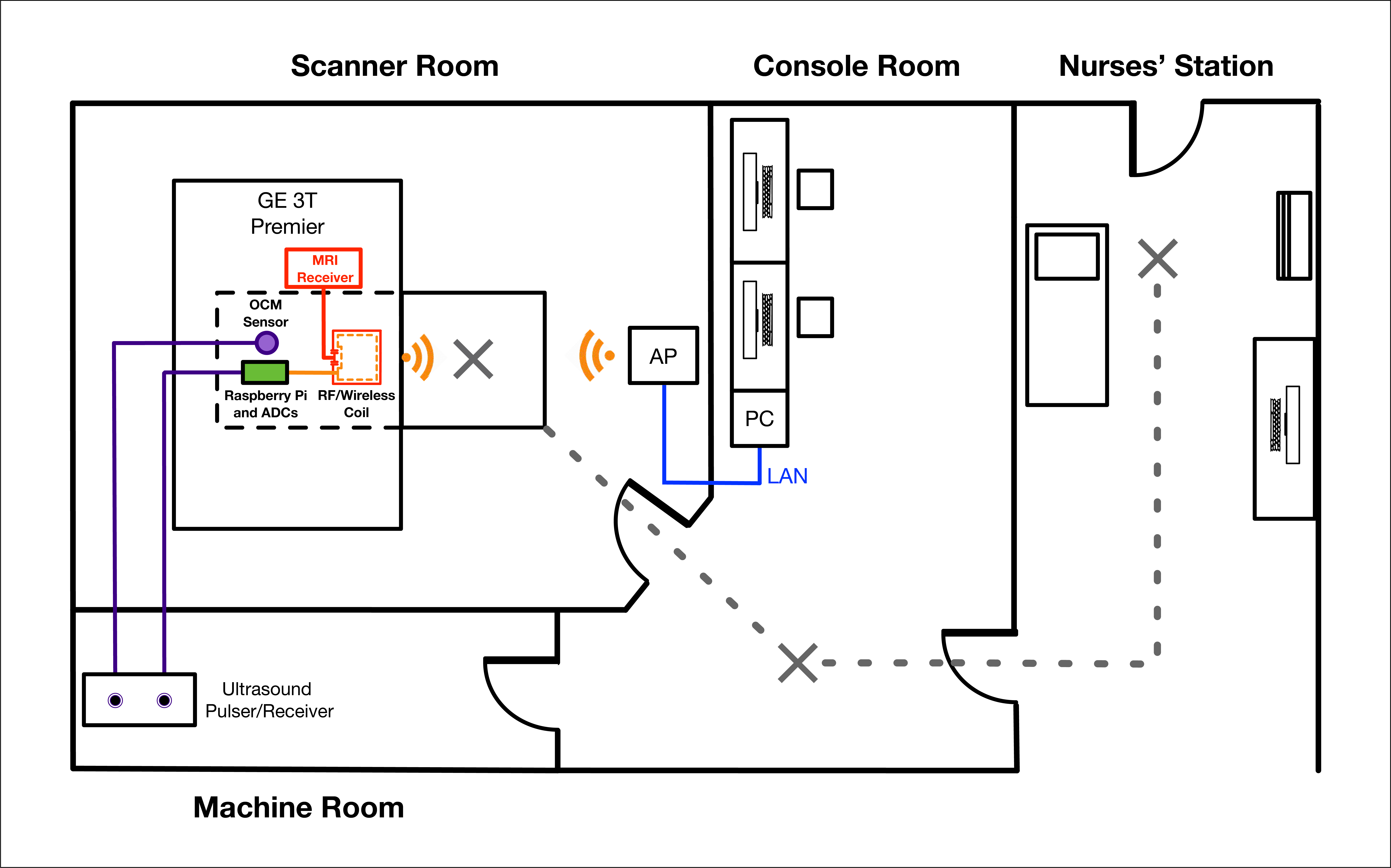
A 7-cm diameter integrated RF/wireless coil was constructed by placing capacitors, C, and an inductor, L, about its perimeter to allow the coil to resonate at 127.7 MHz (for a 3T scanner) and in the Wi-Fi communication band (2.412-2.472 GHz). Additionally, two high-impedance band-stop filters tuned to 127.7 MHz and to the Wi-Fi frequencies were placed 1) between the coil and the Wi-Fi transceiver of a modified battery-powered Raspberry Pi Zero-W; and 2) between the coil and the MRI preamplifier, to RF-isolate the transceiver and preamplifier, respectively (Fig. 1a,b).The 1-MHz analog ultrasound signal from the OCM sensor was digitized by two analog-to-digital converters (ADCs) connected to the Raspberry Pi5 within the scanner bore (Fig. 1a,b). The digitized signal flowed from the ADCs to the Wi-Fi transceiver, and then to the coil, where it was wirelessly transmitted to a computer in the console room via an access point (AP) mounted on the scanner room wall. A pulser/receiver in the machine room fired the OCM sensor and delivered the received signal to the ADCs through two 50-ft coaxial cables, which were long enough to extend outside of the MRI suite (Fig. 2).
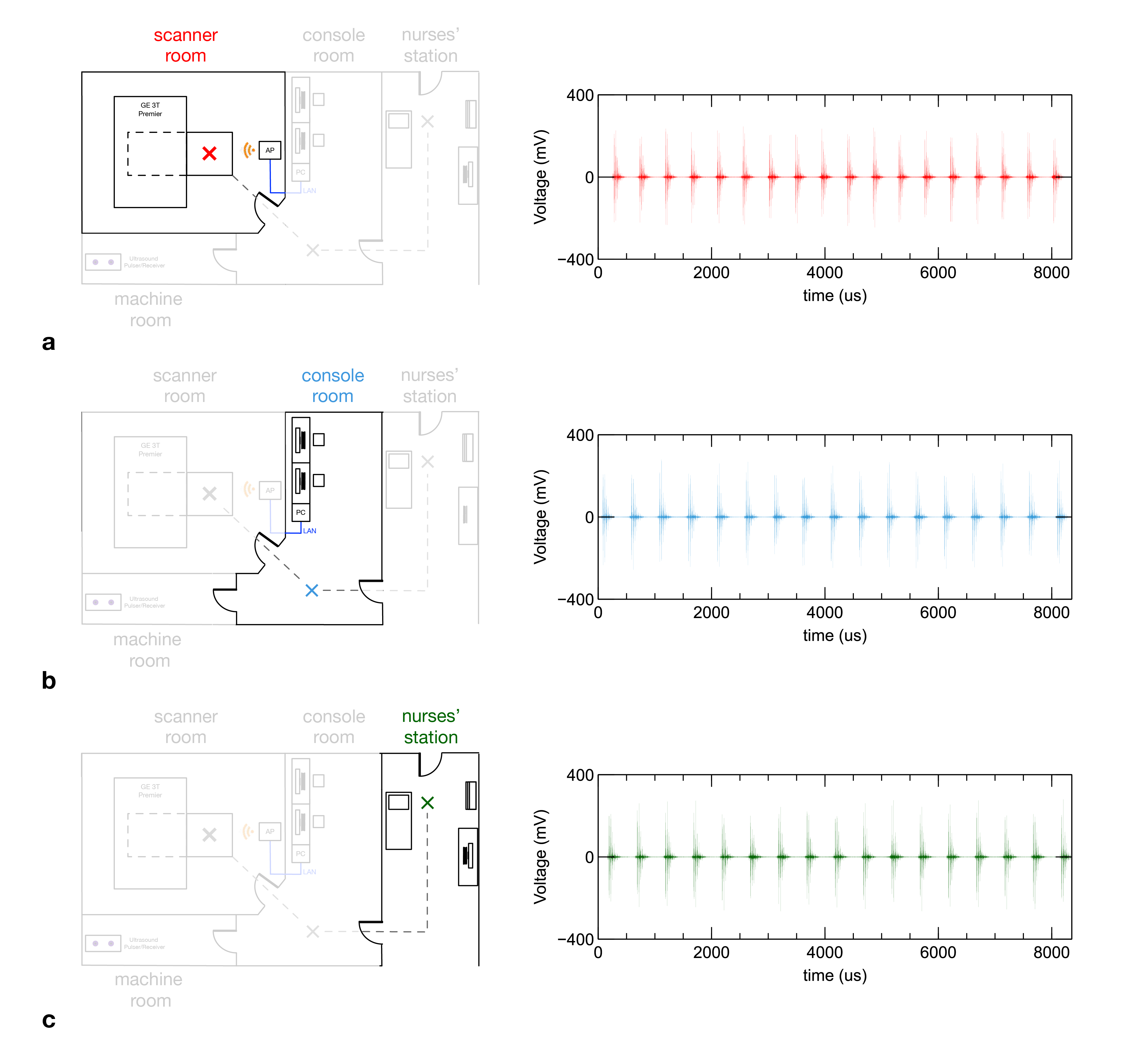
Next, two proof-of-concept experiments were performed to show that the integrated RF/wireless coil can 1) simultaneously perform imaging and wirelessly transmit ultrasound data and 2) wirelessly transmit the ultrasound data from various locations outside the scanner room. First, fast spoiled gradient-echo (FSPGR) images were acquired on a uniform water phantom with the coil, while wirelessly transmitting the analog OCM ultrasound signal data, which was acquired and digitized within the scanner bore, to the AP over a Wi-Fi network. Second, the coil was disconnected from the scanner and both the OCM sensor and the coil were moved 1) from the scanner bore, 2) to the console room, and then 3) to a nearby nurses’ station, while still on the patient’s table and transmitting ultrasound data to the AP (Fig. 2).
Results
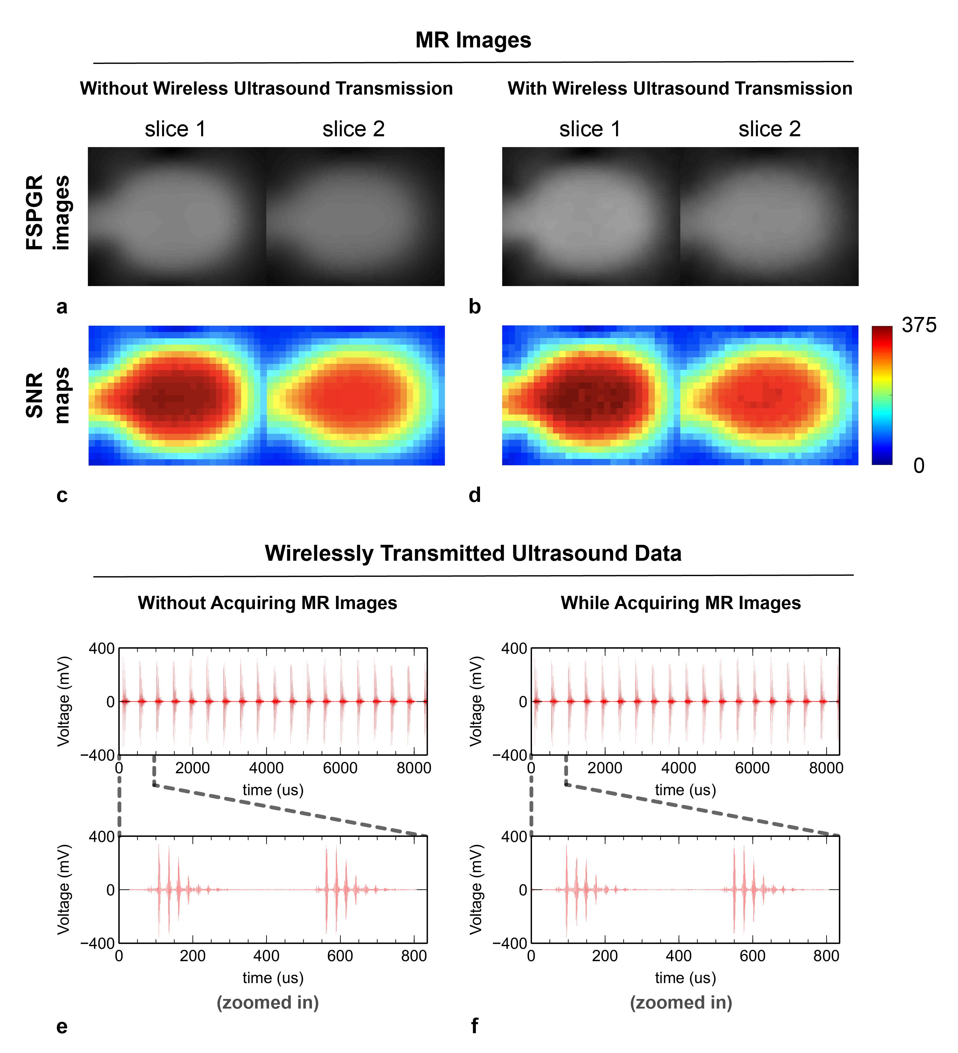
The images acquired with the integrated RF/wireless coil in two representative slices (Fig. 3a,b) show a less than 2% change in SNR (Fig. 3c,d) with and without wireless OCM sensor ultrasound data transmission. Additionally, the ultrasound data wirelessly transmitted from the coil to the computer in the console room was the same with (Fig. 3f) and without (Fig. 3e) imaging, which illustrates the coil’s ability to perform wireless transmission of high-fidelity analog data independently of MRI. Finally, the OCM ultrasound data wirelessly transferred to the AP from the coil located in the: scanner room (Fig. 4a); console room (Fig. 4b); and nurses’ station (Fig. 4c) shows no data loss at any location, which demonstrates the portability of a wireless OCM sensor system.Conclusion
The proof-of-concept experiments presented here showed that the integrated RF/wireless coil can wirelessly transmit ultrasound data 1) with and without acquiring MR images and 2) at locations inside/outside the scanner room. The coil design was optimized for RF reception and wireless data transfer, which can potentially translate to other diagnostic (e.g., PET-MRI) and therapeutic (MR-LINAC) systems. In future work, the wireless OCM system mobility could further be improved by integrating the pulse generator, processor, and ADCs onto a small single printed circuit board that can be placed near and moved with the patient during care.Acknowledgements
This work was in part supported by grants R21 EB024121, R01 NS075017, R01 EB028644, P41EB015898 and R03EB025546 from the National Institutes of Health, GE Healthcare, and the Duke-Coulter Translational Partnership.References
1. Preiswerk F, Toews M, Cheng CC, Chiou JY, Mei CS, Schaefer LF, Hoge WS, Schwartz BM, Panych LP, Madore B. Hybrid MRI‐Ultrasound acquisitions, and scannerless real‐time imaging. Magnetic resonance in medicine. 2017 Sep;78(3):897-908.
2. Darnell D, Cuthbertson J, Robb F, Song AW, Truong TK. Integrated radio‐frequency/wireless coil design for simultaneous MR image acquisition and wireless communication. Magnetic resonance in medicine. 2019 Mar;81(3):2176-83.
3. Truong TK, Darnell D, Song AW. Integrated RF/shim coil array for parallel reception and localized B0 shimming in the human brain. NeuroImage. 2014 Dec 1;103:235-40.
4. Cuthbertson JD, Darnell D, Stormont R, Robb F, Song AW, Truong TK, 2019. A 4-channel iPRES-W AIR coil array for simultaneous MR image acquisition and wirelessly-controlled localized B0 shimming of the spinal cord. Proc ISMRM 27, 1489.
5. Ghosh K, 2017. Echomods: stable modules set, v2.0.0, Zenodo, doi:10.5281/zenodo.377054
Figures

Figure 1: a) Diagram of the integrated RF/wireless coil and OCM sensor setup. The OCM sensor is fired by the pulser/receiver, which sends the received signal to the ADCs. The digitized signal is sent to the Wi-Fi transceiver of a Raspberry Pi and finally to the RF/wireless coil. b) Photo of the experimental setup of the OCM sensor and integrated RF/wireless coil on a soft tissue and water phantom.