1280
Motion detection using reflected signals from an eight channel parallel transmit head coil at 7T1UMCU, Utrecht, Netherlands, 2Philips, Best, Netherlands
Synopsis
Directional couplers are used to measure reflected waves from an eight channel PTx coil to detect head motion at 7T. The method doesn't require any changes to pulse sequences and has no time penalty. A general linear model is used to predict head motion from the signals measured at the couplers.
Introduction
Subject motion remains one of the key sources of image degradation in MRI. This is especially true for the detailed images acquired at high field (7T and above) which require long scan times (10 min). Ideally motion should be detected and corrected for in real time. In this work, we use the method that is based on measuring reflected RF waves from an 8 channel parallel transmit (PTx) coil using directional couplers (DICOs) [1]. The amount of reflection depends on the loading of the coil which in turn depends on the position and orientation of the head in the coil. Scattered RF waves are measured for each RF pulse in the sequence without any time penalty. Unlike recent previous work [2], our implementation doesn’t require any changes to pulse sequences and has no time penalty. Only the scattered waves from the coil are used to detect and estimate changes in the position of the subjects head.Methods
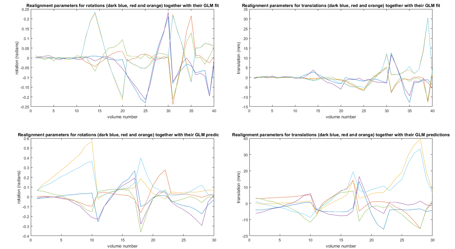
All measurements were performed on an 8 channel PTx 7T scanner (Philips) using the DICOs integrated into the eight 2kW amplifiers (CPC) for motion detection together with an 8 channel transmit coil (NOVA medical). Three single shot EPI sequences with 48 slices per volume (TR 2s) were acquired on a single volunteer. During the first scan (10 volumes) the subject was instructed to keep his head still. During the other two scans, consisting of 30 volumes with a manual start of each volume, the subject moved his head in between the acquisition of the volumes. Both head rotations and translations were performed. DICO data consisted of 8 times 48 forward and reflected RF pulses (asymmetric sinc gauss) per volume with 43 samples per pulse. Only the reflected RF waves were used for motion detection. EPI data was realigned (rigid body) to first volume of the first scan using the McFLIRT package from FSL. DICO signals were processed in Matlab. The maximum of the volume averaged reflected RF pulses (magnitude only) was used as the ‘motion detector’(MD) sample of a volume. The mean of the 10 MD samples obtained from the scan without head motion was subtracted from each of the 70 MD samples. The 40 mean corrected MD samples of the first and second scan (10 without motion and 30 with motion) were fitted to the corresponding rotations and translations obtained from the FSL image registration using a General Linear Model (GLM). The fitted GLM matrix was used to calculate the head motion of the subject based on the MD samples from the DICOs for all 3 scans. The correlation coefficient between the GLM and the FSL based realignment parameters was used to evaluate the quality of the GLM fit (scan 1 no motion and scan 2 motion) and the predicted motion (scan 3 motion).Results
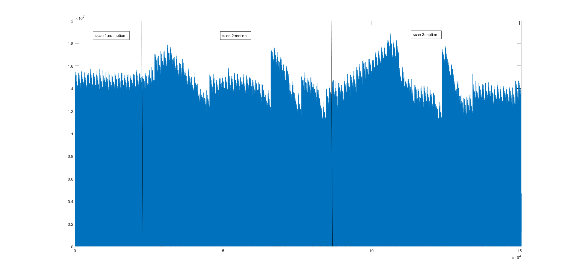
Figure 1 shows the raw reflected signals from a DICO of one of the RF channels. The sensitivity to motion is clearly visible in the raw data. The additional small variation on top of the data is related to the slice dependent change in RF frequency in combination with the high Q-factor of the coil. Figure 2 shows the realignment parameters from FSL and the mean corrected MD signals for scan 1 and 2 used to build the GLM. Figure 3 shows the realignment parameters together with the fitted signal for scan 1 and 2 (correlation coefficient 0.98), and the motion predicted by the model for scan 3 (correlation coefficient 0.87).Discussion
Reflected signals, as measured by DICOs, scattered from an 8 channel head coil can be used to detect head motion. In previous work [2] the full complex S-matrix was used to determine head motion. Here we only use the magnitude of reflected signals. In addition, no changes to the pulse sequence are needed and no additional scan time is required. Both gross rotations and translations can be predicted with reasonable accuracy. By using both the real and imaginary part of the signal the fits can probably be improved. In future work, the method will be used to update the scan orientation based on GLM predictions in real time.Conclusion
The proposed method for detection of subject head motion opens the way to improve high field high resolution image quality without a penalty in scan time or the need for sequence modifications.Acknowledgements
The first author wishes to thank Nico van den Berg for fruitful discussions on motion detection in MRI.References
[1] Buikman D, Helzel T, Roeschmann P. The RF coil as a sensitive motion detector for magnetic resonance imaging. Magn Reson Imaging. 1988;6:281–289.
[2] Papp D. Jaeschke S, Rieger S, Clare S, Hess A. Simultaneuous detection of cardiac, respiratory, and rigid body head motion using the scattering of a parallel transmit RF coil at 7T. Abstract 1168, ISMRM 2018.
Figures
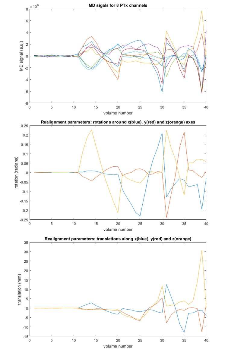
Top: processed signals from the DICOs for all eight transmit channels for scan 1 (no motion; first 10 volumes) and scan 2 (motion; volumes 11-40).
Middle and bottom: rotation (top) and translation (bottom) parameters from FSL McFLIRT realignment of the EPI volumes to the first volume of scan 1.
The data in the 3 panels was used to fit a GLM between the MD signals and the motion parameters.