1279
Improvement of SNR in MRgFUS with Strategic Design of Bath Medium and Transducer Ground Plane
Christopher M. Collins1,2, Ryan Brown1, and Daniel K. Sodickson1
1New York University School of Medicine, New York, NY, United States, 2Center of Advanced Imaging, Innovation, and Research (CAI2R)), New York, NY, United States
1New York University School of Medicine, New York, NY, United States, 2Center of Advanced Imaging, Innovation, and Research (CAI2R)), New York, NY, United States
Synopsis
By adjusting the electrical permittivity of the material in the bath and adding slots to the conductive ground for the ultrasound array in MR-guided focused ultrasound it is possible to go from a situation where the ultrasound array and associated fluid bath detrimentally affect the RF fields for MRI and prohibit effective imaging in the Region of Interest, to where they actually enhance it, thereby improving image quality.
Introduction
MRI-guided focused ultrasound (MRgFUS) is increasingly used to treat a number of medical disorders. The process requires both an array of ultrasound transducers with a common conductive ground and a large bath of fluid (transfer medium) suitable for both transmitting ultrasonic energy into the human body and cooling the surface of the body. Both of these adversely affect the ability of the radiofrequency magnetic fields needed for MRI to produce the images needed for effective MRgFUS.Simulation Methods and Results
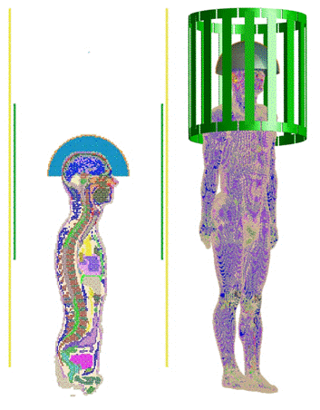
We show that by adjusting the electrical permittivity of the material in the bath, which can be accomplished with specific biologically inert additives (including, but not limited to sugar as found in food ingredients, glycerin, ethylene glycol, or PVP as found in contact lens lubricant, for example) and adding slots to the conductive ground (which could be bridged by filters allowing lower-frequency ultrasound currents to pass, maintaining its function as a ground plate for focused ultrasound) it is possible to go from a situation where the ultrasound array and associated fluid bath detrimentally affect the RF fields for MRI and prohibit effective imaging in the ROI, to where they significantly enhance it, improving the quality of image data in the ROI.Figure 1 shows the geometry used for simulation including birdcage body coil, RF shield, transducer array, fluid medium, and human body. Figure 2 shows results of electromagnetic field simulations of the RF magnetic (B1) fields produced without any ultrasound transducer array, with a conventional array and water bath, with a slotted ground plane in the transducer array and water bath, with a conventional array and lower-permittivity medium, and (in the two left-most images) with both a slotted ground plane and lower permittivity liquid medium.
Analysis of receive sensitivity (B1-/√Pdiss), proportional to SNR, shows a 5.7-fold improvement at the brain center by adding slots to transducer ground and reducing permittivity of the medium.
Discussion
Use of the conventional ultrasound transducer array and transfer medium results in low B1 field intensity and poor image quality in the brain, thus poor ability to perform MRgFUS. With strategic slots in the ground plate of the transducer array and adjustment of the permittivity of the transfer medium, the B1 field is enhanced in the brain, improving image quality and the ability to perform MRgFUS. The improved field distribution has sufficiently strong B1 that it is shown with an additional color scale at far right with five times the maximum value for full appreciation of the improvement in B1 field strength, which is proportional to image signal-to-noise ratio.In a previous work[1], electrical conductivity of the transfer medium (water bath) was increased with addition of salt, effectively damping standing-wave artifacts and producing a more homogeneous B1 field distribution in the imaging region, but also a lower overall B1 field strength, and thus lower overall image signal-to-noise ratio. We show that with strategic slots and reducing permittivity (rather than increasing conductivity) we can greatly improve B1 field strengths and thus image quality in the region of interest.
While it may be possible to achieve the desired permittivity with a variety of methods, the other requirements of the liquid bath – including viscosity, thermal conductivity, and heat capacity adequate for cooling of the outer surface of the head, and acoustic properties effective transmission of ultrasonic energy into the head – also would need to be maintained. Similarly, the producing slots in the ground plane that prohibit RF currents above about 100MHz but still allow for an effective continuous ground plane for focused ultrasound (operating in the high kHz) will require significant further development.
Acknowledgements
This work was supported by National Institutes of Health grant R01EB021277 and was performed under the rubric of the Center for Advanced Imaging Innovation and Research (CAI2R, www.cai2r.net) at the New York University School of Medicine, which is an NIBIB Biomedical Technology Resource Center (NIH P41 EB017183), and was informed by discussions and correspondence with Kim Butts Pauly at Stanford.References
1. Ianniello et al., Magn Reson Med 2018;80(1):413-419.
2. Leung et al., J Therapeutic Ultrasound 2015;3:P27
Figures

Figure
1. Simulation Geometry on mid-sagittal plane (left) including shield (yellow) and
in 3D rendering (right) without shield. Arrangement includes MRI coil (green)
and ultrasound transducer array (gold) with fluid bath medium (blue) placed on
the head for transcranial MRgFUS.

Figure 2. B1- distribution for different designs of transducer array and fluid bath
including (left to right) no transducer or medium, conventional transducer ground
plane and water medium, and different combinations of slotted transducer ground
plane and different mediums. Slotted transducer and medium with lower
permittivity (εr = 40) can
enhance, rather than reduce B1- in the ROI. Birdcage coil driven with 100V and color
scale (bottom) ranges from 0 to 2μT
for all but the right-most frame, which has a max value of 10μT.