1278
A compact vertical 1.5T human head scanner with shoulders outside the bore and window for studying motor coordination1Center for Magnetic Resonance Research, University of Minnesota, Minneapolis, MN, United States, 2Victoria University of Wellington, Wellington, New Zealand, 3Yale University, New Haven, CT, United States, 4Columbia University, New York, NY, United States, 5Centro de Imagens e Espectroscopia por Ressonância Magnética, Universidade de São Paulo in São Carlos, São Carlos, Brazil, 6Arizona State University, Tempe, AZ, United States, 7MGH/Harvard, Boston, MA, United States
Synopsis
A multi-disciplinary team of researchers in a multi-institutional consortium have designed and are building an easily relocatable head-only 1.5T MRI scanner weighing only ~500 kg. The goal is to develop a radically new type of MRI scanner that will enhance brain research, and ultimately, enable the diagnosis of neurological diseases in underserved populations throughout the world where MRI scanners are currently unavailable. To image with this system, pulse sequences have been developed and implemented to generate images using a highly inhomogeneous B0.
Introduction
MRI scanning of people living in remote locations, and the ability to study certain human behaviors, are largely inaccessible by current MRI technology. For example, any behavior involving motion, and especially those involving the upright real-time interaction with objects in natural environments, cannot be studied. Such studies are of enormous scientific interest, for example, in understanding the neuronal basis of motor planning, but also of considerable practical and clinical importance in order to eventually understand and address the motor deficits associated with injury, stroke, or disease which preclude everyday behaviors as important as feeding and reaching. In this project, we are building a compact head-only1.5T MRI scanner requiring only the head to be in the magnet bore, with a window for viewing outside the bore.Hardware Components of the Scanner
A CAD drawing in Fig. 1 shows the magnet, its stand, and chair. This second-generation high temperature superconductor (HTS) magnet is designed to allow the subject’s shoulders to be outside the magnet, with a window for viewing outside the bore. The magnet is cryogen free, operating at 30 K using a single stage pulse-tube cryocooler. The magnet has been designed to cool down rapidly (1-2 days), consuming only 5 kW power during steady state operation. A water-cooled multi-coil (MC) array consisting of 31 DC coils (Fig. 2) is used for dynamic shimming and for producing spatial-encoding fields for imaging (1,2). Each channel of the MC array can be driven independently and dynamically using a homebuilt multi-channel digital driver hardware (3) and commercial amplifiers with +/-5 A (Resonance Research Inc) for concurrent B0 shimming and linear or non-linear imaging. When configured to produce gradient fields, the MC array achieves a maximum of 10 kHz/cm linear field gradient in any of the three orthogonal directions. In addition, parabolic and hyperbolic field shapes up to 500 Hz/cm2 can be produced for enabling non-linear spatiotemporal encoding with methods such as STEREO (4). The magnet cryogenics have been optimized to minimize eddy current coupling between the MC array and the magnet. The digital spectrometer of this MRI system was specifically designed for advanced research capabilities, and includes eight transmit/eight receive RF channels and an additional field lock channel. The RF headcoil is tapered to conform to the tapered magnet bore and has a modular design allowing operation in either circularly-polarized or 8-channel parallel transmission mode.Software and Pulse Sequences of the Scanner
The drastically decreased size of the magnet comes at the cost of greatly diminished B0 uniformity, which after passive shimming is predicted to be ~300 ppm over the adult human brain. Obviously, narrowband pulse sequences like EPI will fail with such an inhomogeneous B0. Thus, for this project to be successful, methods must be identified and/or developed to permit high quality MR imaging using a highly inhomogeneous B0. To accomplish this, only certain classes of pulse sequences are feasible, and in some cases, new ones must be developed to produce the main types of contrast (T1, T2, T2*, diffusion) needed for the proposed research and clinical uses. In addition, the RF pulse and acquisition bandwidths in these pulse sequences must be maximized, within SAR and SNR limitations, to fully excite the brain and to minimize image distortion. Several sequences have been developed and implemented to image with a highly inhomogeneous B0, including short-TE gradient-recalled echo (5) and missing-pulse steady state free precession (MP-SSFP) (6). The user interface developed for this new MRI system consists of software implemented with PyMR, a framework for programming MR systems, which includes an Integrated Development Environment (IDE) to help MR methods designers create sequences with full graphical and unique programming capabilities (7, 8, 9). All that software is part of the operator console that allows high-level post-processing of acquired datasets. Using these features, sequence designers can easily implement the most sophisticated pulse sequences using some of the most modern tools available to software projects.Discussion
There is an urgent need for brain imaging technology that is more portable and less restricting than current MRI scanners. One way to address these issues is to decrease the size of the magnet to make a head-only system which does not confine the body; however, this approach leads to drastically reduced B0 homogeneity which, with current technologies, precludes high resolution imaging. In this project, we have addressed this problem by developing new hardware, as well as new acquisition and reconstruction methods, capable of producing high quality brain images despite extreme B0 inhomogeneity. The goal is to demonstrate the first-ever high-field (1.5T) MRI scanner requiring only the head to be inside the magnet bore and having a large window for viewing outside the magnet bore. The small size, weight, and power requirements of this MRI system will enable it to be transported and sited almost anywhere in the world and the MRI system can be brought to the subject rather than the other way around. By midyear 2020, system integration will begin, and by late 2020, we hope to acquire the first images.Acknowledgements
This work was supported by the National Institutes of Health grants U01 EB025153 and P41 EB015894.References
1) Rudrapatna SU, Fluerenbrock F, Nixon WT, de Graaf RA, Juchem C. Combined imaging and shimming with the dynamic multi-coil technique. Magn Reson Med;81(2):1424-1433, 2019
2) Juchem C, Mullen M, Kumaragamage C, DelaBarre L, Adriany G, Brown PB, McIntyre S, Nixon TW, Garwood M, de Graaf RA. Dynamic Multi-coil technique (DYNAMITE) MRI on human brain. 2019; Montreal, QC, Canada. p 0219
3) Nixon TW, McIntyre S, de Graaf RA. The design and implementation of a 64 channel arbitrary gradient waveform controller. 2017; Honolulu, HI, USA. p 0969
4) Snyder ALS, Corum CA, Moeller S, Powell NJ, Garwood M, MRI by steering resonance through space. Magn Reson Med 72:49–58, 2014
5) Mullen M, Kobayashi N, Garwood M. Two-dimensional Frequency-swept pulse with resilience to both B1 and B0 inhomogeneity. J Magn Reson 299:93-100, 2019
6) Kobayashi N, Idiyatullin D, Adriany G, Garwood, M. Magnetic resonance imaging under highly inhomogeneous B0 fields using missing-pulse steady-state free precession (MP-SSFP). 2017; Honolulu, HI, USA. p 5052
7) Pizetta DC, Lourenço GV, Silva DMDD, Vidoto ELG, Martins MJ, Tannús A 2015, Magnetic resonance system configuration and editing tools, Jaffray DA (Ed.), IUPESM 2015: Wold congress on medical physics & biomedical engineering, Toronto, Canada, Jun 7-12, p667-668.
8) Pizetta DC, Shimada DHD, Falvo M, Souza PVBD, Silva LR, Bittencourt HP, Vidoto ELG, Martins MJ, Tannús A, Universidade de São Paulo 2017, PyMR - a framework for programming magnetic resonance systems, BR-512019001829-0.
9) Pizetta DC 2018. PyMR: a framework for programming magnetic resonance systems, PhD Thesis, Universidade de São Paulo, São Carlos, doi:10.11606/T.76.2019.tde-06052019-103714.
Figures

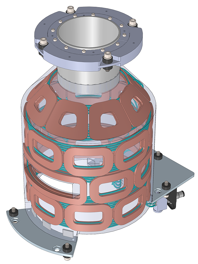

Figure 3: TEM
transceiver coil with eight independently configurable channels, each
with a swappable T/R switch or CMOS circulator board (green).