1268
Improving Image Quality in Transcranial Magnetic Resonance Guided focused Ultrasound Using a Conductive Screen
J. Rock Hadley1, Henrik Odeen2, Robb Merrill2, Sam Adams2, Viola Rieke2, Allison Payne2, and Dennis Parker2
1Radiology and Imaging Sciences, University of Utah, Salt Lake City, UT, United States, 2University of Utah, Salt Lake City, UT, United States
1Radiology and Imaging Sciences, University of Utah, Salt Lake City, UT, United States, 2University of Utah, Salt Lake City, UT, United States
Synopsis
This work uses an RF screen, placed over the top of a human skull phantom, to reduce image banding artifacts that are common in transcranial transducer MRI. The goals of the study are to improve imaging homogeneity over the region of the brain by changing RF field patterns that cause the artifacts, and to find a solution that doesn’t attenuate or distort the ultrasound properties of the transducer. Hydrophone and focused ultrasound heating studies are performed to measure ultrasound screen transparency and MRI studies are performed to evaluate the effects the screen has on homogeneity and artifact reduction.
Introduction
This work investigates a solution to the Radio Frequency (RF) signal banding artifacts that can occur with Magnetic Resonance Imaging (MRI) of the head inside an Insightec Transcranial Transducer (ITT), see Figure 1. We hypothesized that these artifacts are caused by the dielectric properties of the large water bath and the waveguide nature of the boundary conditions of the transducer ground plane. Furthermore, we hypothesized that the banding artifacts could be shifted by the insertion of a conductive screen in the space between the transducer and the patient’s head. The two major goals in this work were to: 1) modify the electromagnetic field behavior of the B1-transmit and receive field inside the transducer to improve field homogeneity and eliminate the banding artifacts in the MR images, and 2) Do so in such a way that the ultrasound transmission into the human skull would not be attenuated or otherwise negatively affected.Methods
A human skull, with a Data Quality Assurance phantom mounted inside, was imaged in the ITT (650 kHz) with a 2D Gradient Echo pulse sequence using a Skyra MRI with body coil (Siemens Medical Solutions). Imaging was done with and without a 24-cm diameter conductive screen positioned over the top of the head as shown in Figure 2. Two different imaging studies were performed using the RF screen. The first study was using the screen in position #1, where it had slipped off the skull, but the images look interesting so the study was continued. the second imaging study was done with the screen in position #2, the originally desired configuration (see Figure 3). The copper screen was made from un-insulated bronze wire that was approximately 0.25-mm in diameter, and the wires formed small mesh squares that were 2mm in diameter. The screen was held in position with string and small rubber spacers that were used to isolate the screen from the skull. Hydrophone measurements were performed with the screen to assess ultrasound transparency. The screen was positioned in the hydrophone tank perpendicular to the propagating beam at 2.5, 4.5 and 6.5-cm from the focus and at 45° to the beam propagation. The transducer was a 256 element semi-rectangular transducer with a 14.4x9.8-cm aperture ultrasound transducer (Image Guided Therapy/Imasonic, France) with a 10-cm focal length and operating at 940 kHz. Heating studies in the ITT transducer were performed with and without the screen in place. For each situation, five different heating runs were performed and the standard deviation of the temperatures and FWHM of the temperature distributions were measured.Results
The screen caused pressure attenuations on the order of 6% for the different flat and angled configurations compared to the water-only hydrophone measurement. Imaging results (see Figure 3) show improved signal homogeneity in the brain region with the screen in position. The Signal-to-Noise Ratio (SNR) was improved on the order of 50% in regions of no artifact and by factors of 5 and more in regions of artifact. Temperature accuracy was improved and more homogeneous throughout the brain when using the screen. The mean and standard deviation of the temperatures and temperature profile widths from the heating studies are shown in Figure 4.Discussion
Figure 4 demonstrates that there was negligible change in the peak temperature with the screen although the temperature distribution (FWHM) was reduced with the screen in place. The increased SNR with the screen in place resulted in improved precision of the peak and FWHM of the temperature distribution measurements. Hydrophone and force balance studies indicate that the heating through the screen should be reduced compared heating without the screen, however, the hydrophone studies don’t account for the lower frequency and different geometry of the transducer. In the skull heating studies, not all of the transducer elements are required to shoot through the screen.Conclusion
In conclusion, for the various imaging parameters of this study, the copper screen: 1) provided significant improvement in field homogeneity throughout the region of the brain by eliminating the banding artifacts; 2) increased SNR by a factor of 1.5 to 6 over the region of the brain, and 3) was essentially transparent to the ultrasound beam. We hypothesize that these improvements occurred because the copper screen changed the field boundary conditions and modified the waveguide nature of the transducer ground plane. Future work will more fully characterize and optimize the RF screen functionality.Acknowledgements
The Mark H. Huntsman Endowed Chair and NIH grant 1R01 EB028316References
No reference found.Figures
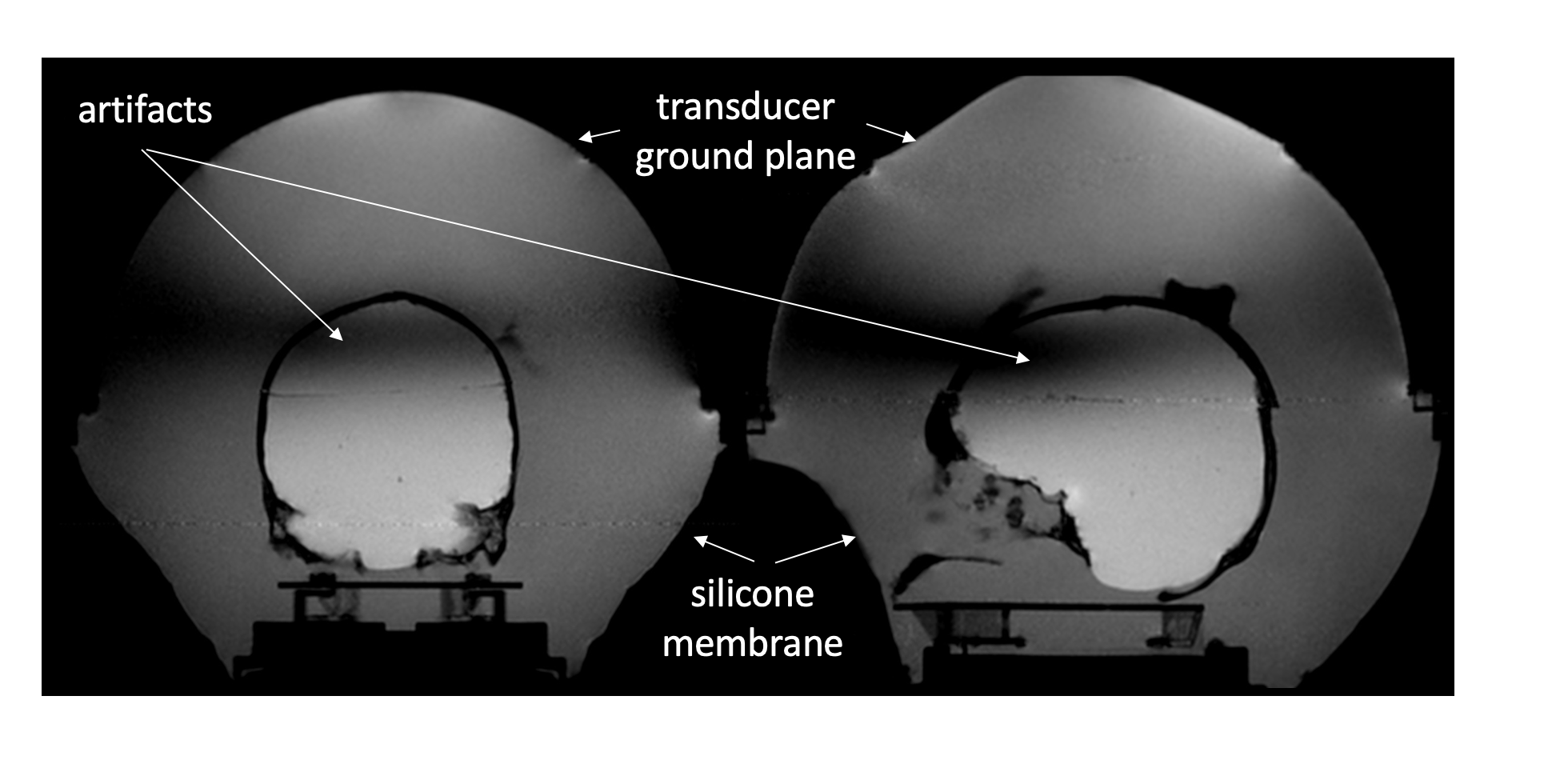
Figure 1. Example imaging artifacts in the region of the
brain that were observed while using an Insightec transcranial transducer,
operating at 650 kHz, with a Siemens 3T Skyra scanner with a proton resonant
frequency of 123 MHz.
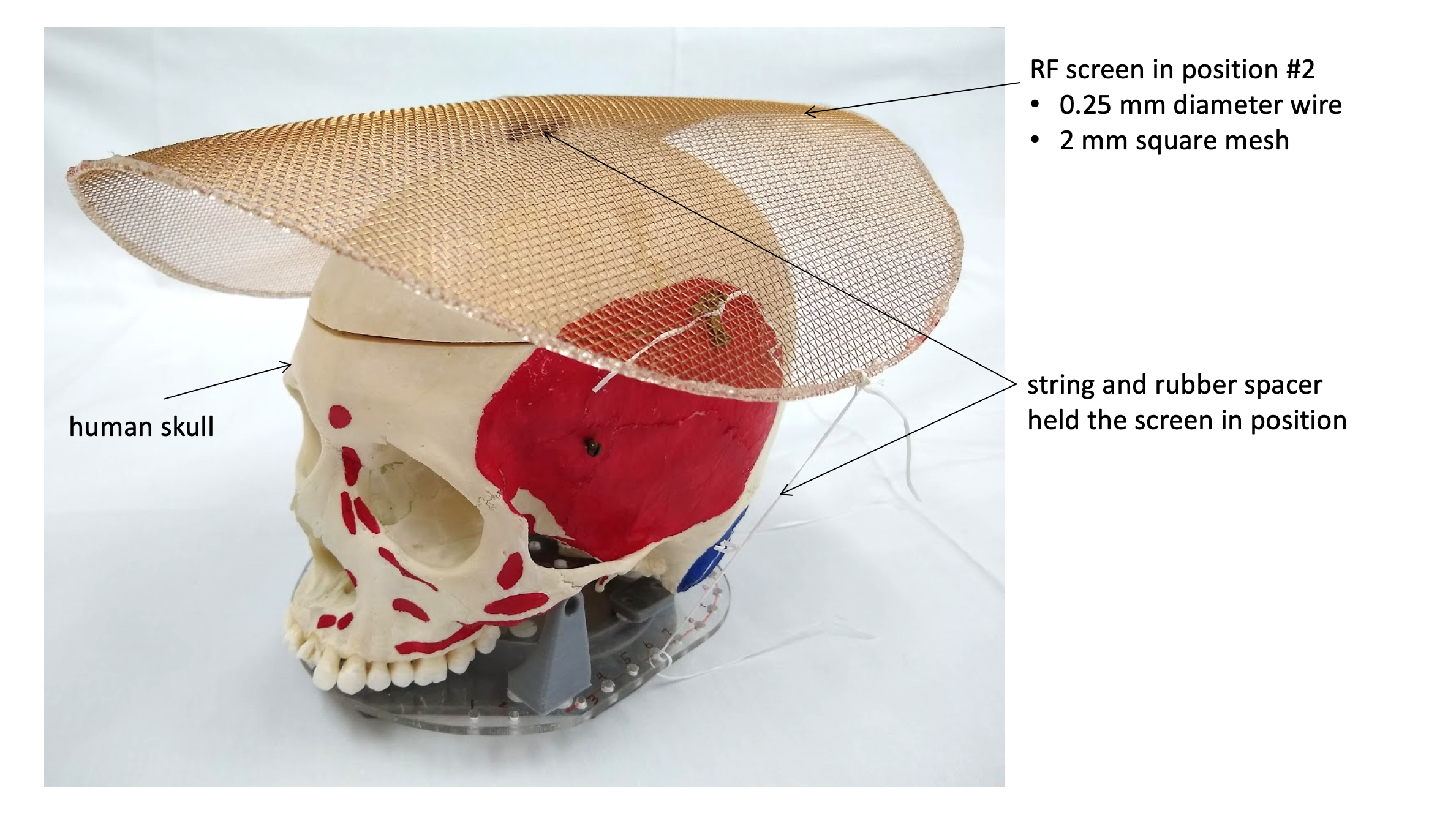
Figure 2. Human skull phantom: A Data Quality Assurance phantom was mounted
inside the skull and the 24cm screen was placed around the top of the
skull. It was held in position with
string, and rubber spacers were used as standoffs to separate the screen from
the skull. The screen was made of bronze
wire that was 0.25mm in diameter and the hole size of the screen was 2mm.
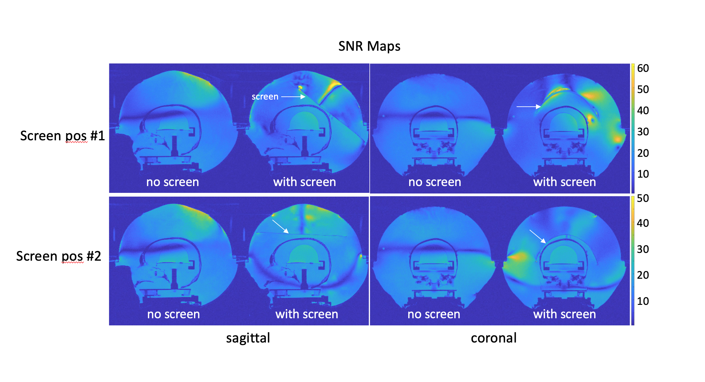
Figure 3. Imaging
results for both screen positions show that the signal homogeneity in the brain
region when using the RF screen was greatly improved compared to images without
the screen, and that the banding artifacts could be removed. These SNR maps also show the SNR gains that
were achieved using the screen. SNR was
improved by a factor of approximately 50% in regions where there was no banding
artifact, and in regions of severe banding artifacts the SNR was increased by a
factor of about 6.
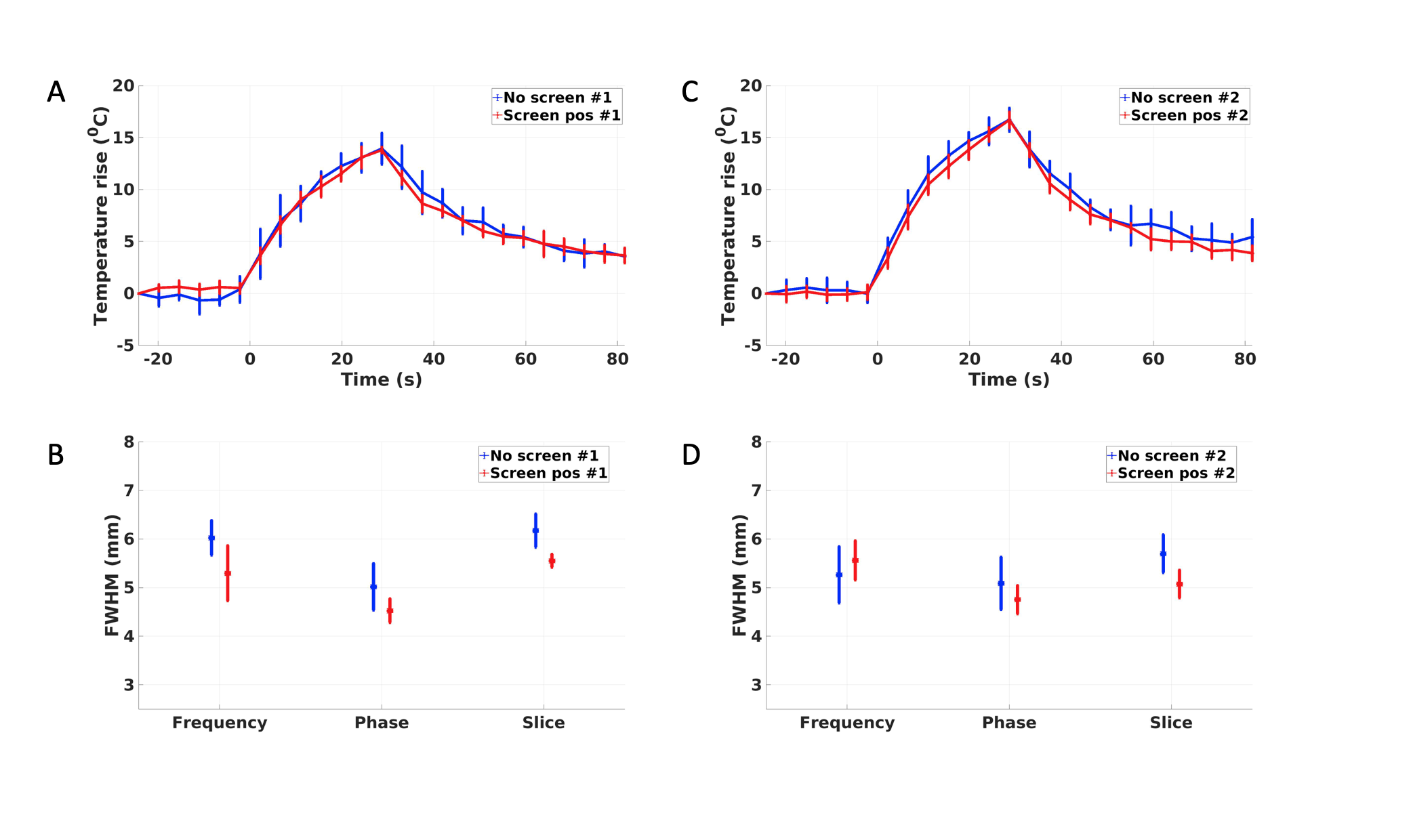
Figure 4. Temperature
profiles and Full Width at Half Max (FWHM) ameasures for the screen in position
#1 (A and B) and position #2 (C and D). studies
for both screen positions show that heating profiles were not noticeably
affected by the screen. A and C) Peak
mean temperatures and standard deviations were essentially the same with and
without the screen in position. B and D)
show that the FWHM was generally smaller when using the screen with lower
standard deviation.