1262
Dual-Stream iPRES-W Head Coil Array for MR Imaging, Wireless Respiratory Tracking, and Wireless Localized B<sub>0</sub> Shimming1Brain Imaging and Analysis Center, Duke University, Durham, NC, United States, 2Medical Physics Graduate Program, Duke University, Durham, NC, United States, 3GE Healthcare Inc., Aurora, OH, United States
Synopsis
The integrated RF/wireless coil design enables MRI imaging and wireless data transfer with the same coil thereby reducing the number of wired connections in the scanner. Here, we implement this design onto a 48-channel head coil array to enable two independent wireless data streams for two separate applications, specifically, wireless 1) control of the DC currents used for B0 shimming and 2) respiratory tracking with a respiratory belt. In vivo experiments in the brain showed that this coil array significantly reduced B0 inhomogeneities (-41%) and EPI distortions while simultaneously streaming respiratory data from the subject without data loss.
Introduction
In MRI, data transfer between the scanner subsystems (e.g., RF coils and shim coils) or peripheral systems (e.g., respiratory and cardiac monitoring) and the computers outside the scanner room currently requires a network of carefully routed wired connections, which take up space within the scanner and require costly RF filters, baluns, and cable traps to preserve data integrity and MR image quality. To reduce the number of wired connections, a novel integrated RF/wireless coil design has recently been developed, in which RF currents at the Larmor frequency (e.g., 127 MHz for 3T scanners) and in a wireless communication band (e.g., 2.4 GHz for WiFi) can flow on the same coil to enable simultaneous MR image acquisition and wireless data transfer for different applications, respectively, without requiring any scanner modifications or additional antenna systems within the scanner bore1,2.Previously, this coil design was combined with the integrated parallel reception, excitation, and shimming (iPRES) coil design3, in which a DC current can also flow on the same coil to enable wireless localized B0 shimming1,4. In this iPRES-W coil design, the coil wirelessly receives commands from outside the scanner room to control an MR-compatible power supply in the scanner bore, which delivers the DC currents for shimming. In this work, the integrated RF/wireless design is implemented onto a 48-channel head coil array to enable two independent wireless data streams for two separate applications, specifically, wireless 1) control of the DC currents used for B0 shimming and 2) respiratory tracking with a respiratory belt.
Methods
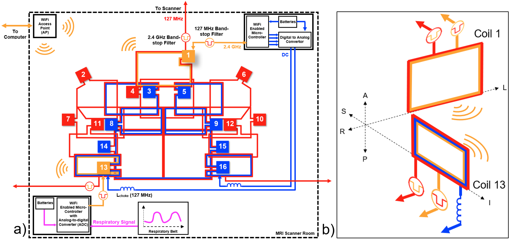
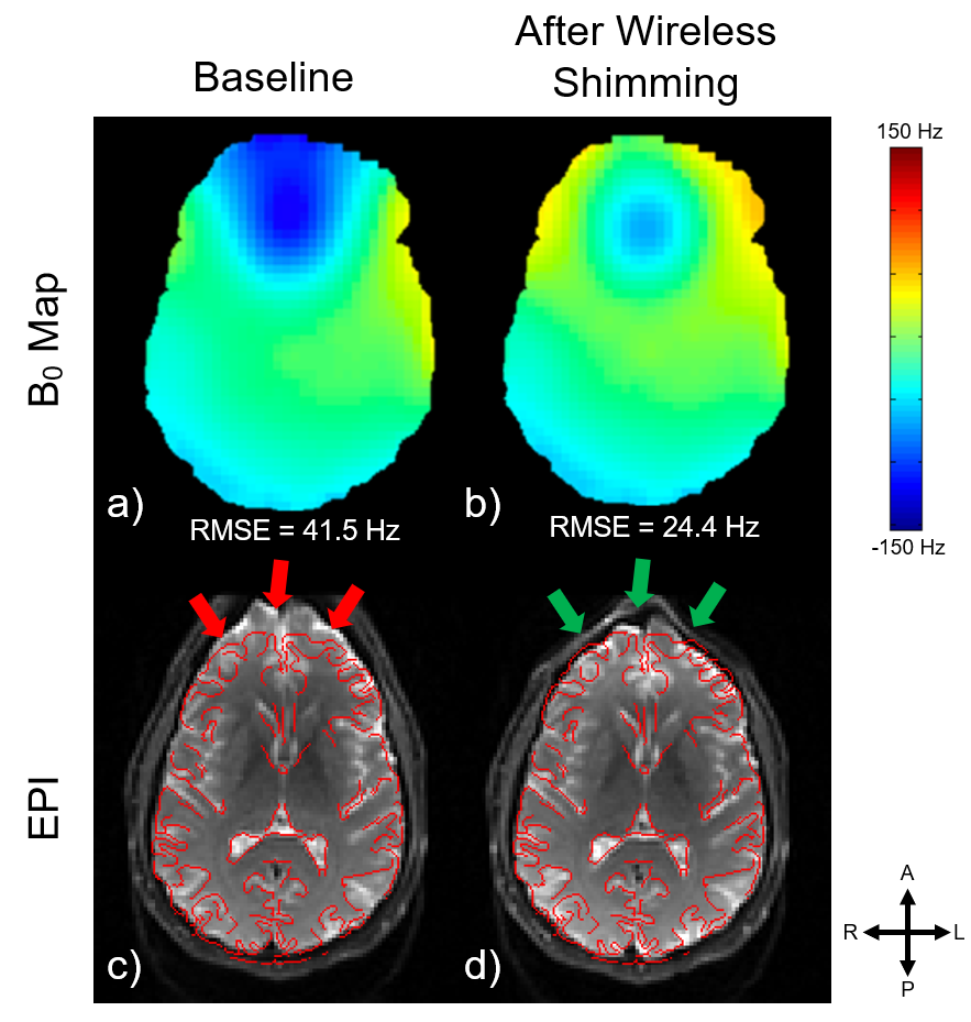
First, the 16 anterior coil elements of the 48-channel head coil array were modified into iPRES coil elements by adding: inductors to bypass the capacitors; an MR-compatible battery pack to provide the DC currents for B0 shimming (range: ±2 A, resolution: 4 mA); and inductive chokes to provide RF-isolation between the coil elements and the battery pack to maintain a high SNR3. Next, two of the iPRES coil elements were modified into iPRES-W coil elements by adding 127-MHz and 2.4-GHz band-stop filters between each of these coil elements and the WiFi micro-controller or the preamplifier, respectively1, to enable two wireless single-input single-output data streams (Fig. 1a, orange). The two iPRES-W coil elements were chosen to be spatially orthogonal to provide a high RF-isolation (S21 ~ -35 dB) between them, which reduces coupling and increases the amount of power radiated from each coil element in the WiFi frequency band (Fig. 1b).One of the iPRES-W coil elements (Fig. 1a, coil 1) was used to wirelessly control the DC currents for B0 shimming. In this work, 8 of the iPRES coil elements were connected to the battery pack and used for shimming (Fig. 1a, blue). The optimal DC currents to shim a slice were determined by minimizing the root-mean-square error (RMSE) between a combination of basis B0 maps acquired on a phantom3 with 1 A separately applied in each iPRES coil element and a B0 map acquired in the brain of a healthy volunteer. The commands to control the DC currents were then wirelessly transmitted from outside the scanner room to the WiFi micro-controller of the iPRES-W coil element via an access point (AP) placed on the scanner room wall. B0 maps and spin-echo EPI images (2 x 2 mm resolution) were acquired on a 3T scanner with and without DC currents to assess the B0 shimming performance.
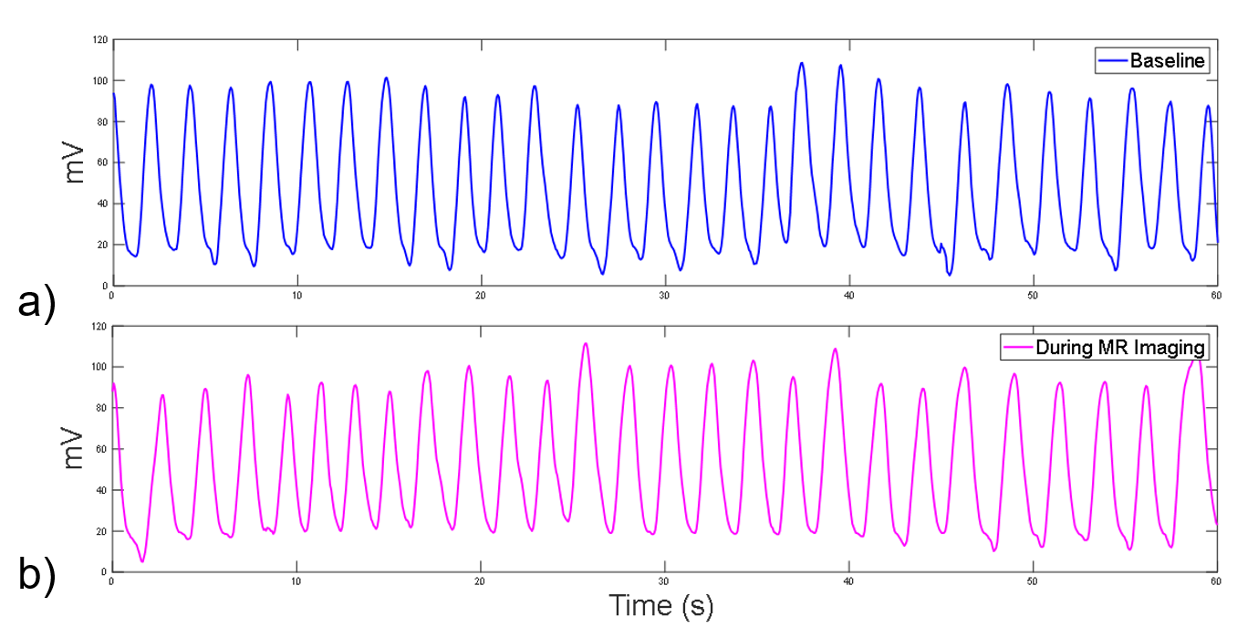
The other iPRES-W coil element (Fig. 1a, coil 13) was used to perform real-time wireless respiratory tracking during MR image acquisition and shimming. The signal from a force-sensitive respiratory belt placed around the subject’s abdomen (Fig. 1a, magenta) was wirelessly transmitted from the iPRES-W coil element to the AP, then recorded on a computer outside the scanner room.
Results
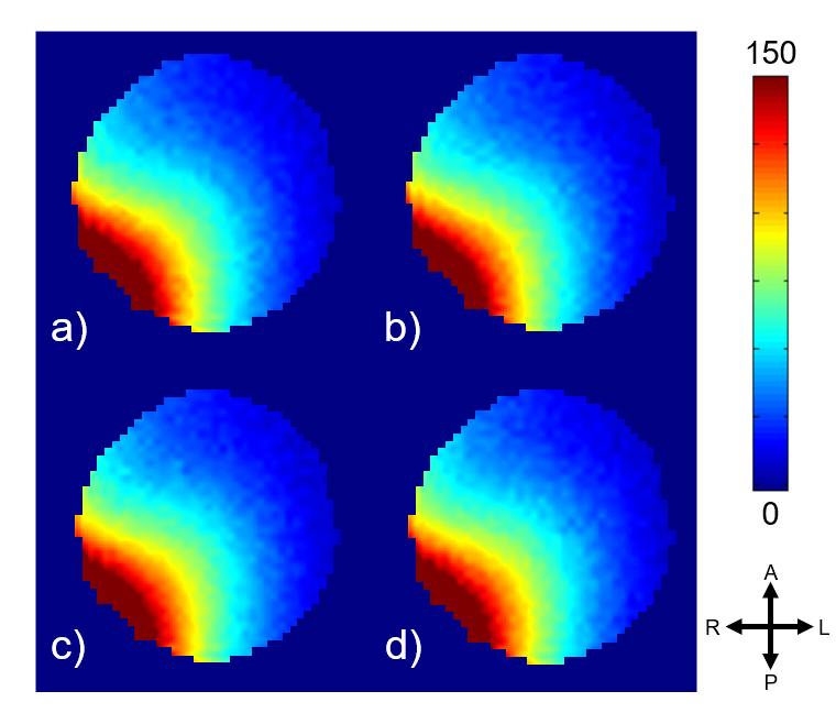
The iPRES-W coil element modifications did not significantly impact the SNR with or without wireless transmission of the respiratory data (Fig. 2). Wireless shimming significantly reduced the B0 field inhomogeneity (-41%) and the geometric distortions in the spin-echo EPI images (Fig. 3, green arrows). The B0 shimming performance can be further improved by implementing iPRES into all coil elements. Additionally, the wirelessly transmitted respiratory signal was similar with or without simultaneous MR image acquisition and wireless localized B0 shimming (Fig. 4).Discussion and Conclusion
These results demonstrate that the dual-stream iPRES-W head coil array can perform simultaneous image acquisition and wireless data transfer for multiple applications, specifically, for wireless localized B0 shimming and respiratory tracking. In future applications, the respiratory signal can be used to perform dynamic shimming with the iPRES coil elements and correct for respiration-induced B0 variations, which has previously been done using wired rather than wireless connections5. In addition, the integrated RF/wireless coil design can be used for wireless data transfer in other applications, such as cardiac monitoring or field monitoring with NMR probes.Acknowledgements
This work was in part supported by GE Healthcare, grants R21 EB024121, R01 NS075017, R01 EB028644, and S10 OD021480 from the National Institutes of Health, and the Duke-Coulter Translational Partnership.
References
- Darnell D et al. Integrated radio-frequency/wireless coil design for simultaneous MR Image acquisition and wireless communication. Magn. Reson. Med. 2019;81:2176-83
- Bresticker J et al. Simulations of Integrated Radio-Frequency/Wireless Coil Designs for Simultaneous MR Image Acquisition and Wireless Communication. Proceedings of the ISMRM, May 2019, Montreal; pg. no. 1541
- Truong TK et al. Integrated RF/shim coil array for parallel reception and localized B0 shimming in the human brain. NeuroImage 2014; 103;235-40
- Cuthbertson J et al. A 4-Channel iPRES-W Coil Array for Simultaneous MR Image Acquisition and Wirelessly-Controlled Localized B0 Shimming of the Spinal Cord. Proceedings of the ISMRM, May 2019, Montreal; pg. no. 1489
- Rios L et al. Integrated AC/DC coil and dipole Tx array for 7T MRI of the spinal cord. Proceedings of the ISMRM, May 2019, Montreal; pg. no. 0220
Figures