1253
First Experiences with a Three-Bore Conduction-Cooled Cryogen-Free Extremity Scanner
Jerome L. Ackerman1,2, Shahin Pourrahimi3, Marcus Donaldson1,2, Nadder Pourrahimi3, Julien Rivoire4, Julien Muller4, Hizami Murad4, Ouri Cohen5, and Isabela Choi1
1Martinos Center, Dept of Radiology, Massachusetts General Hospital, Charlestown, MA, United States, 2Department of Radiology, Harvard Medical School, Boston, MA, United States, 3Superconducting Systems, Inc., Billerica, MA, United States, 4RS2D, Mundolsheim, France, 5Memorial Sloan Kettering Cancer Center, New York, NY, United States
1Martinos Center, Dept of Radiology, Massachusetts General Hospital, Charlestown, MA, United States, 2Department of Radiology, Harvard Medical School, Boston, MA, United States, 3Superconducting Systems, Inc., Billerica, MA, United States, 4RS2D, Mundolsheim, France, 5Memorial Sloan Kettering Cancer Center, New York, NY, United States
Synopsis
We developed an MRI scanner designed for limb scanning based on a novel conduction-cooled three-bore cryogen-free 1.5T magnet. The two outer bores accommodate the unscanned leg for enhanced patient comfort. We describe the issues relevant to this unique magnet and scanner, including our solution to the field instability problem common in conduction-cooled MRI magnets. By recording the field variation as a function of the phase of the cold head cycle, a compensating waveform may be played out via the numerically-controlled oscillator of the scanner to reduce the periodic field excursion from 25 Hz to about 1-2 Hz.
Introduction
We developed a compact, easy to site prototype limb scanner for orthopedic and metabolic bone disease applications. Using a prototype (Alpha) tilted, conduction-cooled, cryogen-free, highly shielded 1.5T magnet with three bores allows the scanner to be installed in an extremely compact space, eliminates the need for a quench duct or helium refills, and offers a more comfortable patient experience. This presentation reports our experience in recovering after a quench, the reduced cooling efficiency operating the magnet at a tilt, and suppressing the field shift caused by the periodic cycling of the cold head. Because conduction-cooled magnets may have a small cyclic field variation arising from the periodic cold head regenerator displacement or temperature cycling, or from mechanical vibrations, these magnets may suffer “motion” artifacts caused by the field fluctuation rather than actual motion with respect to the gradient. Results of testing of the Alpha magnet have been used to design a Beta magnet with improved performance and patient comfort.Methods
The magnet was designed and produced by Superconducting Systems under NIH grant R44AR065903 and sited at MGH to be incorporated into a scanner.1 The console is based on an RS2D Chameleon 3 spectrometer (comprising a digital RF transceiver, pulse sequencer, gradient drivers, shim supplies and RFPA), a Ningbo Yici (China) EC04 gradient coil, three PCI (Performance Controls, Inc., Montgomeryville, PA, USA) GA-300 amplifiers powered with a Sorensen-Ametek (Berwyn, PA, USA) SGA330X15C-1EAA DC power supply. The magnet was cooled with a Sumitomo (SHI Cryogenics, Allentown, PA, USA) SRP-082B2 pulse tube cold-head with F70LP helium compressor. RF coils were either designed and built in-house or were modifications of commercial devices retuned to 1.5T. The magnet diode temperature sensor was read with a Lake Shore Cryotronics (Westerville, OH, USA) 211A monitor. The scanner host computer records magnet temperature, compressor diagnostics, gradient and water temperatures via serial ports.Results
Tilting the magnet modestly considerably enhances patient comfort for arm and leg scanning, but can subject internal magnet structures to undue strains and displacements, and would be particularly challenging were the coil to be cooled in a liquid helium bath. Pulse tube refrigerators exhibit lower vibration than Gifford-McMahon refrigerators but suffer reduced efficiency when operated at an angle from vertical. We selected a modest tilt of 7 degrees, and found that the coil temperature increased by only 0.2 K to about 4.8 K. Although a spontaneous quench of this magnet has never occurred, quenches due to interrupted compressor electrical power or cooling do occasionally occur. A quench will result within about 30 minutes of an interruption, but is uneventful as there is no helium release. The magnet temperature rises to about 50 K, and requires about 12 hours of cooling to resume operation at 5 K or less. The cold head-induced periodic field fluctuation is about 25 Hz peak-to-peak, and with a period of about 2 Hz causes severe “motion” artifacts. Hypothesizing that the fluctuation was due to a cyclic magnetization of the regenerator, we tried placing a superconducting shield around the cold head, but this failed to eliminate the fluctuation. External actively powered compensation coils about the cold head also failed; in this case the 2 Hz magnetic waveform did not penetrate the superconducting shield. The spectrometer is equipped with a memory that reads out a modulation of the spectrometer’s numerically controlled oscillator (NCO) which generates the base operating frequency. It runs asynchronously with respect to the pulse sequence. The uncompensated B0 waveform was measured as a function of the phase of the cold head pressure cycle, synchronized via the compressor’s pressure sensor. This waveform was used to modulate the NCO, reducing the field fluctuation to about 1-2 Hz peak-to-peak. Preliminary tests of the field variation while the magnet was in motion (tilted up and down by several degrees at about a 1 Hz rate, or pushed horizontally back and forth by ~50 cm) showed excellent stability of the NMR spectrum.Discussion
We found that the periodic field fluctuation that may occur with conduction cooled MRI magnets, where the cryocooler must have tight thermal (and therefore possibly significant mechanical) coupling to the main coil, can be compensated with feedforward compensation using the measured uncompensated field waveform. This worked for our magnet, which exhibited primarily a spatially uniform periodic field shift. If the field spatial uniformity also fluctuates, a more complex higher order correction will be necessary, and cannot be accomplished purely by the means reported here (i.e., frequency offset).The static tilt of the system as it is currently configured is not a barrier to magnet operation, and the motion tests suggest that it may be possible to use such a system for dynamic biomechanical studies in which the entire magnet and subject move together, thereby suppressing motion artifacts due to relative motion of the subject with respect to the magnet and gradient coil.
Conclusions
A prototype extremity MRI scanner based on a highly compact, conduction-cooled, three-bore cryogen-free 1.5T tilted superconducting magnet is feasible. By proper compensation of cold head induced field fluctuations, a highly stable magnetic field may be achieved. It is possible to translate and tilt the magnet while in operation.Acknowledgements
Funding was provided by grant R44AR065903 from the National Institute of Arthritis and Musculoskeletal and Skin Diseases. I. C. received funding from the Fulbright Foundation.References
-
Pourrrahimi
S, Ackerman JL, Williams JEC, Pourrahimi N, Kaplan A. A compact affordable three-bore
cryogen-free superconducting magnet for extremity imaging, ISMRM, Honolulu, HI,
April 22-27, 2017.
Figures
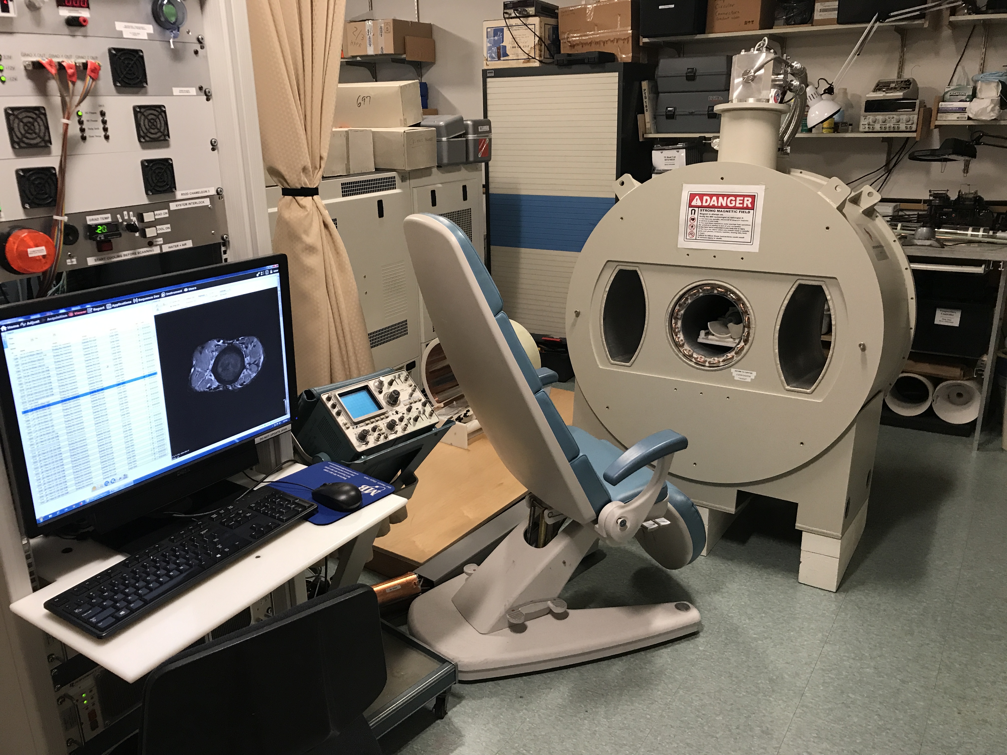
Figure 1. Tiltable cryogen-free three-bore
compact superconducting magnet. The central bore is active, and either of the
side bores comfortably accommodates the knee not under examination. The active
shield coils surround the side bores as well as the central bore. The magnet is supported at a 7 degree tilt
(downward away from the patient). The fringe
field shielding is highly effective; note the close proximity of the CRT
oscilloscope and workbench to the magnet.
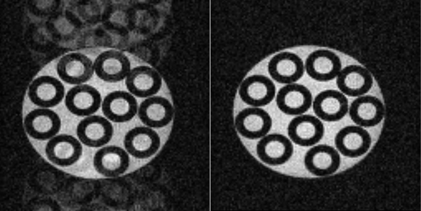
Figure 2. Gradient echo image of a phantom,
35 mm field of view, 128 x 128 matrix, TE 11 ms, TR 200 ms, 25 s scan. Left: field
compensation off. Right: field compensation on.