1251
Three-dimensional in-vivo human Imaging on a 50 mT low-cost portable Halbach Array1C.J. Gorter Center for High Field MRI, Leiden University Medical Center, Leiden, Netherlands, 2Circuits and Systems, Delft University of Technology, Delft, Netherlands
Synopsis
We show the first 3D in-vivo images acquired from our custom-built 50 mT low-cost Halbach based portable MRI scanner. 3D Images of a knee were acquired with a ~2 mm isotropic resolution using a 3D turbo spin echo sequence in less than 12 minutes. Gradient non-linearity induced image distortions are minimal within the central ~10 cm of the magnet bore length, but require correction beyond this point. These results represent the latest step towards our goal of creating a fully portable MRI scanner targeting pediatric neuroimaging in the developing world for less than 30 000 euros.
Introduction
MR imaging has become a vital tool in clinical settings, yet the high hardware cost as well as both technical and resource challenges in keeping MRI scanners fully operational has resulted in a distinct lack of access to this technology for a significant fraction of the world’s population [1]. In this work we present progress on developing a low-cost permanent magnet based MRI scanner targeted at imaging paediatric hydrocephalus at a 3 x 3 x 5 mm resolution in low-resource settings. We previously presented the design of a highly homogenous Halbach array for generating the required B0 field [2]. In this work we have incorporated gradient coils designed using a modified target-field approach [3] to acquire in-vivo images of the knee.Method
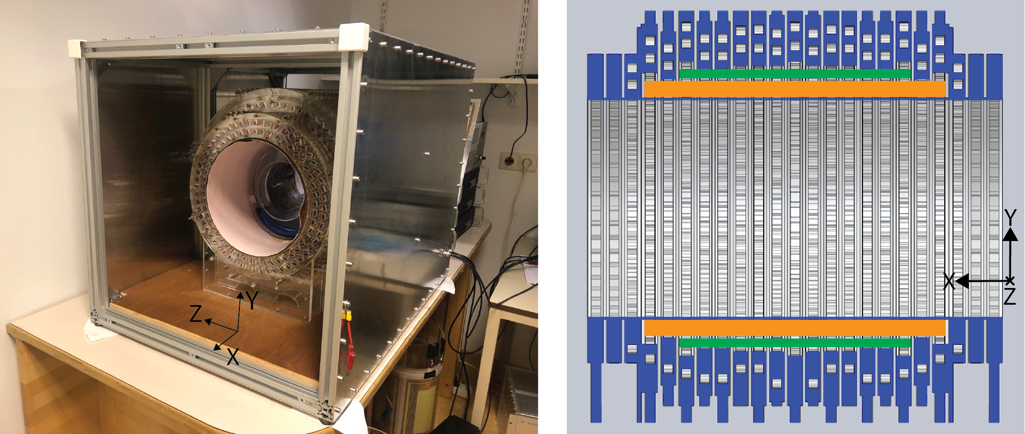
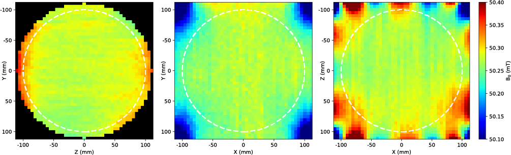
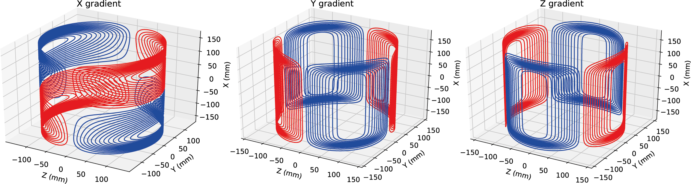
The B0 field is produced by a 50 cm long, Halbach array with a 27 cm inner diameter (figure 1), a B0 field strength of 50 mT and a homogeneity of 2400 ppm over a 20 cm sphere in the isocentre of the magnet [2], see figure 2. Gradient coils were designed using the Target Field Method adapted to produce their encoding fields commensurate with the transverse B0 direction of the Halbach array, see figure 3. Gradients coils were constructed using 1.5 mm diameter copper wire pressed in to 3D printed formers. The efficiencies of the X, Y and Z gradient coils are 0.59, 0.95 and 1.02 mT/m/A, respectively. The inductance of the X gradient is 180 µH with a resistance of 0.37 Ω, the inductance of the Y and Z gradients are both 225 µH with a resistance of 0.4 Ω. The gradients were driven with two AE Techron 7224 and one AE Techron 2105 amplifiers (Elkhart, IN, USA). A Magritek Kea2 spectrometer (Aachen, Germany) was used to generate gradient wave forms and RF pulses as well as digitising the generated echoes. The RF pulses generated by the spectrometer were amplified by a custom built 1 kW RF amplifier (adapted from [4]) and transmitted to a 15 cm long, 15 cm diameter solenoid used as an RF transceiver through a T/R switch built in to the spectrometer. The magnet was placed inside a 80 cm long faraday cage constructed using aluminium plates and extrusion profiles. To eliminate noise introduced in to the system by the human body, the part of the body that extended out of the Faraday shield was wrapped in a conductive fabric (Holland Shielding Systems, Dordrecht, the Netherlands). A phantom was constructed of 8 mm diameter, 30 mm long tubes filled with water, evenly spaced 17 mm apart on a rectangular grid to map geometric distortions in the images due to gradient non-linearity.Phantom images were acquired using a 2D spin echo sequence with the following parameters: FoV: 200 x 200 x 30 mm for the transverse images and 256 x 256 x 30 mm for the coronal images, acquisition matrix: 128 x 128, TR/TE: 6000 ms/100 ms, acquisition bandwidth: 20 KHz, no signal averaging, acquisition time: 12 minutes 48 seconds. In-vivo knee images were acquired from a healthy volunteer using a T1 weighted 3D RARE sequence with the following parameters: FoV: 256 x 256 x 256 mm, acquisition matrix: 128 x 128 x 128, TR/TE: 130 ms/15 ms, acquisition bandwidth: 20 KHz, echo train length: 3, no signal averaging, acquisition time: 11 minutes 50 seconds. The RF pulses were non-selective. The acquired k-space data was filtered using a squared sine bell filter and reconstructed using the Numpy v1.16 FFT library in Python 3.7.
Results
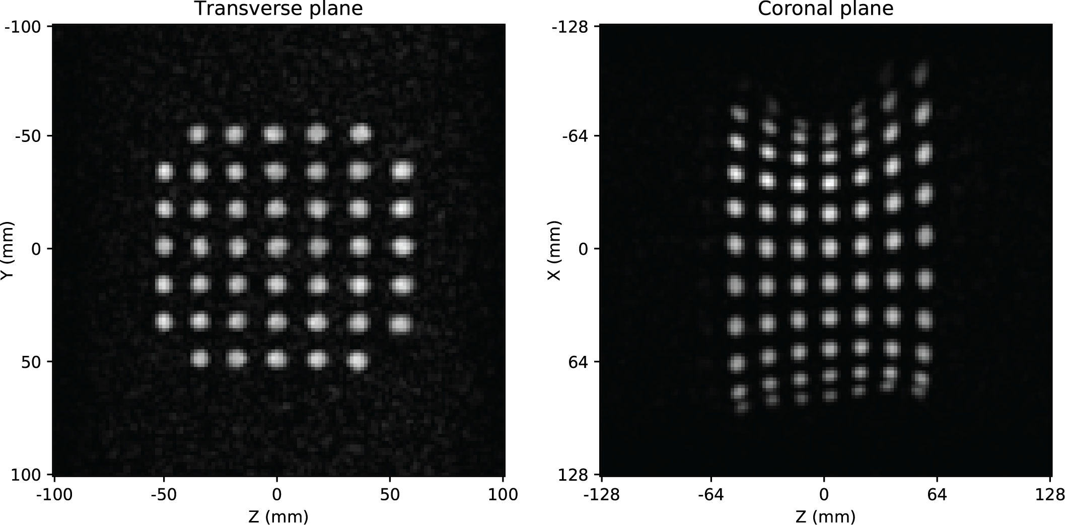
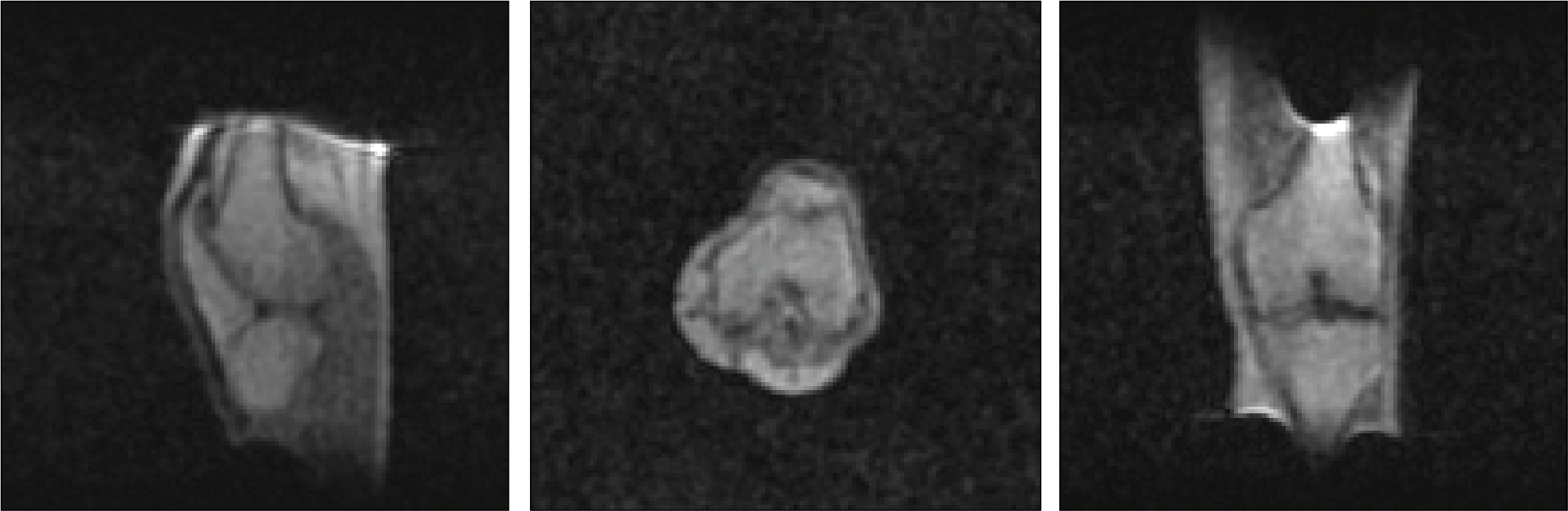
Figure 4 shows images acquired of the geometric phantom. No significant geometric distortions are visible in the central transverse plane, however, clear warping is visible in the coronal plane as the distance from the isocentre increases, closely matching simulations of the fields generated by the gradient coils, and with relative magnitudes similar to those on conventional clinical scanners. Figure 5 shows in-vivo images acquired of the knee. Major structures in the knee can be differentiated with clear contrast between muscle, fat and cartilage. Image distortions, particularly visible in the coronal image, closely match those seen in Figure 4.Discussion
In-vivo images have been acquired with an SNR of 26, sufficient to clearly delineate different structures in the knee joint and show the feasibility of neuroimaging in paediatrics at, or exceeding, the targeted 3 x 3 x 5 mm resolution. Images are shown without correction for gradient non-linearities (the main cause of artefacts in these images). Simulations indicate temperature induced B0 drift causes by heat radiated from the body, typically less than 1 kHz an hour, only causes minimal blurring of the reconstructed images with a full-width-half-maximum (FWHM) of the point spread function (PSF) of around 1.4 pixels, comparable to the FWHM of the PSF caused by T2 relaxation in the muscle during the RARE echo train. Future work in this project will focus on implementing standard gradient distortion algorithms based on local Jacobian scaling factors [5], as well as switching the gradient amplifier and spectrometer to low-cost open source alternatives to create a fully open-source imaging system for neuroimaging of infants in low-resource settings for less than 30 000 euros.Acknowledgements
This work is supported by the following grants:Horizon 2020 ERC FET-OPEN 737180 Histo MRI, Horizon 2020 ERC Advanced NOMA-MRI 670629, Simon Stevin Meester Award and NWO WOTRO Joint SDG Research Programme W 07.303.101.References
[1] World Health Organization ‘Baseline country survey on medical devices, 2014 update’, March 9, 2016, http://gamapserver.who.int/gho/interactive_charts/health_technologies/medical_equipment/atlas.html?&indicator=i1 Accessed November 5, 2019
[2] O’Reilly et al. “Three-dimensional MRI in a homogenous 27 cm diameter bore Halbach array magnet”, J Magn Reson. 307 (2019).
[3] Turner “A target field approach to optimal coil design”, J. Phys. D, 19(8), 1986
[4] Blücher et al "COSI Transmit: Open Source Soft- and Hardware Transmission System for traditional and rotating MR", Proc ISMRM, 2017, #184
[5] S.J. Doran, L. Charles-Edwards, S.A. Reinsberg, M.O. Leach, "A complete distortion correction for MR images: I. Gradient warp correction," Physics in medicine and biology 50, 1343-1361 (2005).
Figures