1239
Repetitive TMS increases whole brain metastability and dynamic integrity in Essential Tremor1NEUROLOGY, NIMHANS, Bengaluru, India, 2Coma Science Group, Universitè de Liège, Liège, Belgium, 3NI & IR, NIMHANS, Bengaluru, India
Synopsis
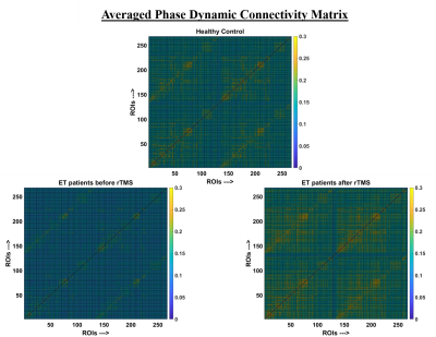
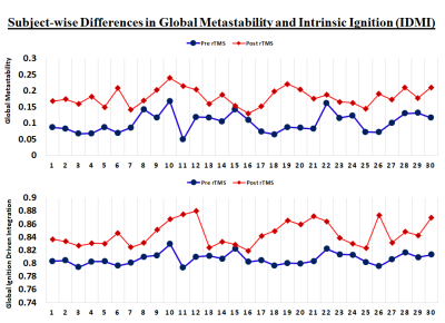
We assessed the effect of repetitive transcranial magnetic stimulation (rTMS) on whole brain dynamics using fMRI. MRI was acquired before and after a single session of rTMS in 30 patients of Essential Tremor and 20 age matched healthy controls. Whole brain dynamic synchronization - “metastability”, and propagation of integration - “intrinsic ignition”, were studied in order to assess brain network topological fluctuations following rTMS. Single subject and group wise whole brain metastability, integration and ignition driven integration was found improve with a single session of rTMS.
INTRODUCTION
Essential tremor (ET) is a highly prevalent movement disorder characterized predominantly by an action tremor. Although disruptions in brain connectivity and topological architecture have been previously reported in movement disorders [1, 2], abnormalities pertaining to neural communication and information processing are uncertain. Various parameters may be utilized to evaluate these changes. Metastability i.e. whole-brain dynamic synchronization measures the synchronization between the fluctuation of nodes over time, and intrinsic ignition which reflects the ability of a given brain region to propagate neuronal activity to other regions, giving rise to different levels of integration. These measures may also play a role in understanding brain communication and information processing. In order to ascertain changes in network dynamics following a single session of repetitive transcranial magnetic stimulation (rTMS) in ET, we utilized a computational modeling approach to assess whole-brain metastability, integration, and ignition driven integration.METHODS
Thirty patients of ET, diagnosed as per the 1998 consensus criteria of tremor [3], and twenty, age and gender-matched healthy controls were recruited from the neurology outpatient department. The standard protocol followed for all patients of ET was as follows – clinical evaluation, estimation of resting motor threshold (RMT), resting-state functional MRI (rsfMRI) (r1), rTMS, and a second rsfMRI (r2) within 10 minutes of the rTMS. HC only underwent a single session of rsfMRI.RMT was recorded from the primary motor area. The following parameters were utilized for the acquisition of both sessions of rsfMRI: TR 2000 ms, TE 35 ms, Voxel Size 3x3x4 mm3; Low frequency rTMS was given over the left primary motor area (M1), by delivering 900 stimuli (90% of RMT) at 1 Hz for 15 minutes.
A standard pre-processing protocol was employed for the rsfMRI. Neural time series were extracted for 256 nodes [4]. Phase dynamics of each network were computed using the Hilbert transform [5]. Metastability was measured using the Kuramoto order parameter, which indicates the synchronization between different brain regions across time. The Kuramoto order parameter measures the global level of synchronization of ‘n’ oscillating signals [6]. Parallel to metastability, brain integration, and intrinsic ignition were also measured. Ignition is computed by quantifying the elicited level of integration for each intrinsic ignition event at a single region, averaged over all events [7]. The group differences were compared between ET (pre rTMS) and HC, ET (pre rTMS) and ET (post rTMS). Bonferroni correction was applied to account for multiple comparisons.
RESULTS
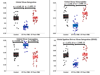
The comparison of r1 and rsfMRI of HC revealed significantly lower metastability and intrinsic ignition in patients with ET. Whereas, no significant differences were observed in a comparison of r2 and rsfMRI of HC. Significant differences in metastability and intrinsic ignition were observed when r1 and r2 were compared, with r2 showing higher levels.A single session of rTMS was found to increase whole-brain synchrony in patients with ET. Integration and intrinsic ignition were also found to be higher after rTMS at a group level. These trends were highly significant at an individual subject level and were found to approach the values obtained in HC.
DISCUSSION
ET is suggested to be a network-level disorder with predominant involvement of the cerebello thalamo cortical network. This is due to an abnormality in the number of Purkinje cells which leads to the reduction in GABA and subsequent lack of inhibition. The effect of multiple sessions of rTMS to transiently reduce disease severity in ET by increasing inhibition has been previously reported. However, the actual functional effect of rTMS, i.e. the changes in dynamic functional connectivity due to rTMS have not been reported. In the present study, we observed significantly lower metastability and intrinsic ignition in patients with ET in comparison to HC. These abnormalities may be a by-product of the disease pathophysiology in ET and could be indicative of the extent of the abnormality. A single session of rTMS was found to reset these parameters by bringing them close to the baseline, i.e. healthy controls. The lack of significant differences between r2 and rsfMRI of HC lends support to this notion. In addition, the significant difference observed between r1 and r2 also showed a definite increase in these parameters. The effects of a single session of rTMS are reported to last for approximately 30 minutes owing to which rTMS utilized as a therapeutic aid has to be given in multiple sessions.CONCLUSION
Dynamic functional connectivity has been seldom utilized in movement disorders and the effect of rTMS on this connectivity has not been previously reported. Our results provide novel evidence pertaining to the utility of metastability and intrinsic inhibition as measures to estimate the effect of rTMS.Acknowledgements
We acknowledge the funding support by the Department of Science and Technology, Cognitive Science Research Initiative (CSRI), INDIA and Indian Council Of Medical Research (ICMR), INDIA.References
1. Lenka, A., et al., Role of altered cerebello-thalamo-cortical network in the neurobiology of essential tremor. Neuroradiology, 2017. 59(2): p. 157-168.
2. Bharath, R.D., et al., A Single Session of rTMS Enhances Small-Worldness in Writer's Cramp: Evidence from Simultaneous EEG-fMRI Multi-Modal Brain Graph. Front Hum Neurosci, 2017. 11: p. 443.
3. Deuschl, G., P. Bain, and M. Brin, Consensus statement of the Movement Disorder Society on Tremor. Ad Hoc Scientific Committee. Mov Disord, 1998. 13 Suppl 3: p. 2-23.
4. Shen, X., et al., Groupwise whole-brain parcellation from resting-state fMRI data for network node identification. Neuroimage, 2013. 82: p. 403-15.
5. Ponce-Alvarez, A., et al., Resting-state temporal synchronization networks emerge from connectivity topology and heterogeneity. PLoS Comput Biol, 2015. 11(2): p. e1004100.
6. Deco, G. and M.L. Kringelbach, Metastability and Coherence: Extending the Communication through Coherence Hypothesis Using A Whole-Brain Computational Perspective. Trends Neurosci, 2016. 39(3): p. 125-135.
7. Deco, G. and M.L. Kringelbach, Hierarchy of Information Processing in the Brain: A Novel 'Intrinsic Ignition' Framework. Neuron, 2017. 94(5): p. 961-968.


