1217
Implementing multi-echo balanced SSFP with a sequential phase-encoding order at 7T1State Key Laboratory of Brain and Cognitive Science, Beijing MRI Center for Brain Research, Institute of Biophysics, Chinese Academy of Sciences, Beijing, China, 2University of Chinese Academy of Sciences, Beijing, China, 3Center for Biomedical Imaging Research, Department of Biomedical Engineering, School of Medicine, Tsinghua University, Beijing, China, 4Siemens Shenzhen Magnetic Resonance Ltd, Shenzhen, China, 5CAS Center for Excellence in Brain Science and Intelligence Technology, Chinese Academy of Sciences, Beijing, China, 6Laboratory of FMRI Technology (LOFT), Stevens Neuroimaging and Informatics Institute, University of Southern California, Los Angeles, CA, United States, 7Beijing Institute for Brain Disorders, Beijing, China
Synopsis
In the past decade, passband bSSFP has emerged as an alternative method to the widely-used GE-EPI in fMRI studies at high-fields. Multiline bSSFP with an interleaved phase-encoding order was further proposed to accelerate bSSFP fMRI. However, it intrinsically suffers from high spatial frequency ghosts which blur the image. In this study, we developed a multi-echo bSSFP sequence using a sequential phase-encoding order, combined with the GRAPPA technique for ghost elimination. In vivo experiments demonstrated that this sequence could shorten the imaging time and provide high-quality structural and functional MR images of the human brain at 7T with sub-millimeter resolution.
Introduction
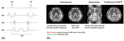
Most functional MRI (fMRI) studies employ T2*-weighted gradient echo-echo planar imaging (GE-EPI). Alternatively, a novel fMRI method based on passband balanced SSFP (bSSFP) was proposed in 20051. It is immune to several drawbacks of GE-EPI at ultra-high fields and more selective to signals from small vessels close to the source of neural activities via its spin-echo like BOLD contrast2,3. To accelerate bSSFP fMRI, multiline bSSFP with an interleaved phase-encoding order was further proposed, which employed highly segmented EPI readout4,5. However, EPI readout always leads to a Nyquist ghost due to hardware imperfections which misalign k-space lines in opposite readout directions. Moreover, k-space modulations caused by amplitude and phase variations among different echoes within an echo train further distort the original point spread function (PSF), which splits the Nyquist ghost into multiple ghosts. Interleaved phase-encoding order is always used in segmented imaging because it has a PSF with low sidelobes that results in ghosts with low intensity. However, high-frequency sidelobes of the PSF intrinsically result in high-frequency ghosts in phase-encoding direction. In this study, we explored the feasibility of multi-echo bSSFP with a sequential phase-encoding order for fast structural and functional MRI.Methods
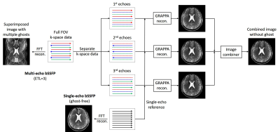
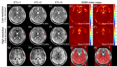
This study was performed on a 7T MRI system (Siemens, Erlangen, Germany) with a Nova Medical volume transmit/32 channel receive head coil. Image reconstruction and post-processing were performed in Matlab (The MathWorks, Natick, MA) and AFNI6. As shown in Fig. 1a, the 2D multi-echo bSSFP was modified from bSSFP by applying bipolar readout gradients to fill multiple k-space lines within one TR. Extra blips, each with a moment equals Δky , were added in phase-encoding direction to encode k-space lines in a sequential order.For structural MRI, we acquired single-slice multi-echo bSSFP images with low (1.0x1.0x3.0mm3) and high resolution (0.5x0.5x3mm3) using the following parameters: FOV=200x200mm2, echo train length (ETL)=1/3/5, TE=1.97/3.5/5.1ms and 2.56/4.65/6.75ms, TR=3.94/7.0/10.2ms and 5.12/9.3/13.5ms, slice-acquisition time=864/573.56/541.8ms and 2107.04/1345.45/1227.61ms, bandwidth=801Hz/pixel and 595Hz/pixel for low- and high-resolution images respectively. The k-space data of multi-echo bSSFP (ETL=3/5) were reconstructed using GRAPPA7 following the method shown in Fig. 2, whose performance was evaluated by structural similarity (SSIM) index maps8 and ghost-to-signal ratio (GSR) in percent9 shown in Fig. 3. More details about the reconstruction can be found in our another abstract.
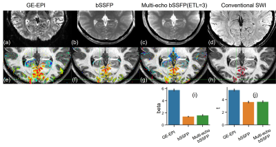
For fMRI, a radial flickering checkerboard visual stimulus was applied with a block paradigm alternating between a task (16s) and rest (10s) period repeated for 16 times. The BOLD fMRI data were successively acquired with GE-EPI (FOV=200x200mm2, TR/TE=2000/34ms, FA=70°, slice-thickness=1mm, bandwidth=1136Hz/pixel), bSSFP (FOV=200x155.8mm2, TR/TE=3.8/1.9ms, FA=35°, slice-thickness=3mm, 3 slices, bandwidth=1002Hz/pixel) and multi-echo bSSFP (ETL=3, FOV=200x186.6mm2, TR/TE=6.5/3.25ms, FA=35°, slice-thickness=3mm, 4 slices, bandwidth=1002Hz/pixel) with the same in-plane resolution of 1.0x1.0mm2 and volume-acquisition time of 2s. Anatomical sequences included T1-weighted MP2RAGE (0.7x0.7x0.7mm3) and conventional SWI (0.3x0.3x2mm3). The fMRI data were corrected for geometric distortion in phase-encoding direction (EPI only) and head motion, and fitted with GLM using AFNI. Activation maps were thresholded at p<0.05 (uncorrected) and interpolated to 0.5mm iso for visualization purpose.
Results
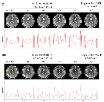
As shown in Fig. 3a-3i, multi-echo bSSFP images showed good structural consistency with T1-MPRAGE and T2-FLAIR images except frontal lobes with severe B0 inhomogeneity. According to SSIM index maps, except banding-affected regions, multi-echo bSSFP (ETL=3/5) images showed very high structural similarities with single-echo bSSFP. Besides, the proposed reconstruction method robustly removed ghost artifacts of multi-echo bSSFP with relatively low GSR values, especially in low-resolution cases.As shown in Fig. 4, higher FAs increased the signal magnitude of grey matters and cerebrospinal fluid, resulting in better SNR and CNR.
As shown in Fig. 5d-5f, regions with large signal changes of GE-EPI were coincide with large draining veins, whereas the signal changes of bSSFP did not necessarily cover them, which is consistent with previously reported results1. Besides, as shown in Fig. 5f-5j, the activation map of multi-echo bSSFP was very similar to that of bSSFP, and exhibited comparable beta-value and t-value to bSSFP.
Discussion
Compared with traditional bSSFP, the time-saving sequential multi-echo bSSFP suffers from more banding artifacts due to longer TRs, which, on the other hand, also lead to a lower SAR limit. Therefore, the SNR loss in reconstructed images due to the use of GRAPPA for ghost cancellation may be compensated by using higher FAs.Unlike interleaved multiline bSSFP, the sequential multi-echo bSSFP doesn’t need reference scans to do phase correction for the Nyquist ghost, or echo time shift for ghost reduction that lengthens TR and results in obvious chemical shift10. Furthermore, it shows reduced distortion and blur in geometry. Although this new sequence needs single-echo reference data and multi-channel coil for robust GRAPPA reconstruction, multi-echo bSSFP with an ETL of 3 is relatively robust and time-efficient in this study.
The fMRI experiments demonstrated that multi-echo bSSFP was immune to spatial distortion and signal dropouts of GE-EPI at ultra-high fields and showed an inherent insensitivity to BOLD signal changes from large veins. Higher FAs and resolution may be applied in future studies. Thus, this new method could provide high-resolution fMRI for visual cognitive research.
Conclusion
In this study, we developed a time-efficient multi-echo bSSFP sequence using a sequential phase-encoding order and validated its feasibility for 2D high-resolution structural and functional MR imaging at 7T.Acknowledgements
This work was supported in part by the Ministry of Science and Technology of China grant (2015CB351701), the National Nature Science Foundation of China grants (81871350, 31730039), and the Strategic Priority Research Program of Chinese Academy of Science (XDB32010300).References
1. Bowen CV, Menon RS, Gati JS. High field balanced-SSFP fMRI: a BOLD technique with excellent tissue sensitivity and superior large vessel suppression. In Proc Intl Soc Mag Reson Med 2005 (Vol. 119).
2. Scheffler K, Hennig J. Is TrueFISP a gradient-echo or a spin-echo sequence? Magn Reson Med 2003;49:395-397.
3. Bieri O, Scheffler K. Effect of diffusion in inhomogeneous magnetic fields on balanced steady‐state free precession. NMR Biomed 2007:1–10.
4. Miller KL, Smith SM, et al. High-resolution FMRI at 1.5T using balanced SSFP. Magn Reson Med 2006;55:161–170.
5. Ehses P, Scheffler K. Multiline balanced SSFP for rapid functional imaging at ultrahigh field[J]. Magn Reson Med 2018;79(2):994-1000.
6. Cox RW. AFNI: software for analysis and visualization of functional magnetic resonance neuroimages. Comput Biomed Res 1996;29:162–173.
7. Griswold M A, Jakob P M, et al. Generalized autocalibrating partially parallel acquisitions (GRAPPA). Magn Reson Med 2002;(6):1202-1210.
8. Wang Z, Bovik A C, et al. Image quality assessment: from error visibility to structural similarity[J]. IEEE transactions on image processing 2004;13(4):600-612.
9. Poser BA, Barth M, et al. Single-shot echo-planar imaging with Nyquist ghost compensation: interleaved dual echo with acceleration (IDEA) echo-planar imaging (EPI). Magn Reson Med 2013;69:37-47.
10. Kellman P, McVeigh E R. Phased array ghost elimination. NMR in Biomedicine 2006;19(3): 352-361.
Figures
