1205
Clinical Value of an Almost Automated Fast Free-breathing Cardiac Magnetic Resonance Workflow
Keyan Wang1, Michaela Schmidt2, Jing An3, and Xiaoming Bi4
11st affiliated hospital of zhengzhou university, Zhengzhou, China, 2Siemens Healthcare GmbH, Erlangen, Germany, 3Siemens Shenzhen Magnetic Resonance Ltd, Shenzhen, China, 4Siemens Healthineers, Los Angeles, CA, United States
11st affiliated hospital of zhengzhou university, Zhengzhou, China, 2Siemens Healthcare GmbH, Erlangen, Germany, 3Siemens Shenzhen Magnetic Resonance Ltd, Shenzhen, China, 4Siemens Healthineers, Los Angeles, CA, United States
Synopsis
The application of cardiac magnetic resonance (CMR) is limited in patients with arrhythmia or poor breath holding ability. In this study, clinical value of a rapid free-breathing workflow was assessed by employing real-time compressed-sensing for cardiac function and motion-corrected LGE embedded in a workflow engine for cardiac assessment. Results indicated that the proposed free-breathing fast workflow allowed in a short acquisition time to assess the morphological features of the heart and left ventricular function, especially in patients with severe heart failure.
introdction
Cardiac Magnetic Resonance (CMR) has become an important tool for non-invasive diagnosis of heart diseases, risk stratification and prognosis assessment. The conventional imaging protocolsinclude localizer, morphology, cine, and late gadolinium enhancement (LGE) imaging. Cine and LGE images are typically acquired by segmenting readout into multiple heartbeats to achieve sufficient spatial and/or temporal resolution. Unfortunately, such segmented data acquisition is prone to motion artifacts particularly in CMR patients with arrhythmia or limited breath-holding capability. Recent studies have reported newly developedcompressed sensing (CS) cine1 and motion correction (MOCO) LGE2 techniques that eliminated motion artifacts using single-shot data readout in combination with advanced reconstruction. The aim of this study was to develop a free-breathing CMR workflow using these latest techniques combined with scan automation like auto slice positioningand automatic adaption of scan parameters to heart rate and size of the patient and explore the clinical value of a method for the evaluation of heart diseases.Methods
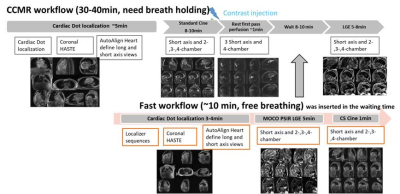
One hundred and fifty-eight patients with heart diseases were prospectively enrolled. All patients underwent conventional cardiac magnetic resonance (CCMR) and the proposed fast workflow on a 3T scanner (MAGNETOM Skyra, Siemens Healthcare, Erlangen, Germany). Key protocols included in the CCMR workflow were segmented cine and late gadolinium enhancement (LGE) acquired in multiple breath-holds. Fast workflow mainly included real-time CS cineand prototype MOCO PSIR LGE acquired under free-breathing. Both workflows were performed using Cardiac DOT Engine and shown in Figure 1.Image quality (IQ) of both workflows was evaluated based on a five point Likert score. Acquisition time, IQ, morphological features and quantitative cardiac function were compared between the two workflows. Results were sorted into 4 groups based on ejection fraction (EF): (1) group 1 (n=74), EF>50%; (2) group 2 (n=11), EF 45%–50%; (3) group 3 (n=23), EF 35%–44%; (4) group 4(n=50), EF<35%. Statistical analysis was performed to compare these two workflows for each group.Results
All patients completed both workflows. The acquisition time of the fast workflow was significantly shorter than that of CCMR[(10.8±0.6) min vs. (35.5±2.9)min, P=0.001].The overall IQ of CCMR was significantly higher than that of the fast workflow[(4.39±0.833)vs.(3.41±0.741),P=0.001],[(4.34±1.021)vs.(3.37±0.786),P=0.001], [(4.06±1.336) vs.(3.43±0.778),P=0.006]) in the group of 1,2 and 3. However, it was significantly lower than that of the fast workflow [(1.73±0.96) vs. (3.69 ± 0.73), P = 0.001]in the group 4. For consistency comparison, 111 patients with good IQ for both workflows were included. There were no significant differences in the characterization of myocardial, papillary muscle, ventricular aneurysm, thrombus, and delayed enhancement (P>0.05), and the consistency was high (Kappa, 0.79-1.0), but for the valves , the detection percentage of valves’ movement by cine in CCMR was significantly higher than that of CS in the fast workflow [92.8% (103/111) vs. 62.2% (69/111), P=0.001], and the two methods showed low consistency (Kappa, 0.142). There were no significant difference in the evaluation of the left ventricular function for CCMR and faster workflow(P>0.05),and the consistency was high (Kappa, 0.730-0.933). The diagnosis consistency of the two workflows was 0.899.Conclusion
The fast workflow was able to evaluate the morphological features and left ventricular function of the heart in 10 minutes while patients could breathe freely. It is a promising method for speeding up scan time, increasing patient comfort and getting robust image quality in vulnerable patients like those with severe heart failure.Acknowledgements
No acknowledgement found.References
1. Forman C et al ISMRM 2017, pp. 4669, 2017
2. Piehler KM, Wong TC, Puntil KS et al (2013). Cir Cardiovasc Imaging 6:423-432

