1190
DW-EPI distortion reduction using Multi-shot EPI (MUSE) and Reverse Polarity Gradient (RPG) in Head & Neck Region
Maggie M Fung1, Amaresha Konar Shridhar2, Arnaud Guidon3, Amita Shukla-Dave2,4, and Vaios Hatzoglou4
1MR Apps & Workflow, GE Healthcare, New York City, NY, United States, 2Department of Medical Physics, Memorial Sloan Kettering Cancer Center, New York City, NY, United States, 3MR Apps & Workflow, GE Healthcare, Boston, MA, United States, 4Department of Radiology, Memorial Sloan Kettering Cancer Center, New York City, NY, United States
1MR Apps & Workflow, GE Healthcare, New York City, NY, United States, 2Department of Medical Physics, Memorial Sloan Kettering Cancer Center, New York City, NY, United States, 3MR Apps & Workflow, GE Healthcare, Boston, MA, United States, 4Department of Radiology, Memorial Sloan Kettering Cancer Center, New York City, NY, United States
Synopsis
The purpose of this study is to investigate the distortion correction performance and ADC value consistency of the single shot EPI (SSEPI), multi-shot EPI (MUSE) and reverse polarity gradient (RPG) method in phantom, and head & neck cancer patients. We observed improved distortion correction performance in MUSE, and best distortion correction in MUSE plus RPG method. Improved anatomical details, reduced artifacts and improved perceived clinical utility were also observed in MUSE (with and without RPG) as compare to SSEPI. ADC values remained consistent between these techniques.
Purpose:
Head and Neck Diffusion Weighted Echo Planar imaging is often challenging due to the high B0 in-homogeneity and tissue susceptibility, which leads to geometric distortion. Multi-shot EPI with multi-flexed sensitivity encoding (MUSE) [1] has been shown to reduce geometric distortion by decreasing effective echo spacing. In addition, reverse polarity gradient (RPG) method, a retrospective approach to correct distortions by exploiting the symmetry of the artifact in the forward and reverse phase-encoding (PE) trajectories has also been shown to reduce distortion in brain [2] and pelvis applications [3,4]. However, a combination of these methods has not been assessed in the head and neck region. The purpose of this work was to compare the distortion correction performance, and ADC measurements of single shot-EPI (SS-EPI), MUSE, and MUSE with RPG in head and neck region.Methods:
Phantom study was performed using the ACR phantom and QIBA ice-water phantom to assess the distortion and ADC consistency. T2w FSE, SSEPI and MUSE EPI were acquired using parameters listed below. Geometric dimensions and landmark coordinates of the ACR phantom were measured to assess distortion. ADC values were measured in 7 vials in the QIBA ice-water phantom to compare the ADC consistency.Patient studies were performed under an IRB approved protocol. Eleven head and neck cancer patients were recruited with informed consent. The study was performed on a GE 3T 70cm bore scanner (MR750w, GE Healthcare, USA) using the 21 channel Head and Neck array (GE Healthcare, USA).The following MR sequences were performed:
- T2w FSE anatomical imaging: FOV: 18-22cmx18-22cm, Matrix: 320x224, TR/TE:2500-4861ms/97.8-106.6ms, BW:83kHz, Slice thickness:3mm, number of slices:37-50, NEX: 2, ETL=19-21, scan time: 1:14-3:10min.
- SSEPI: FOV: 26cmx26cm, Matrix: 128x128, TR/TE:6000ms/70.9-75.1ms, single spin echo, number of shot:1, ASSET acceleration: 2, BW:250kHz, slice thickness:5mm, number of slices:14-24, b-value=0s/mm2 (2 NEX), 1000s/mm2 (12 NEX), diffusion encoding: All 3 directions, scan time: 1:42min.
- MUSE EPI diffusion weighted imaging: FOV: 26cmx26cm, Matrix: 128x130, TR/TE:6000ms/71.2-77.4ms, single spin echo, number of shot:2, ASSET acceleration:1.5, BW:250kHz, slice thickness:5mm, number of slices: 14-26, b-value=0s/mm2(2 NEX), 1000s/mm2 (12 NEX), diffusion encoding: All 3 directions, scan time: 3:24min. RPG was turned ON such that images with and without RPG were generated.
Qualitative Assessment: An experienced head and neck radiologist performed image quality rating based on anatomic detail, lack of susceptibility-induced artifacts and perceived clinical utility. 5-point scale was used: 1-non-diagnostic, 2-poor, 3-satisfactory, 4-good, and 5-excellent. Results were compared with paired t-test.
Results:
ACR phantom results: We observed reduced geometric distortion in MUSE & MUSE+RPG as compared to SSEPI (Figure 1 (A)). We measured the length between two end points of the grid line at different angles (0°, 60°, 90°, 120°). These lines on MUSE+RPG show similar values as compared to T2w reference. Similarly, the X,Y coordinates of three points at the grid were compared between the DWI images & T2w reference. In this case, MUSE+RPG coordinates are same as the T2 reference image co-ordinate values.QIBA phantom results: ADC values were consistent in the 3 DWI methods (Figure 1 (C)). Increased hyper-intensity were observed in the edge of the vials in SSEPI and MUSE due to susceptibility induced mismatch between b0 & b1000 images (Figure 1 (B) yellow arrows). This in turn accounts for some small differences in ADC values in SSEPI.
Patient study results: We observed reduced geometric distortion in MUSE & MUSE+RPG as compared to SSEPI. Figure 2 & 3 shows the subsequent improvement in spatial accuracy of MUSE, and MUSE+RPG. We observed statistically significant increase of CC values (between T2w FSE & DWI) when MUSE is used (0.755±0.052, p<0.05) and when MUSE+RPG is used (0.800 ± 0.0677 (p<0.05), compared to SSEPI (0.713 ± 0.056) (Figure 4 A). Qualitative image rating from radiologist also showed statistically significant improvement in anatomical details, lack of artifacts and perceived clinical utility in MUSE and MUSE+RPG, as compare to SSEPI (Figure 4B). ADC values were consistent between SSEPI, MUSE and MUSE+RPG in both tumor and normal masseter muscle (Figure 5).
Discussion:
From the phantom and patient study, we observed that the use of MUSE & MUSE+RPG can help improved the delineation of tumor, especially if the lesion in an area with high susceptibility. In our study, we opted to keep the in-plane resolution the same across the 3 protocols in order to fairly assess the techniques. However,with the distortion reduction capability of MUSE & RPG, the in-plane resolution theoretically can be further increased, and it might have more positive impact on diagnostic confidence.Conclusion:
We demonstrated the feasibility of using MUSE and RPG in DW-EPI for imaging of the head and neck region. The use of MUSE and RPG reduced the distortion compared to conventional SSEPI techniques, while maintaining similar ADC values. Future work will focus on further improving the resolution of MUSE and validating this technique in a larger patient cohort.Acknowledgements
No acknowledgement found.References
[1] Chen, Guidon et al, NeuroImage 2013 May 15: 72: 41-47 [2] Holland, Dale et al, NeuroImage 50 (2010) 175–183 [3] Fung et al, ISMRM 2018 Proceeding 1638, [4] Rakow-Penner et al. Magn Reson Imaging 2015;33(9):1178-1181Figures
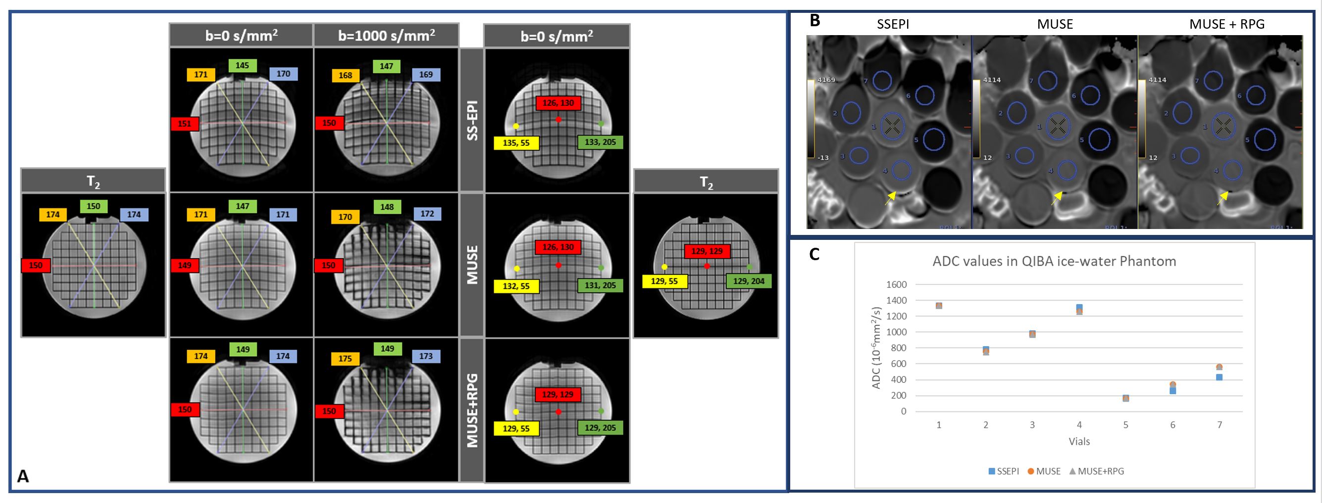
(A) Geometric distortion measurements of the
3 methods using T2w image as a reference on the ACR phantom. 4 lines are drawn
to connect between the end points of the grid line at a particular angle (0°,
60°, 90°, 120°) to measure the length. In
addition, three points on a center horizontal line is selected and compared
with the T2w image to measure the change in the co-ordinate values.
(B & C) ADC values of the 7 vials in
the QIBA phantom are in general consistent between the 3 methods, except in vials
with high susceptibility at the border.
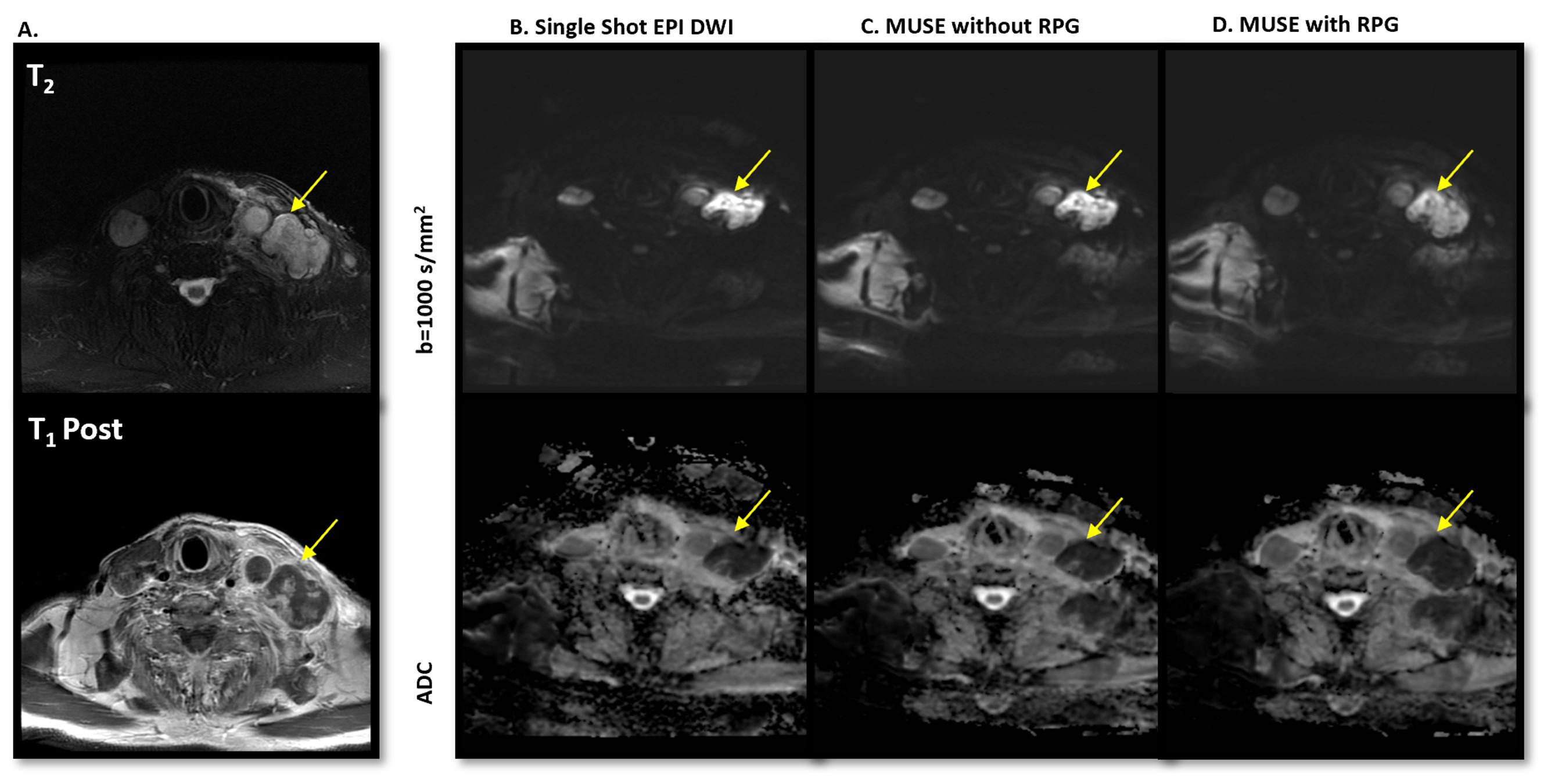
Demonstration of reduction in geometric
distortion with the use of MUSE and MUSE+RPG. A. T2w & T1w Post-Contrast image,
B. SS-EPI, C. MUSE and D. MUSE with RPG. Improved spatial accuracy was observed
in the metastatic lymph node (yellow arrow).
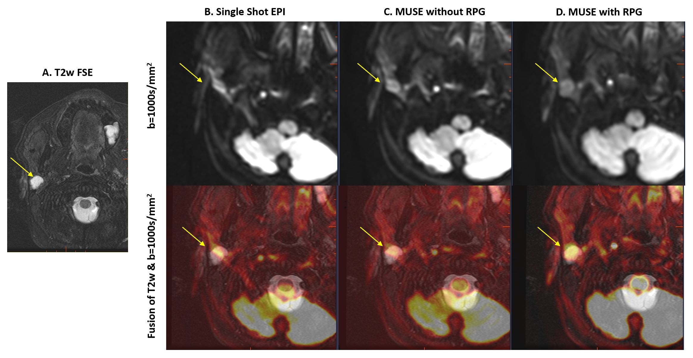
Fusion between T2w reference & b1000 in
a patient with right parotid gland lesion (yellow arrow). Severe distortion was
observed in SS-EPI, and improved geometric accuracy and lesion definition were
observed in MUSE and MUSE+RPG.

A. Correlation coefficient between T2w and the
3 types of DWI images. A statistically significant increase in correlation can
be observed when MUSE was used, and further increase can be observed when
MUSE+RPG was used (also statistically significant). B. Qualitative image
quality rating also showed statistically significant (* denote p<0.05)
improvement in MUSE & MUSE+RPG as compared to SSEPI in all 3 categories.
MUSE+RPG had improved rating in all 3 categories as compared to MUSE but was not
statistically significant.
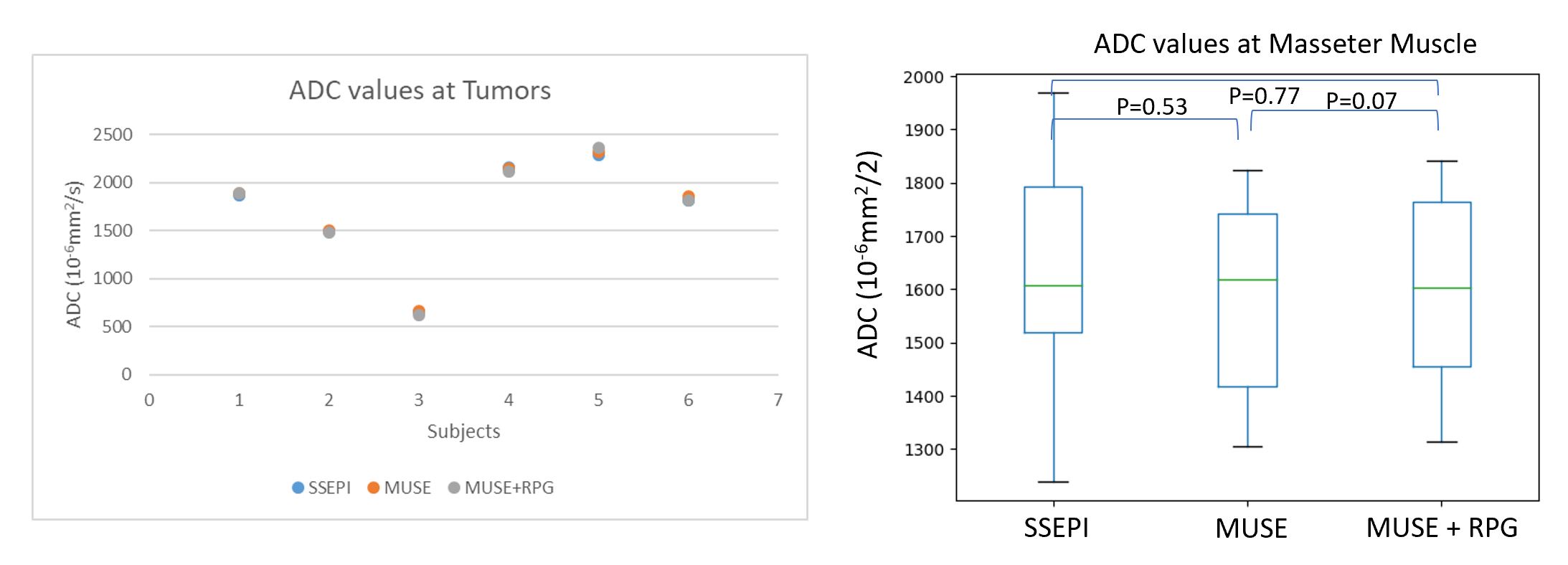
ADC measurements in patient cohort. Left: ADC
values in the tumor region (present in 6 out of 11 subjects) is consistent
between 3 methods. Right: ADC values in normal masseter muscle (included in the
imaging range in 10 out of 11 subjects) is also consistent between 3 methods.
We also observed more variation in ADC values in normal masseter muscle in
SSEPI, due to increase in spatial distortion.