1176
Regional and longitudinal changes of multiple MRI parameters correlate with behavioral impairment and recovery after spinal cord injury1Vanderbilt University Institute of Imaging Science, Vanderbilt University Medical Center, Nashville, TN, United States, 2Radiology and Radiological Sciences, Vanderbilt University Medical Center, Nashville, TN, United States, 3Biomedical Engineering, Vanderbilt University, Nashville, TN, United States
Synopsis
Quantitative magnetization transfer (qMT) and diffusion tensor imaging (DTI) may detect and track compositional and structural changes in spinal cords before and after injury and during repair. This study aims to systematically evaluate the abilities of the qMT-derived pool size ratio (PSR) and DTI-derived diffusion parameters to assess injury-associated regional changes in spinal cords of monkeys, and to correlate them to specific sensorimotor behaviors. An overall goal is to evaluate the relationships between longitudinal changes in different regional MRI measures and sensorimotor behavioral impairment and recovery following spinal cord injury over a long period of time (months).
Purpose
This study aims to evaluate the feasibility of using quantitative magnetization transfer (qMT) and diffusion tensor imaging (DTI) to assess changes in the composition and microstructure of the spinal cords of non-human primates induced by injury. Specific goals are to measure changes in different MT and diffusion measures in individual subjects, both regionally and longitudinally, in order to evaluate their sensitivity for detecting tissue damage and their correlations with behavioral deficits and recovery after cervical spinal cord injury.Methods
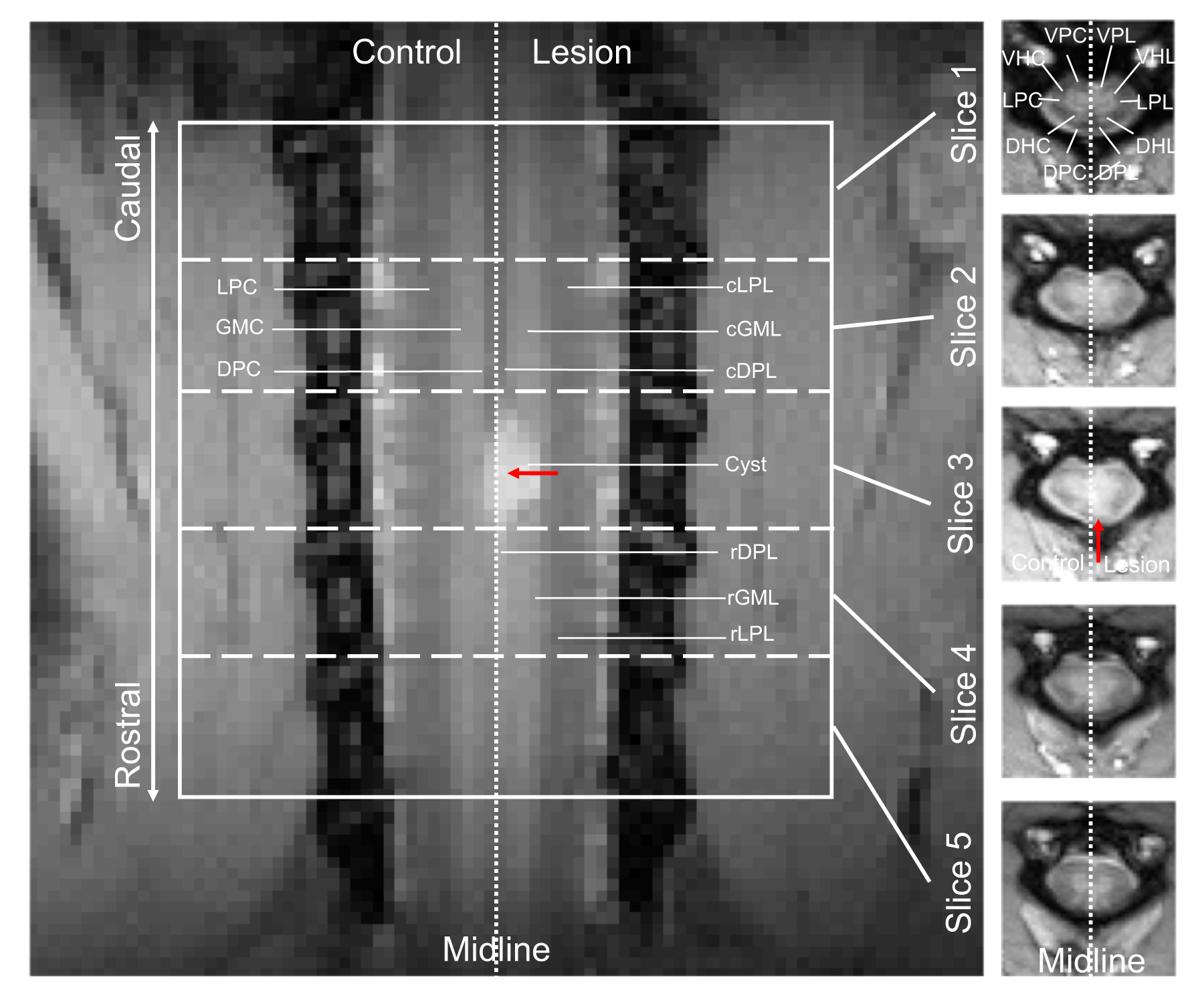
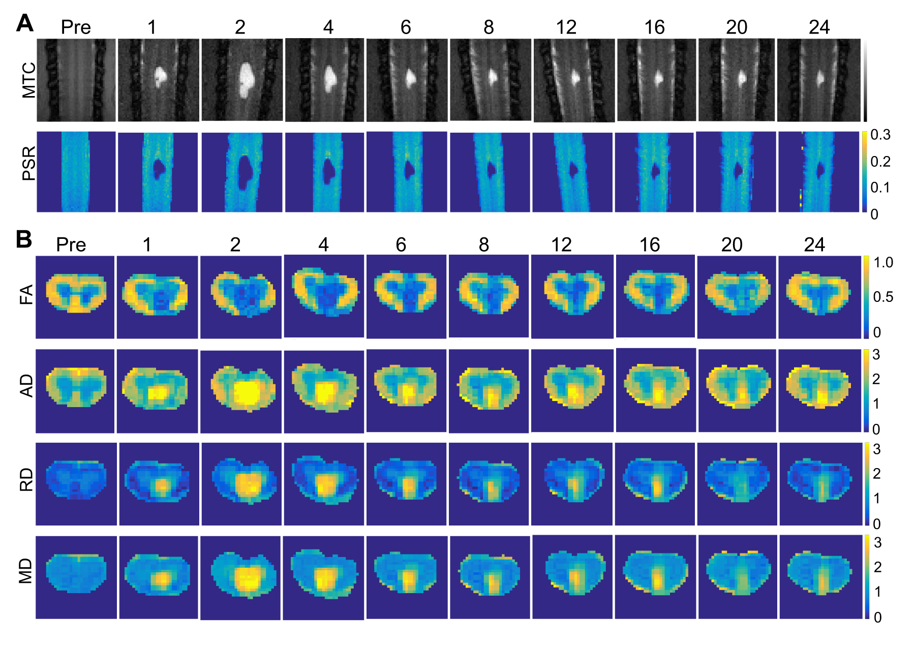
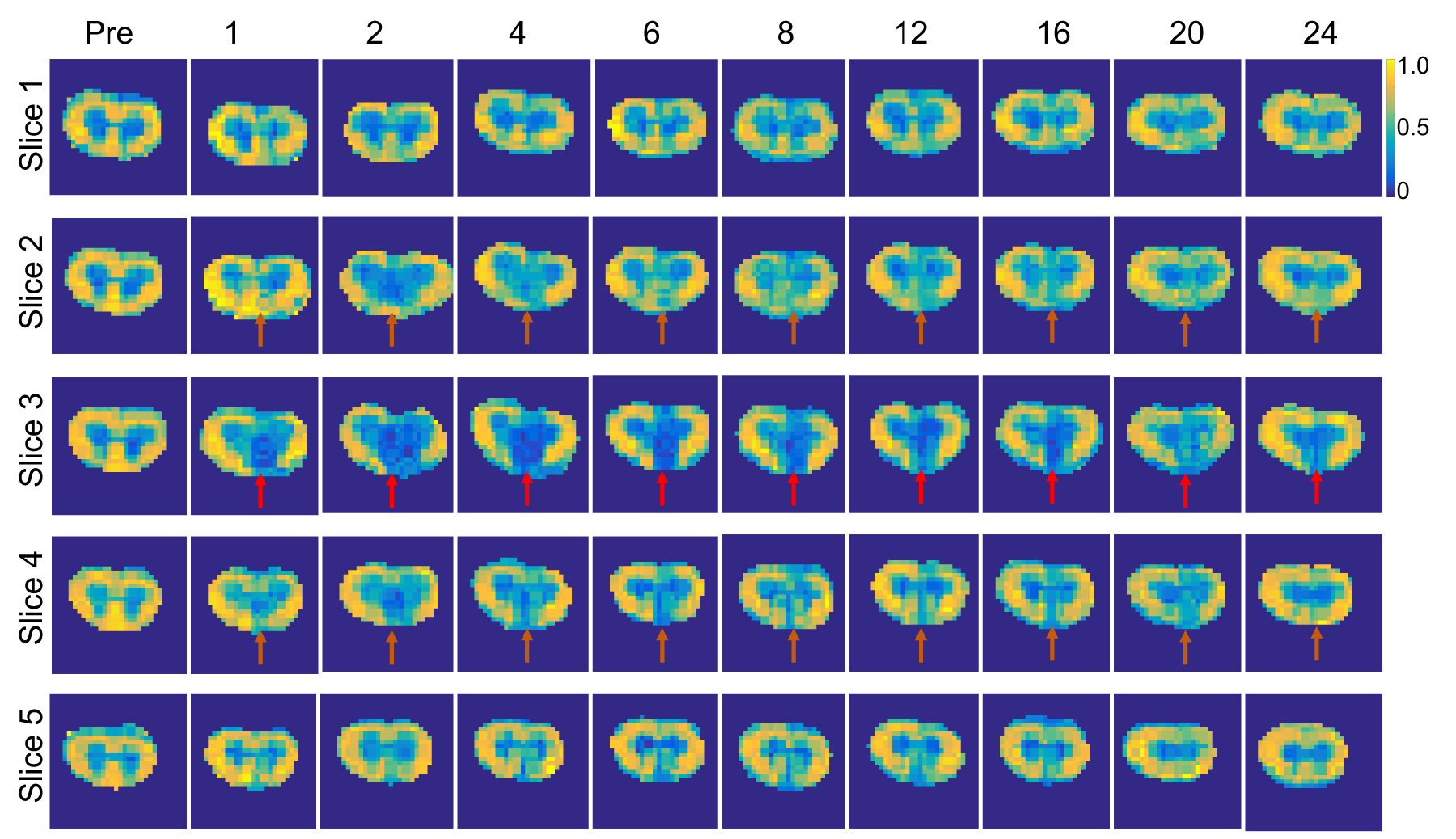
MRI scans were performed of anesthetized monkeys at 9.4T, before and after a unilateral section (~2 mm) of the dorsal column tract (Fig. 1). Diffusion data were obtained using a spin-echo diffusion sequence with echo planar imaging readout (TR/TE = 3000/33 ms, 4 shots, resolution = 0.33x0.33 mm2, slice thickness = 3 mm, 5 slices, b-value at 1000 s/mm2, 30 directions). One non-diffusion weighted scan was also acquired. MT data were obtained from a coronal slice, where the dorsal columns and dorsal horns reside, using a 2D MT-weighted spoiled gradient echo sequence (TR 24 ms, flip angle 7°, resolution 0.31x0.31x1 mm3). At least 10 different RF offsets between 1 and 100 kHz and two saturation powers (θsat = 220° and 820°, pulse width = 12 ms) were used. Animal behaviors were assessed before and after injury on hand-use and food retrieval tasks, and MRI scans were acquired longitudinally for up to 6 months.1 DTI parameters including the fractional anisotropy (FA), axial diffusivity (AD), radial diffusivity (RD), and mean diffusivity (MD) were quantified. Quantitative MT parameters including pool size ratio (PSR) were derived based on the model of Henkelman and Ramani.2,3 Regions of interest were defined as in Figure 1. Correlations between MRI and behavioral measures were calculated using the Pearson correlation function. P < 0.05 was considered statistically significant.Results
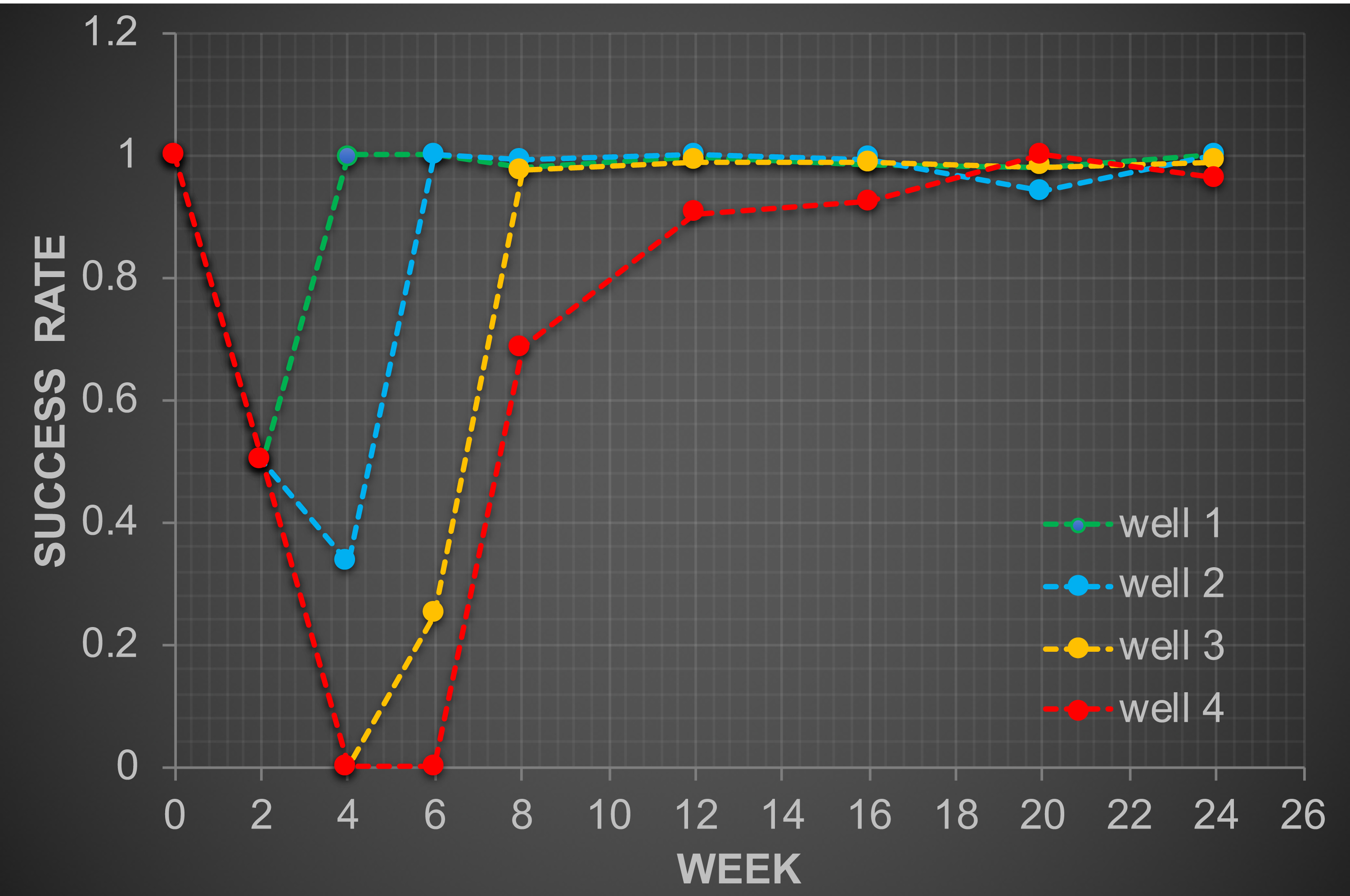
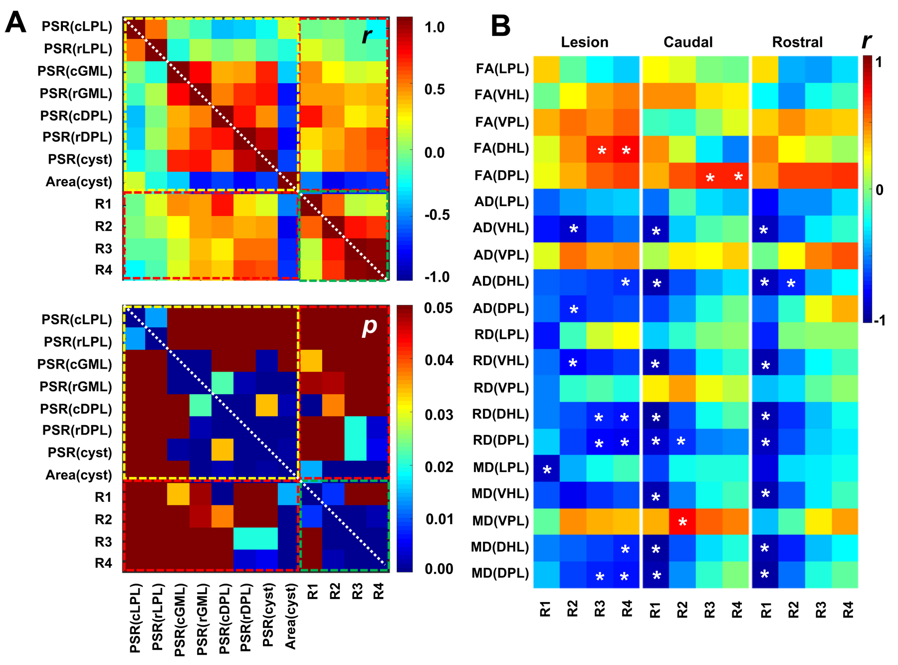
QMT and DTI delineated normal white matter (WM) and gray matter (GM) in the cervical spinal cord of non-human primates (Fig. 2) with high contrast. Regions of WM on the non-injured side (LPC, VPC and DPC) showed significantly higher PSR, FA and AD than GM (VHC and DHC), but lower RD than GM. The DTI-derived measures FA, AD and RD were strongly correlated with each other as expected. QMT and DTI measures detected unilateral changes at the site of the lesion, especially in the dorsal pathway (Fig. 2). A significant decrease in PSR was observed both rostral and caudal to the lesion site (p < 0.05), while FA also decreased (Fig. 3). Although both AD and RD increased in the dorsal pathway at the lesion site, RD showed higher contrast than AD compared to the non-lesion control side after spinal cord injury. In the representative subject shown, the slice rostral to the lesion showed a more severe deficit than caudally (Figs. 2-3). This is also confirmed in the grouped results across subjects. Both MT and DTI parameters were sensitive for detecting axonal damage and recovery in the dorsal column pathway after unilateral dorsal column lesion (Figs. 2-3). The behavioral measures of success rate (R) in retrieving a food pellet from wells at different depths (0.14 cm, 0.28 cm, 0.38 cm, and 0.64 cm for wells 1 to 4 respectively), confirmed drastic behavioral impairments and later recovery after injury (Fig. 4). The most severe deficits (Fig. 4), indicated by significant drops in success rate, were highly associated with the maximum cyst size and minimum PSR and FA of injured tissues (Figs. 2-3). Longitudinally, evident recovery from injury was revealed by regional changes of the MRI parameters, which correlated with behavioral measures (Fig. 5). While the area of the cysts detected by MT showed significant correlation with success rates from all four wells longitudinally (Fig. 5A), the PSR of cysts showed significant correlations with success rates only from wells 3 and 4. PSR of dorsal horn GM in the spinal segments adjacent to the lesion site on the lesion side (cGML, and rGML) showed significant correlations with success rates from wells 1 and 2. While PSR of the dorsal column caudal to the lesion (cDPL) showed significant correlations with success rates from wells 1 and 2, PSR of dorsal column rostral to the lesion (rDPL) showed significant correlations with success rates from wells 3 and 4. Multiple regression based on multiple regional PSR values predicated behavioral deficits better than single regional PSR measures. For the spinal segment that suffered injury, DTI measures from the dorsal horn on the lesion side consistently showed significant correlations with success rates from well 4 and/or well 3 (Fig. 5B). DTI measurements, including diffusivities of dorsal horn and dorsal column in the segments adjacent to the lesion correlated strongly with success rates from well 1 and/or well 2.Conclusion
QMT and DTI can provide efficient and sensitive means to detect and characterize axonal damage after injury and to monitor its recovery over time. The longitudinal changes of MRI measures of specific regions such as dorsal horn GM and dorsal column WM were highly correlated with behavioral impairment and recovery, indicating their key roles in mediating sensorimotor recovery following a targeted dorsal column section.Acknowledgements
We thank Dr. Zou Yue of the Vanderbilt University Institute of Imaging Science for her acquisition of behavioral data. We thank Mrs. Chaohui Tang and Mr. Fuxue Xin for their assistance in animal preparation and care in MRI data collection. We also thank Mr. Ken Wilkens and Dr. Xinqiang Yan for customizing coils for cervical spinal cord imaging. This study is supported by DOD grant W81XWH-17-1-0304, and NIH grants NS092961 and NS078680.References
1. Wang F, Zu ZL, Wu RQ, Wu TL, Gore JC, Chen LM. MRI evaluation of regional and longitudinal changes in Z-spectra of injured spinal cord of monkeys. Magn Reson Med 2018;79(2):1070-1082.
2. Wang F, Li K, Mishra A, Gochberg D, Chen LM, Gore JC. Longitudinal assessment of spinal cord injuries in nonhuman primates with quantitative magnetization transfer. Magn Reson Med 2016;75(4):1685-1696.
3. Ramani A, Dalton C, Miller DH, Tofts PS, Barker GJ. Precise estimate of fundamental in-vivo MT parameters in human brain in clinically feasible times. Magn Reson Imaging 2002;20(10):721-731.
Figures