1156
Intervertebral Disc Elastography: Physiological Strain, Stiffness, and Relaxometry in Axial Compression and Bending1Biomedical Engineering, Rensselaer Polytechnic Institute, Troy, NY, United States, 2Biomedical Engineering, Purdue University, West Lafayette, IN, United States, 3Mechanical Engineering, University of Colorado Boulder, Boulder, CO, United States, 4Engineering Mechanics, Dalian University of Technology, Dalian, China, 5International Research Center for Computational Mechanics, Dalian University of Technology, Dalian, China, 6Center for Biomedical and Healthcare Engineering, MINES Saint-Étienne, Saint-Étienne, France, 7Ecology and Evolutionary Biology, University of Colorado Boulder, Boulder, CO, United States
Synopsis
IVD degeneration is the most recognized cause of low back pain, characterized by the decline of tissue structure and mechanics. MRI relaxometry is one quantitative measure of IVD degeneration, yet MRI metrics of mechanics have not been fully explored. We quantified patterns of IVD strain and mechanics during physiological compression and bending. Strains patterns depended on the loading mode, and shear modulus in the nucleus pulposus was typically an order of magnitude lower than the annulus fibrosis, except in bending, where the apparent stiffness depended on the loading direction. Strain and material properties provide new possible biomarkers for IVD degeneration.
Introduction
Low back pain is the leading cause of chronic disability in industrialized Western societies [1]. IVD degeneration, even its earliest form, is characterized by a breakdown of the extracellular matrix; the associated loss of water and proteoglycan [2] is typically assessed by MRI. The mechanical function of the IVD is closely associated with the integrity and content of the matrix [3]; therefore, its responses under load could provide a more direct assessment of tissue health. MRI approaches, including dualMRI (displacements under applied loading by MRI) [4], have measured internal strains of the IVD under compression. The primary objective of this study was to measure MRI-based strain maps. Additionally, we utilized inverse modelling to calculate the shear modulus throughout the IVD. Finally, we discuss spatial correspondences of mechanical and relaxometry measures as potential assessment for IVD degeneration.Methods
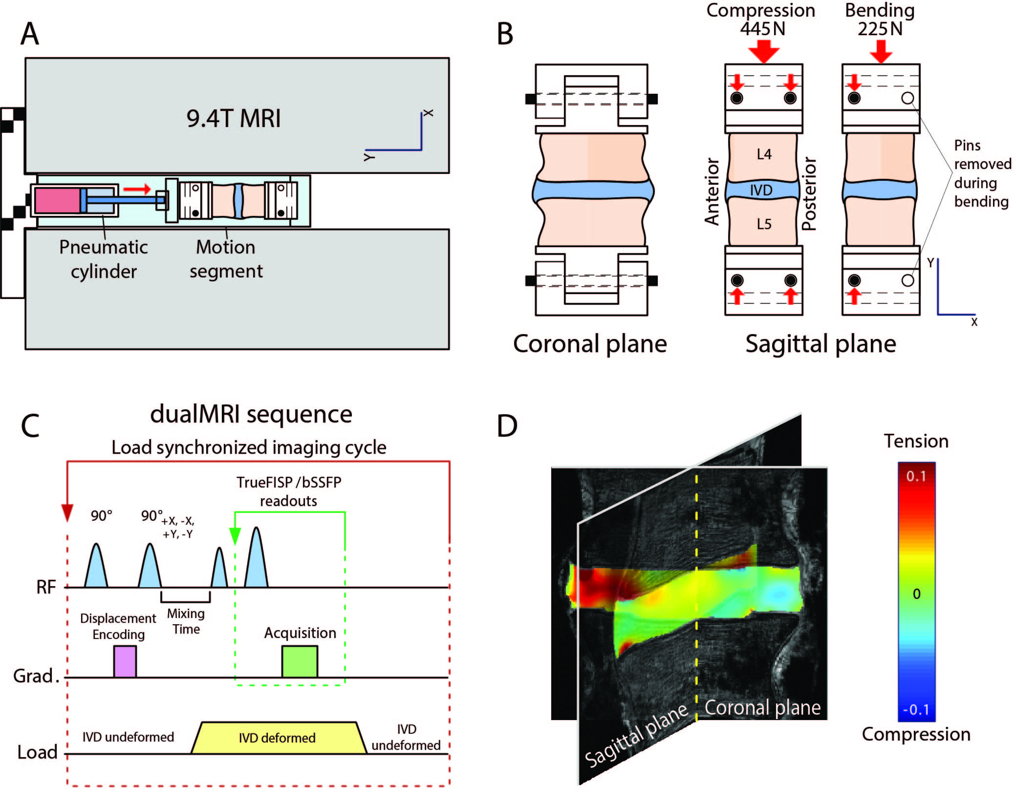
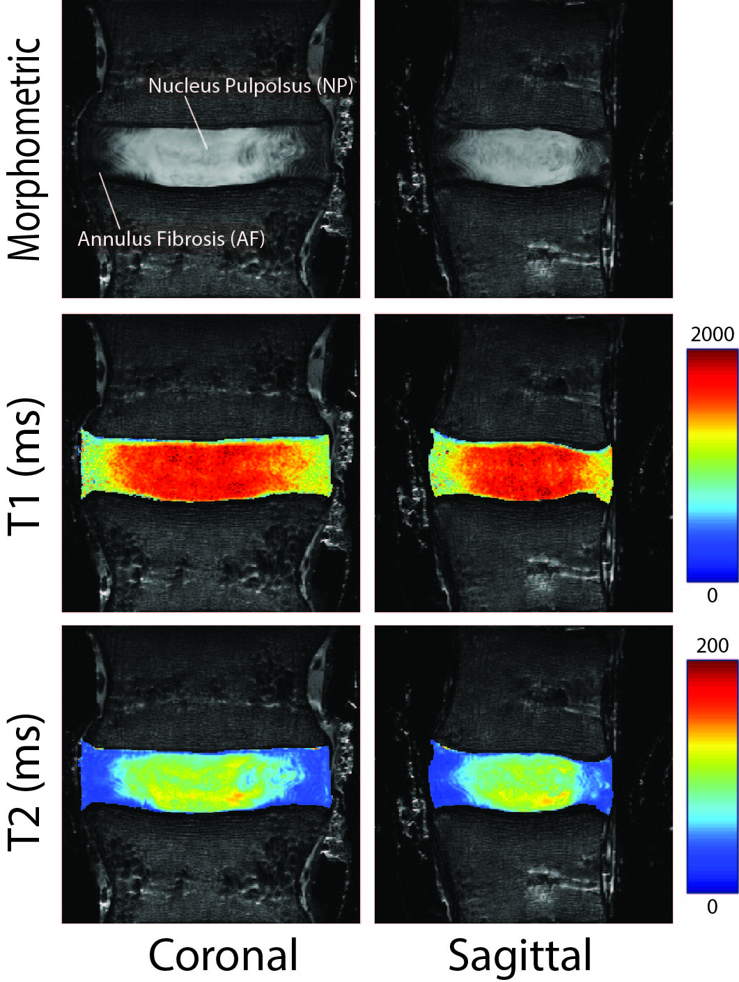
Specimen Preparation: Human lumbar segments (L4-L5) were procured from donors (n=3, 1 female, 35±13yrs, range: 22-48yrs, height: 172±12cm, weight: 92±17kg). Segments were secured to a sample holder, which permitted quick interchange of loading modes from axial-compression to bending by removal of a support pin, for connection into an MRI-compatible electro-pneumatic loading system placed inside a 9.4-Tesla horizontal bore imaging system (Bruker GMBH, Ettlingen, Germany; Figure 1).T1 and T2 Mapping: Relaxometry was performed in both sagittal and coronal planes (Figure 2), matching the planes of subsequent load-synchronized imaging. Scan parameters were: field of view=64×64mm2, spatial resolution=250×250µm2, slice thickness=2mm. For T1 mapping, a fast spin echo (FSE) acquisition was used with multiple repetition times (TR=100, 300, 500, 1000, 2000, 4000ms) and a 10-ms echo time (TE). For T2 mapping, FSE parameters included TE=20, 60, 100, 141, 181, 221, 261, and 301ms and TR=4000ms. T1 and T2 mapping was obtained using monoexponential fitting.
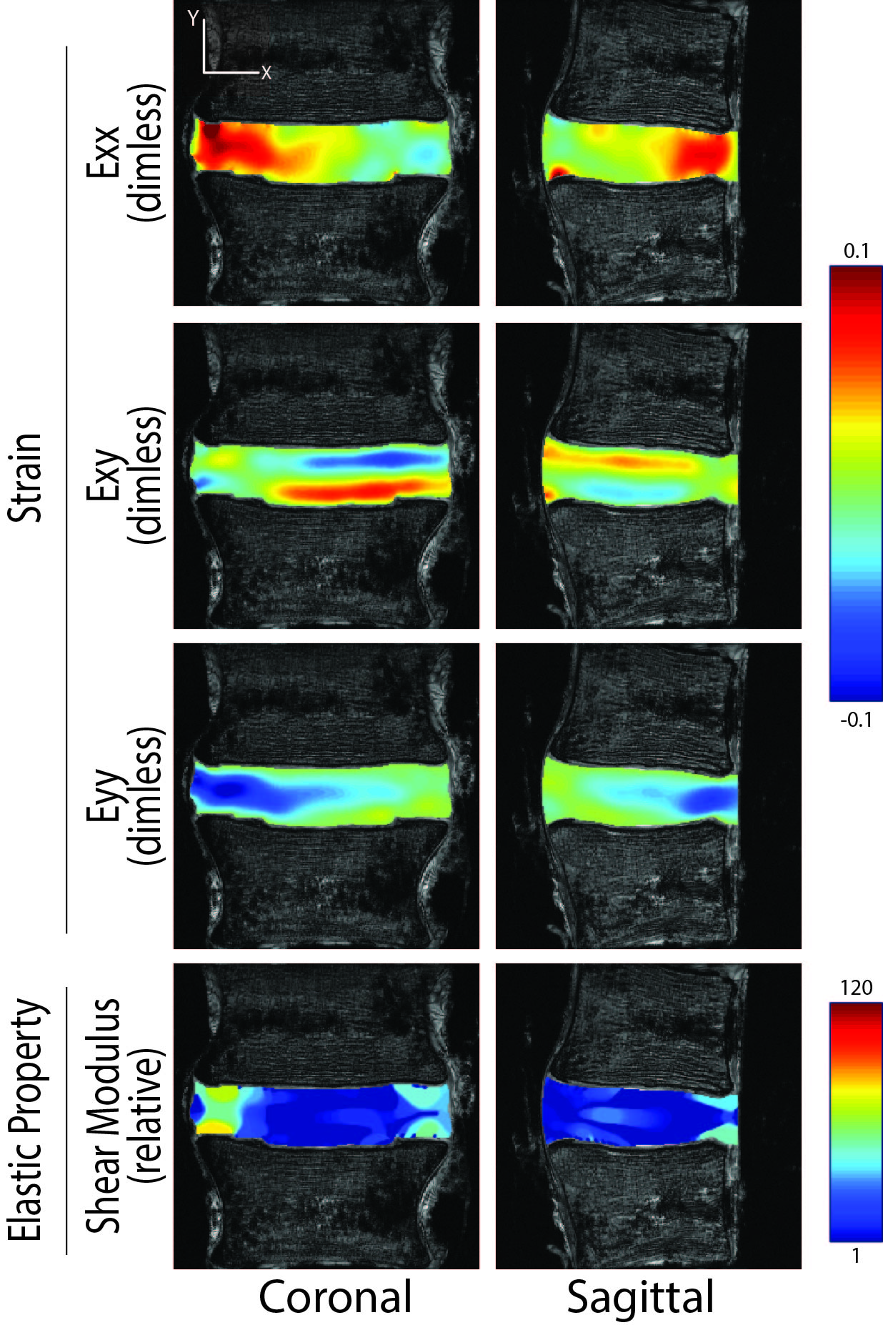
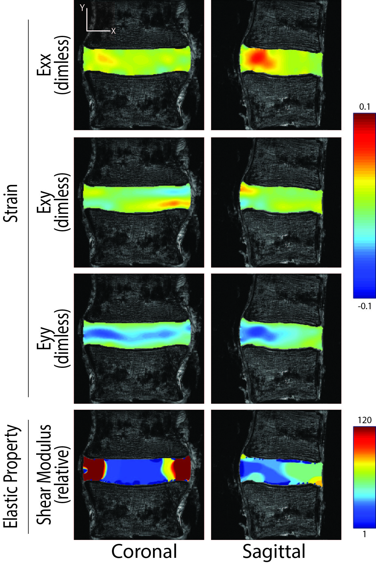
Strain Mapping: Using dualMRI, 2D Green-Lagrange strains (Exx, Eyy, Exy) representing transverse, superointerior and shear directions, respectively, were measured in coronal and sagittal planes under cyclic compression and bending of the IVD applied by the loading system described. For compression, 445 N along the superoinferior axis for 2 seconds, every 5 seconds, simulated a typical force during normal gait [5]. For bending, a 3.0-N∙m moment in the anterior direction was consistent with typical moments within the lumbar spine under non-strenuous movements [7]. dualMRI was achieved with displacement encoding of 0.32 rad/mm [6] and phase cycling to eliminate artifacts. Acquisition of images of the same resolution and field of view as relaxometry was accomplished with balanced steady state free precession (bSSFP, TE/TR=1.607ms/3.215ms, flip angle=25°). Custom software (Matlab) was used to calculate displacements and strains [6].
Inverse Modeling: We mapped the shear modulus of human IVD utilizing an iterative inverse approach, minimizing the gap between measured and computed displacement fields throughout the region of interest in L2 norm [8] and the finite element method. To avoid overfitting, we introduced a regularization term in the cost function to smooth the reconstructed elastic property distribution. The optimization problem was solved by the quasi-Newton method, and the iterative process terminated when the difference of the objective function values or the associated gradients between two neighboring iterations were less than the machine precision.
Statistical Analysis: All data is presented as mean±standard deviation of the mean.
Results
Regions of elevated T1 and T2 values were observed in the center of the IVD in both coronal and sagittal planes (Figure 2), consistent with the nucleus pulposus. All dualMRI-based strains, under compression and bending, exhibited heterogeneous patterns in both coronal and sagittal planes (Figures 3 and 4). The cohort means for Exx, Eyy and Exy were 0.018 ± 0.006, -0.031 ± 0.006 and 0.002 ± 0.002, respectively. Under compression, maximum Exx and Eyy in coronal and sagittal planes showed no apparent pattern. However, under bending, locations of strain maxima were more predictable. In the coronal plane, maxima were located at the midline of the disc, and, in the sagittal plane, at the posterior aspect. Patterns of shear modulus demonstrated a soft nucleus pulposus, except in bending, where an apparent stiffening was observed in anterior joint.Discussion
Strain and material property mapping are important metrics to assess intratissue mechanics under physiologically-relevant loading. Relaxometry cannot be used as a reliable surrogate for the mechanical behavior (i.e. strain) of the IVD, but more closely matched patterns of shear modulus, where elevated T1 and T2 values corresponded to the softer nucleus pulposus. In this study, dualMRI was employed as the gold standard in assessing the strain under large, physiological loading. IVD bending (compared to compression) resulted in more similar strain patterns among the samples and more symmetric strain in coronal plane about the midline, suggesting that bending is a more stable form of loading in IVD motion segments. Moreover, using dualMRI and inverse modelling, calculation of shear modulus was possible and represent the material properties expected during daily activities, unlike conventional MR shear wave elastography, which estimates material properties under high frequency deformations. Our results suggest that dualMRI and shear modulus metrics are well suited for the detailed assessment of intratissue mechanics in the IVD under physiologically relevant loading, allowing for detection or functional monitoring of early tissue damage in future studies.Acknowledgements
This work was supported, in part, by NIH R01 AR063712 and R21 AR066665, and NSF CAREER 134735.References
[1] Murray CJ, Atkinson C, Bhalla K, Birbeck G, Burstein R, Chou D, et al. The state of US health, 1990-2010: burden of diseases, injuries, and risk factors. JAMA. 2013;310(6):591-608.
[2] Antoniou J, Steffen T, Nelson F, Winterbottom N, Hollander AP, Poole RA, et al. The human lumbar intervertebral disc: evidence for changes in the biosynthesis and denaturation of the extracellular matrix with growth, maturation, ageing, and degeneration. The Journal of clinical investigation. 1996;98(4):996-1003
[3] Inoue N, Espinoza Orias AA. Biomechanics of intervertebral disk degeneration. Orthop Clin North Am. 2011;42(4):487-99, vii.
[4] Chan DD, Neu CP. Intervertebral disc internal deformation measured by displacements under applied loading with MRI at 3T. Magnetic resonance in medicine. 2014;71(3):1231-7.
[5] Cappozzo A. Compressive loads in the lumbar vertebral column during normal level walking. Journal of orthopaedic research. 1984;1(3):292-301.
[6] Chan DD, Neu CP. Transient and microscale deformations and strains measured under exogenous loading by noninvasive magnetic resonance. PLoS One. 2012;7(3):e33463.
[7] Adams MA, Dolan P. A technique for quantifying the bending moment acting on the lumbar spine in vivo. Journal of Biomechanics. 1991;24(2):117-26.
[8] Assad A Oberai et al 2003 Solution of inverse problems in elasticity imaging using the adjoint method Inverse Problems 19 297
Figures