1146
Estimating contraction of individual muscles during isometric wrist torque using multi-muscle MR elastography (MM-MRE)1University of Delaware, Newark, DE, United States
Synopsis
In this study, we propose to use a technique called MM-MRE to identify states of contraction of individual forearm muscles based on the measurement of muscle shear wave speed. Using a custom protocol and passive driver device, we scanned four subjects through 45 MRE scans each in three wrist positions during five torque application states. We found significant correlations between increased wave speed and higher applied torques, in both agonist and antagonist motions. The results indicate that MM-MRE is an effective measurement tool for analyzing the contractile state of individual forearm muscle during isometric contractions of the wrist joint.
Introduction
Magnetic resonance elastography (MRE) of skeletal muscle has the ability to assess muscle activity in vivo by measuring shear wave speed, which reflects muscle force output1–3. Multiple techniques are currently used to noninvasively measure muscle activity, including surface electromyography4,5 and ultrasound shear wave elastography6,7. However, these are limited in acquisition scope (ex. deep muscles, field-of-view), and thus cannot be used to study hand and wrist motions which require force from many muscles in the forearm. We propose to overcome this limitation through multi-muscle MRE (MM-MRE), which combines a large, 3D field-of-view to capture activity of all forearm muscles simultaneously along with measurement of applied torque in a variety of positions to evaluate individual contributions to wrist torque. We previously showed the repeatability of the measurements of the proposed technique and initial correlations between muscle activity and shear stiffness. Here, we develop and use a faster MM-MRE sequence and report activation effects in individual forearm muscles during isometric contraction.Methods
Participants and Imaging Protocols: Four healthy participants (3/1 M/F; ages 22-27) completed the MM-MRE protocol on a Siemens 3T Prisma scanner. Participants laid prone and headfirst in the bore of the scanner with their right arm in a forward position in a custom actuation device (Figure 1A). The device incorporates a small flexible receiver coil wrapped around the forearm and has a passive pneumatic driver that connects to the Resoundant actuator system for vibrating the tissue. The device also incorporates wrist and hand support, which is connected to a force sensor to measure the wrist joint torque. Forearm MRE images were acquired with an echo-planar imaging sequence; imaging parameters included: TR/TE = 1314/41 ms; 128 mm field-of-view; 64x64 matrix; 15 slices; 2x2x3 mm3 resolution; 80 Hz vibration; 4 phase offsets; single gradient polarity. The total time for each scan was 21 seconds. The entire MM-MRE scanning protocol included 45 scans:· five applied torques: flexion and extension, at both 0.5 and 1.0 N*m of torque, and rest (no torque);
· three wrist positions: +15°, 0°, -15° from neutral;
· three repeats of each activation at each wrist position.
Participants were cued to the correct application of torque through a visual feedback system and held the isometric contraction for entire 21 s scan. T2-weighted anatomical scans at each wrist position were also acquired for region of interest generation.
Data Analysis and Statistics: We used a nonlinear inversion algorithm (NLI)8,9 to estimate the apparent viscoelastic shear stiffness from the measured displacement fields (Figure 1B and 1C), and report the square of the shear wave speed (SWSS), which are directly related to shear stiffness by the material density. The anatomical scans were used to manually segment the fourteen forearm muscles analyzed in this study (Figure 2), which were then registered to the MRE data to generate individual muscle SWSS measures. For each contraction, muscles were divided between agonists (i.e. their contraction results in an increase in torque in the cued direction), and antagonists (i.e. their contraction results in a decrease in torque in the cued direction but may be useful for task stabilization). We used a mixed-model ANOVA, accounting for subject, wrist position, and muscle, to test for differences in SWSS levels between torque applications, with post-hoc Tukey tests to examine individual relationships.
Results
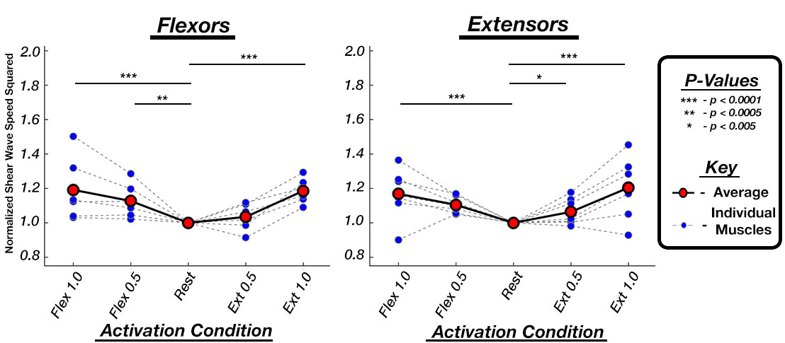
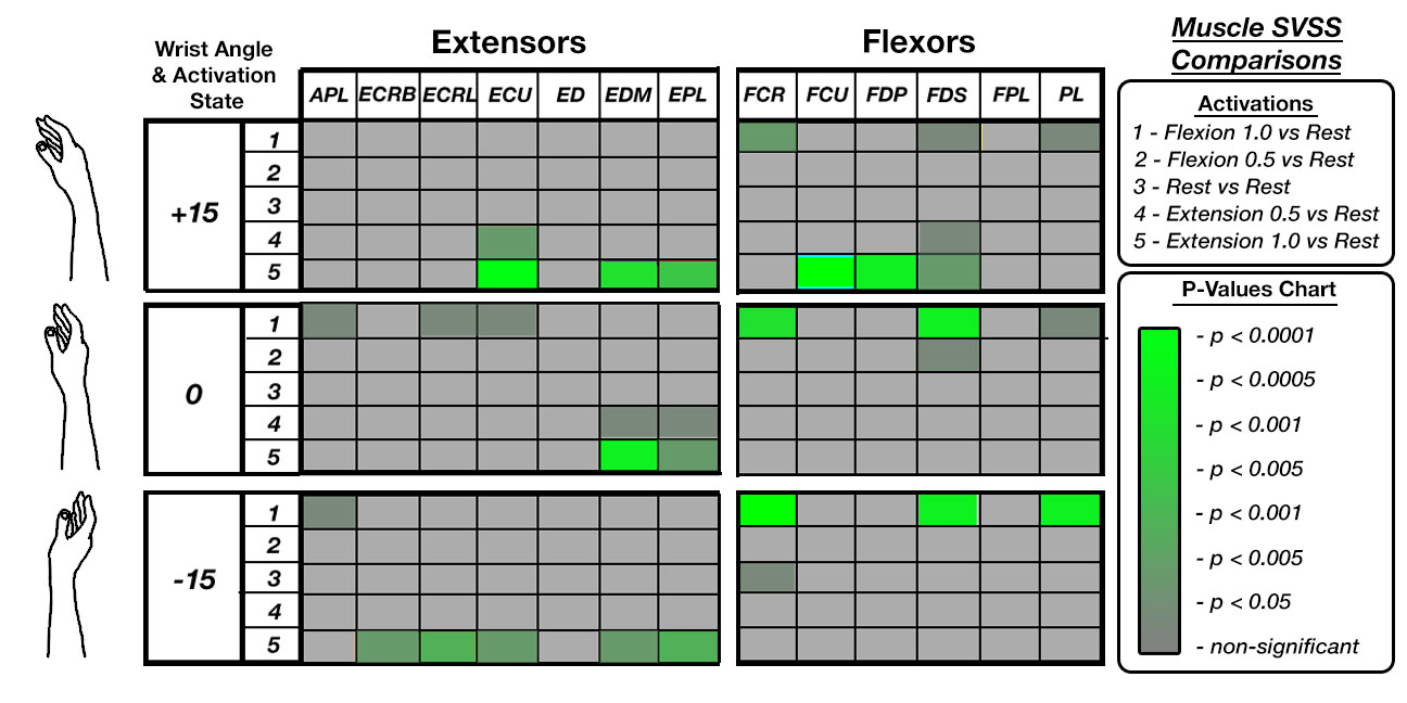
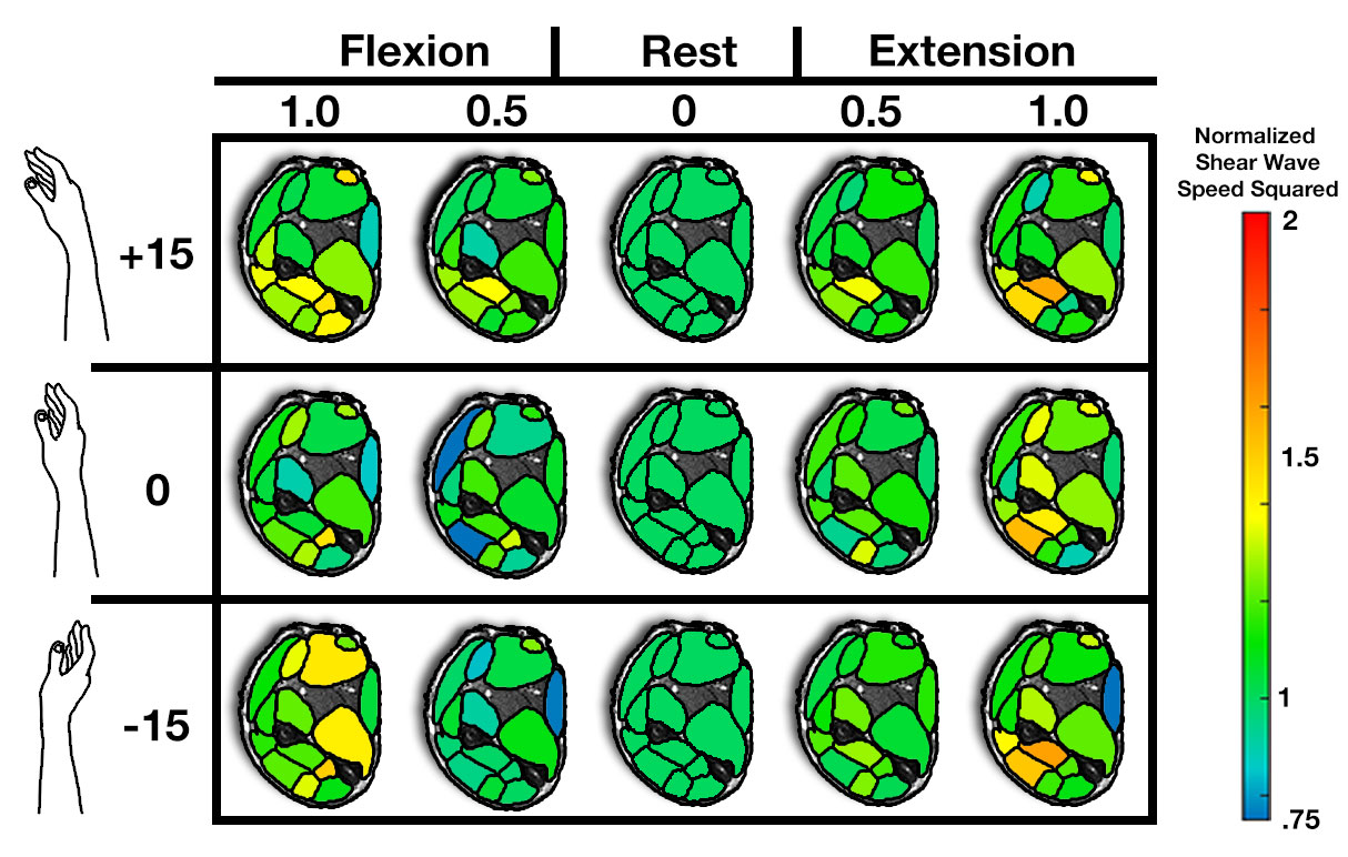
We compared the SWSS values of the individual muscles at the five different activation states (Figure 3). SWSS increased significantly in agonist muscles at both 0.5 and 1.0 N*m torque (average SWSS increases of 13.4% and 26.6%, respectively). Muscles also increased in SWSS when acting as antagonists with SWSS increases of 12.6% at 0.5 N*m and 16.7% at 1.0 N*m torque. Figure 4 shows responses of individual muscles to different torque applications, with several extensor muscles showing increased SWSS during extension, and several flexors increasing SWSS during flexion. Further, different wrist positions, expected to require engagement of muscles differently when applying wrist torques, showed different patterns of contraction amongst both extensors and flexors. Figure 5 illustrates the change in SWSS across all forearm muscles for different torque applications and wrist positions, normalized to rest.Discussion and Conclusions
In this work, we introduced a fast and reliable MM-MRE technique that improved upon our previous method10,11 by reducing acquisition time from 95 to 21 seconds per contraction, increasing reliability in the measurements and greatly reducing muscle fatigue. Using this technique, we observed increases in SWSS of both the flexor and extensor muscles when they are operating as agonists and as antagonists, with increases greatest during higher applications of muscle torque. We also demonstrated the ability to quantify responses in individual muscles across the forearm and how these differed based on wrist position. Together, these results show that MM-MRE can detect differences caused by activity and force generation in multiple muscles simultaneously. Ultimately, this data, when combined with a musculoskeletal model, can be used to estimate force from each individual muscle during hand and wrist motions. Future studies will examine muscle redundancy and how it is impacted in stroke patients. Overall, the results of this study indicate that MM-MRE is an effective measurement tool for analyzing the SWSS in all of the forearm muscles simultaneously during isometric contraction.Acknowledgements
This work was supported by NSF grant CBET-1911683.References
[1] Dresner, M. A., Rose, G. H., Rossman, P. J., Muthupillai, R., Manduca, A., and Ehman, R. L., 2001, “Magnetic Resonance Elastography of Skeletal Muscle,” J. Magn. Reson. Imaging, 13(2), pp. 269–276.
[2] Manduca, A., Oliphant, T. E. E., Dresner, M. A. A., Mahowald, J. L. L., Kruse, S. A. A., Amromin, E., Felmlee, J. P. P., Greenleaf, J. F. F., and Ehman, R. L. L., 2001, “Magnetic Resonance Elastography: Non-Invasive Mapping of Tissue Elasticity,” Med. Image Anal., 5(4), pp. 237–254.
[3] Bernabei, M., lee, S. S. M., Perreault, E. J., and Sandercock, T. G., 2019, “Shear Wave Velocity Is Sensitive to Changes in Muscle Stiffness That Occur Independently from Changes in Force,” J. Appl. Physiol.
[4] Kellis, E., and Baltzopoulos, V., 1998, “Muscle Activation Differences between Eccentric and Concentric Isokinetic Exercise.,” Med. Sci. Sports Exerc., 30(11), pp. 1616–23.
[5] Roberts, T. J., and Gabaldón, A. M., 2008, “Interpreting Muscle Function from EMG: Lessons Learned from Direct Measurements of Muscle Force.,” Integr. Comp. Biol., 48(2), pp. 312–20.
[6] Davis, L. C., Baumer, T. G., Bey, M. J., and Holsbeeck, M. van, 2019, “Clinical Utilization of Shear Wave Elastography in the Musculoskeletal System,” Ultrasonography, 38(1), pp. 2–12.
[7] Andonian, P., Viallon, M., Le Goff, C., de Bourguignon, C., Tourel, C., Morel, J., Giardini, G., Gergelé, L., Millet, G. P., and Croisille, P., 2016, “Shear-Wave Elastography Assessments of Quadriceps Stiffness Changes Prior to, during and after Prolonged Exercise: A Longitudinal Study during an Extreme Mountain Ultra-Marathon,” PLoS One, 11(8), p. e0161855.
[8] McGarry, M. D. J., Van Houten, E. E. W., Johnson, C. L., Georgiadis, J. G., Sutton, B. P., Weaver, J. B., and Paulsen, K. D., 2012, “Multiresolution MR Elastography Using Nonlinear Inversion,” Med. Phys., 39(10), pp. 6388–6396.
[9] Johnson, C. L., McGarry, M. D. J., Gharibans, A. A., Weaver, J. B., Paulsen, K. D., Wang, H., Olivero, W. C., Sutton, B. P., and Georgiadis, J. G., 2013, “Local Mechanical Properties of White Matter Structures in the Human Brain,” Neuroimage, 79, pp. 145–152.
[10] Smith, D.R., Zonnino, A., Delgorio, P.L., Duda, R., Sergi, F., Johnson, C.L., “Magnetic Resonance Elastography of the Human Forearm during Isometric Contractions”, 2019 Summer Biomechanics, Bioengineering, and Biotransport Conference, Seven Springs, Pennsylvannia, June 25-28, 2019.
[11] Smith, D.R., Zonnino, A., Delgorio, P.L., Duda, R., Sergi, F., Johnson, C.L., “Imaging Forearm Muscle Activity with Magnetic Resonance Elastography”, 2019 Biomedical Engineering Society Meeting, Philadelphia, Pennsylvannia, October 16-19, 2019.
Figures