1132
Feasibility of using a 3-axis multi-channel TMS coil array for B0 shimming of the brain at 3T
Jason P. Stockmann1,2, Lucia Navarro de Lara1, Larry Wald1,2, and Aapo Nummenmaa1,2
1A. A. Martinos Center, Massachusetts General Hospital, Charlestown, MA, United States, 2Harvard Medical School, Boston, MA, United States
1A. A. Martinos Center, Massachusetts General Hospital, Charlestown, MA, United States, 2Harvard Medical School, Boston, MA, United States
Synopsis
We explore the local field control capability of a 48ch TMS coil array to perform B0 shimming in the brain. The array uses sixteen 3-axis TMS coils built using three independent orthogonal windings that provide added flexibility to orient the resultant magnetic dipole and thus facilitate tailoring the B0 shim field. We propose adapting the TMS coils to carry DC shim currents during EPI fMRI time series acquisitions to reduce artifact levels and improve the utility of TMS-fMRI for studying brain circuits.
Introduction
Transcranial magnetic stimulation (TMS) of the cortex during functional MRI scans provides a powerful tool to reveal the causal relationships in the patterns of activity between different brain regions non-invasively. In TMS-fMRI experiments, the TMS pulses are delivered to cortex in between volume acquisitions in an echo planar imaging (EPI) time series, thus enabling a depiction of blood-oxygen level dependent changes caused by the TMS stimulus both directly under the coil as well as secondary activations in functionally and anatomically connected regions. To allow different cortical areas to be stimulated sequentially or simultaneously, a 48-channel TMS array built from sixteen 3-axis TMS coils is currently under development for next-generation neuromodulation experiments inside a 3T MRI scanner [1]. Figure 1 shows the 48 independently driven channels which will provide the ability to steer and shape the stimulus rapidly without mechanically moving any of the TMS coils.The success of TMS-fMRI will depend critically on the quality of the EPI fMRI time series data, which in turn will require (a) high SNR and (b) low EPI artifact levels. The first limitation will be addressed by building a 24ch RF receive array that can be used in conjunction with the TMS array to greatly increase sensitivity compared to receiving with the scanner body coil. A remaining major challenge is to address main field (B0) inhomogeneity caused by tissue susceptibility interfaces inside the head, leading to localized B0 offsets in the brain that cause distortion, blurring, and signal voids in gradient-echo EPI. Also, the close proximity of TMS coils to the head results in localized B0 offsets that can introduce additional artifacts. In this work, we explore the feasibility of using TMS coils for B0 shimming and other local field control applications.
In this work, we propose driving shim currents in the 48ch TMS channels to perform high-spatial order, dynamically-switchable B0 shimming of the brain to improve image quality. Used this way, the TMS array is analogous to multi-coil arrays of dedicated shim loops that have been shown to dramatically improve B0 homogeneity in the brain [2]. The use of TMS coils both for neurostimulation and for B0 shimming extends the idea of using the same set of loops for RF signal detection and DC shim currents [3,4].
The TMS array is well-suited for B0 shimming for several reasons. First, the mechanically robust housing that is designed to withstand torques much higher than those caused by switching a few amperes of shim current. Second, TMS pulses or B0 shim updating does not create significant eddy currents due to the distance of the coils from cold bore structures. Third, the TMS coils are designed to withstand currents over 5 kA current pulses and thus could easily carry a few amperes of shim current without heating up. Fourth, the 3-axis design of the array provides the ability to steer the magnetic dipole in any direction, providing flexibility for tailoring the B0 field inside the head.
Methods
To assess the potential usefulness of the 48ch TMS array for B0 shimming, simulations were performed on brain B0 field maps acquired on 2 healthy volunteers. The two volunteers had contrasting head geometries leading to very different residual B0 profiles after 2nd-order shims were applied on the scanner. Both global and dynamic slice-optimized shimming were simulated. The 48 B0 field offsets generated by each TMS channel were simulated using Biot-Savart’s law and optimal shims for each brain were computed subject to practically current limits (3A/ch and 30A/total). For comparison, B0 shim simulations were also performed using B0 field maps from an experimental 32ch 3T AC/DC shim array.Results
Figure 2 shows the simulated B0 field maps in the three cardinal planes for the X, Y, and Z-oriented loops in a single 3-axis TMS assembly. The maps show the distinct, complementary profile of the fields for the channels, providing a rich basis set for B0 shimming.Figure 3 shows simulated B0 shimming on the two healthy volunteers. Compared to conventional global 2nd order shimming, the 48ch TMS array provides 20% and 50% improvement in the standard deviation of B0 for global and slice-optimized shimming. Compared to the 16ch Z-coil array alone, the addition of the 32 X and Y channels improves shim performance by 16% and 43% for global and slice-optimal shimming, respectively.
Discussion
The 48ch TMS coil meets or exceeds the performance of a 32ch AC/DC coil for dynamic shimming. For global shimming, the 32ch AC/DC coil performs slightly better, most likely due to the presence or a greater number of loops around inferior region of the head compared to the 16 TMS array.Figure 4 shows that the 48ch TMS-shim array may provide a remedy for B0 perturbations in the head that are introduced by the TMS coils themselves. Simulated global shimming by the 48ch array almost completely eliminates the measured B0 offset created by the presence of a 3-axis coil adjacent to a phantom.
Conventional TMS amplifiers are designed to play brief (< 1ms) high-current (~5kA) pulses for neuromodulation. To provide stable shim currents to the TMS loops throughout the image acquisition, the amplifier hardware will need to be adapted.
Acknowledgements
Funding support from NIH NIBIB R00EB015445, R01MH111829, R00EB021349.References
- Navarro de Lara L, Mascarenas A, Paulson D, Makarov, Sergey Stockmann J, Wald L, Nummenmaa A. Designing a multichannel TMS/MRI system for 3 T: a 7-channel RF receive-only coil array prototype. Int. Soc. Mag. Res. Med. In: Int. Soc. Magn. Res. Med. ; 2019:1590.
- Juchem C, Nixon TW, McIntyre S, Boer VO, Rothman DL, De Graaf RA. Dynamic multi-coil shimming of the human brain at 7 T. J Magn Reson. 2011;212(2):280-288. PMID: 21824794.
- Truong T-K, Darnell D, Song AW. Integrated RF/shim coil array for parallel reception and localized B0 shimming in the human brain. Neuroimage. 2014;103C:235-240. PMID: 25270602.
- Stockmann JP, Witzel T, Keil B, Polimeni JR, Mareyam A, LaPierre C, Setsompop K, Wald LL. A 32 channel combined RF and B0 shim array for 3T brain imaging. Magn Reson Med. 2015.
Figures
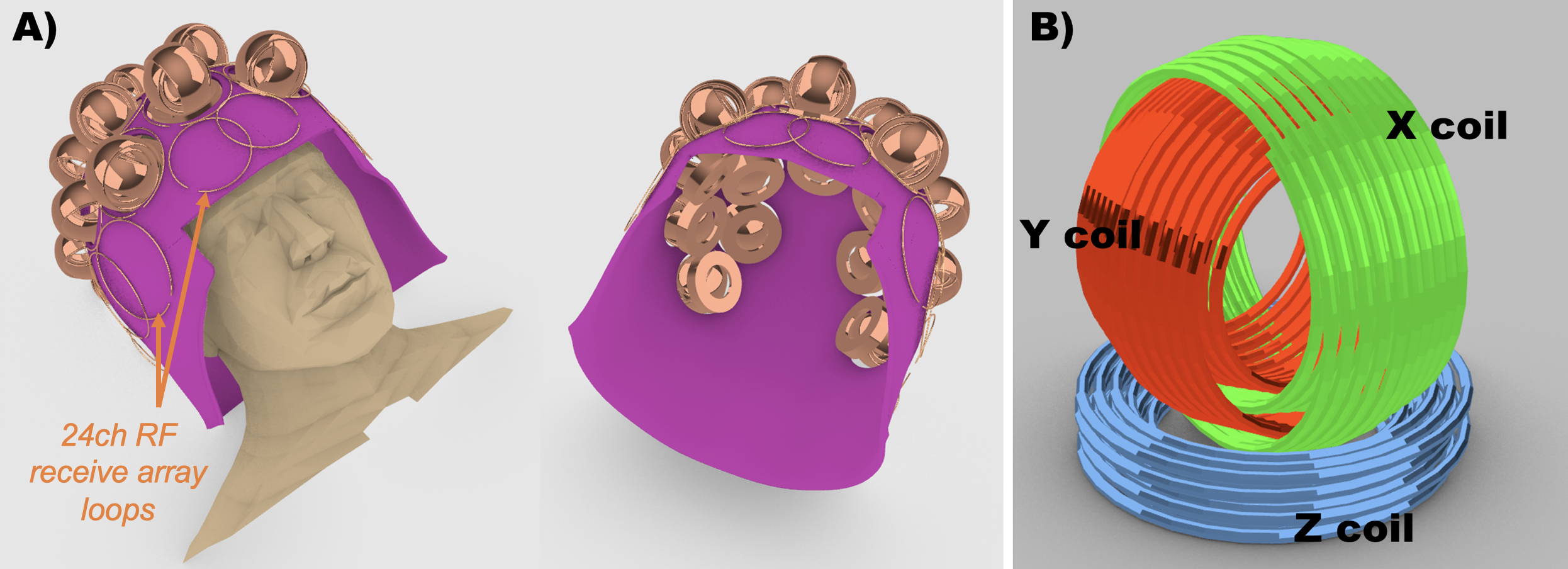
Figure 1.
The multi-channel TMS arrays uses sixteen 3-axis TMS coils arranged over the cortex. An RF receive array is built onto the helmet surface. Holes in the helmet allow TMS coils to slide
onto the head surface for efficient delivery of TMS to the cortex. The dynamic field control provided by the
sixteen 3-axis TMS coils allows the stimulus to be rapidly steered across
cortex. We exploit these same degrees of
freedom to generate magnetic fields for high-spatial order B0 shimming of the
brain.

Figure 2.
Simulated B0 field maps in the three cardinal planes for the three
orthogonal loops comprising each TMS element (normalized to 1 amp of
current). The maps show the spatially
distinct and complementary field profiles provided by the three
independently-driven coils. Together the
three loops provide the ability to steer the magnetic dipole in a desired
direction, allowing flexibility for tailoring B0 field offsets inside the head
for shimming applications.

Figure 3. (A) The 48ch TMS-shim and 32ch AC/DC shim geometries used for brain B0 shimming simulations. (B-C) Global and slice-optimized shimming with the 48ch array provides 20% and 50% improvement versus conventional 2nd order shimming, respectively. Compared to only using the "Z" TMS loops on the head surface, the addition of “X”
and “Y” loops greatly improves shim performance. The 48ch coil outperforms a 32ch AC/DC shim array for dynamic shimming and is comparable for global shimming.

Figure 4. (A) Prototype 3-axis TMS coil for initial
testing. (B) When the coil is placed on
a spherical phantom inside the scanner, it generates a local B0 offset that is
large enough to create artifacts in EPI scans (TE=30ms, 3mm iso.
voxels), see yellow arrow. (C) The cause of the artifact
is a localized B0 offset caused by the susceptibility interface between air and
the materials in the TMS coil assembly.
(D) Global B0 shimming with the 48ch TMS array provides a remedy for
this problem, reducing the standard deviation of B0 over the volume from 7 Hz
to 1 Hz.