1128
Magneto-phosphenes in head-only gradient coils
Colin M McCurdy1, Amgad M Louka1, William B Handler1, and Blaine A Chronik1
1The xMR Labs, Department of Physics and Astronomy, Western University, London, ON, Canada
1The xMR Labs, Department of Physics and Astronomy, Western University, London, ON, Canada
Synopsis
Magneto-phosphenes are caused by induced potentials in the retina, that result in visual stimulation, appearing as flashing lights. In the MR environment, magneto-phosphenes have been encountered with higher gradient strengths and longer slew times than are typically encountered in MRI. However, in a prototype head-only gradient coil we were able to repeatably induce magneto-phosphenes in four subjects. We then tested the effects of slew times, external light, and eye direction on the subject’s perception of magneto-phosphenes, finding that slew times had little effect but dimming lights and changing eye direction raised thresholds in most cases.
INTRODUCTION
Magneto-phosphenes are caused by induced potentials in the retina, that result in visual stimulation, appearing as flashing lights. In the MR environment, magneto-phosphenes have been encountered with higher gradient strengths and longer slew times than are typically encountered in MRI1. As these have no known harmful effects2 and they are rarely observed in typical MRI operation, they have not been considered as a requirement for testing gradient exposure. However, head-only gradient coils are capable of greater gradient strengths than typical full body ones and magneto-phosphenes may be possible. For a prototype head-only gradient coil in our labs, subjects have reported flashing lights occurring at gradient output lower than the onset of peripheral nerve stimulation for specific gradient waveforms3. Characterizing the conditions for which these visual phenomena occur can be explored, to make it possible to avoid the effect and enhance patient comfort.METHODS
Four subjects that had previously reported experiencing magneto-phosphenes in a different study of PNS thresholds3 for a head-only gradient coil were recruited to participate in a study to investigate magneto-phosphenes. Subjects were positioned in a prototype head-only gradient coil such that they were in as far in as possible without contacting the coil, which was known to be the position associated with the lowest stimulation thresholds. The waveform used was representative of a gradient diffusion waveform (Figure 1) consisting of repeated pairs of unipolar trapezoidal pulses. All three gradient axes (XYZ) were set to 20% of the maximum strength (129 mT/m, or a geometric sum of 223 mT/m) and incremented at 2.5% of the maximum strength every 1.5 seconds, to be stopped when the subject reported either peripheral nerve stimulation or magneto-phosphenes. The polarities of each gradient axis were chosen individually based on which combination resulted in magneto-phosphenes. Tests were repeated three times at slew times of 0.25, 0.3, and 0.35 ms. The test at the lowest rise time that did not cause peripheral nerve stimulation was then repeated in dim ambient light. Then, in regular lighting, the subject was asked to direct their eyes in 5 different directions and the test was repeated. The directions were: towards the back end of the gradient coil, towards the subjects’ feet, to the subjects left, to the subjects right, and directly up (i.e. at the top of the gradient coil). Additional rise times were tested between 0.1 and 0.7 ms to determine if there was a maximum or minimum in our testing range that would result in magneto-phosphenes.RESULTS
All subjects reported magneto-phosphenes in a repeatable manner. Two of the subjects experienced peripheral nerve stimulation before magneto-phosphenes on shorter rise times; however, as the rise time was increased, all subjects experienced magneto-phosphenes without peripheral nerve stimulation (Figure 2). All gradient strengths are reported as a geometric sum with equal weighting. Dimming lights resulted in the following changes in threshold for each subject: +12.99 mT/m, +9.29 mT/m, +16.70 mT/m, and -18.57 mT/m. Each difference was calculated from the mean of three repeats for each subject. Directing the eyes away from the top of the gradient coil resulted in a higher threshold in most cases (Figure 3).DISCUSSION
Within the studied rise times (0.1 to 0.7), there appeared to be minimal differences in the reporting of magneto-phosphenes. However, all subjects reported a gradual increase of the visual stimulus that increased with gradient excursion. When the eyes were relaxed and directed towards the top of the gradient coil the threshold for magneto-phosphenes was the lowest. Moving the eyes away from that orientation generally increased the threshold, with a few cases where it reduced the threshold. Reducing the light in the room also seemed to increase the threshold for three of four subjects, suggesting that the stimulus is more difficult to perceive with dimmed lights. It is possible that the optic nerve is stimulated in such a manner that visual stimulus is momentarily blocked, which would be consistent with the effect being more noticeable in brighter ambient lighting.CONCLUSION
Although no risks are known to be associated with magneto-phosphenes2, these experiments show that in head-only gradient coils this effect can be experienced before the threshold of peripheral nerve stimulation, and it may be advisable to inform subjects being scanned on systems with head-only gradient coils that they may experience this sensation.Acknowledgements
NSERC and the Ontario Research FundReferences
- Setsompop, K., et al. (2013). Pushing the limits of in vivo diffusion MRI for the Human Connectome Project. NeuroImage. https://doi.org/10.1016/j.neuroimage.2013.05.078
- IEC 60601-2-33:12, Medical electrical equipment, Particular requirements for the basic safety and essential performance of magnetic resonance equipment for medical diagnosis.
- McCurdy CM, et al. (2019) Spoiler, Crusher, and Diffusion Gradient Pulses Yield Higher PNS Thresholds than Long Pulse Trains in Head-only Gradient Coils. Proceedings of the 27th Annual Meeting of ISMRM. Montreal, Canada, 2019 (Abstract 7121).
Figures
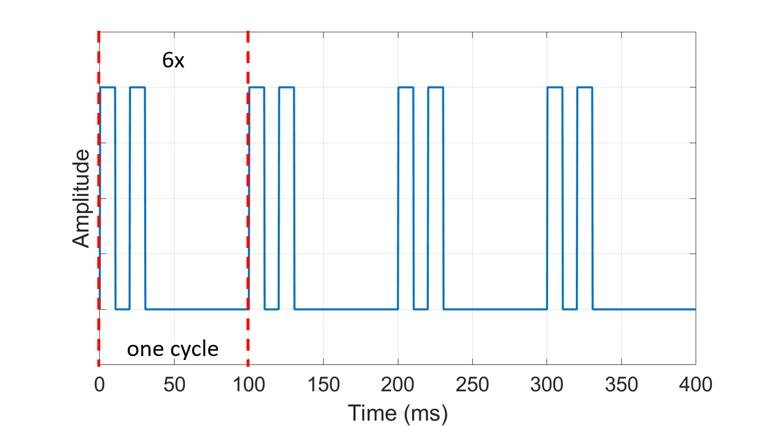
Figure 1. Gradient waveform run on all gradient axes simultaneously (XYZ). The waveform consists of 6-cycles of trapezoids with 10 ms flat tops, 20 ms betweenthe trapezoid pair, and 100 ms between the start of each pair.
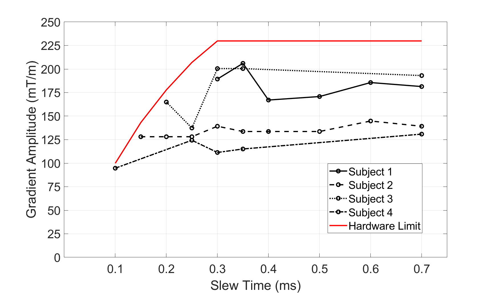
Figure 2. Effect of slew time on magneto-phosphene threshold. Individual subjects are connected by a line, and the hardware limits from the gradient amplifier is in red. All points are the average of three repeats and the geometric sum of the gradient strength on each axis.

Figure 3. Threshold change for onset of magneto-phosphenes arising from a change in eye looking direction. Values are the gradient strength of all three axes combined as a geometric sum.