1099
Feasibility of Rapid Quiescent-Interval Slice-Selective MRA of the Carotid Arteries Using Radial Sampling and Deep Learning Reconstruction1Radiology, NorthShore University HealthSystem, Evanston, IL, United States, 2The University of Chicago Pritzker School of Medicine, Chicago, IL, United States, 3Radiology, Northwestern University Feinberg School of Medicine, Chicago, IL, United States, 4Neurology, NorthShore University HealthSystem, Evanston, IL, United States
Synopsis
Ungated quiescent-interval slice-selective (QISS)-based magnetic resonance angiography (MRA) of the extracranial carotid arteries normally carries scan times of approximately 7 minutes. This work evaluated the feasibility of 3-fold accelerated single-shot QISS MRA in under three minutes using radial k-space sampling and a patch-based deep learning image reconstructive strategy.
Introduction
Multi-shot ungated quiescent-interval slice-selective (QISS) magnetic resonance angiography (MRA) has recently been found to provide better image quality than 2D time-of-flight (MRA) for evaluation of the extracranial carotid arteries, and can serve as a nonenhanced alternative to contrast-enhanced MRA.1,2 However, the current QISS protocol takes ≈7 minutes to cover the full lengths of the extracranial carotid arteries. Shorter protocols would be desirable to improve patient comfort and reduce table times. In this work we evaluated whether a 3-fold reduction in scan time might be feasible with single-shot QISS, while also probing the impacts of Cartesian and radial k-space sampling as well as the utility of a deep learning-based image reconstruction approach.Methods
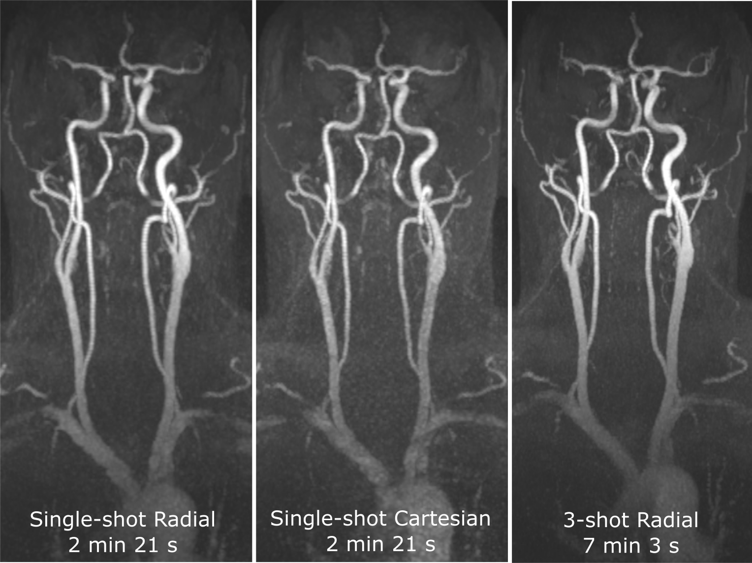
This study was approved by our institutional review board and all subjects provided written informed consent. Seven healthy volunteers and four patients with recent stroke or transient ischemic attack were imaged on a 3 Tesla MRI system (MAGNETOM Skyrafit, Siemens Healthineers, Erlangen).Image Acquisition: Rapid QISS MRA of the extracranial carotid arteries was done using prototype single-shot Cartesian and radial k-space sampling trajectories in scan times of 2 min 21 s, while comparing results to prototype 3-shot implementations carrying scan times of 7 min 3 s. Other imaging parameters were as follows: fast low-angle shot (FLASH) readouts with TR/TE/flip angle=15 ms/2.1 ms/30 degrees, 128 slices, 2 mm slice thickness, -0.6 mm slice gap, in-plane resolutions of 1.44 mm x 1.44 mm and 1.08 mm x 1.08 mm for single-shot and 3-shot imaging, golden radial view angle increment.
Qualitative and Quantitative Analysis: In volunteers, image quality in 8 carotid arterial segments was scored by two radiologists using a 4-point scale (1: non-diagnostic, 2: fair, 3: good, 4: excellent). Temporal signal-to-noise ratio (tSNR), apparent arterial-to-background contrast-to-noise ratio (CNR), and arterial-to-background contrast were measured using the respective equations of Sa/σa, (Sa-Sm)/σm, and Sa/Sm-1, where Sa (σa) and Sm (σm) denote the means (standard deviations) of arterial and muscle signals.
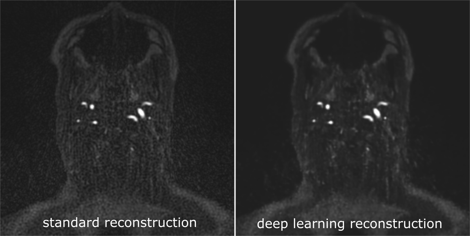
Deep Learning Image Reconstruction: An image patch-based deep learning image reconstruction strategy was trained and applied to single-shot images to see if it could improve image fidelity using the 3-shot result as the reference standard. Using a leave-one-out training and application strategy, the deep neural network, based on the well-known U-net architecture3, was trained using spatially registered 3-shot and 1-shot data. Image fidelity in offline-reconstructed training data was evaluated using the structural similarity index (SSIM); results were obtained with deep learning reconstruction were compared with the prominent image denoising approach of block matching and 3D filtering (BM3D).4 The trained deep neural network was thereafter applied to prospectively-acquired single-shot and 3-shot source images, which were evaluated in a blinded fashion by two radiologists using a two-alternative forced choice test.
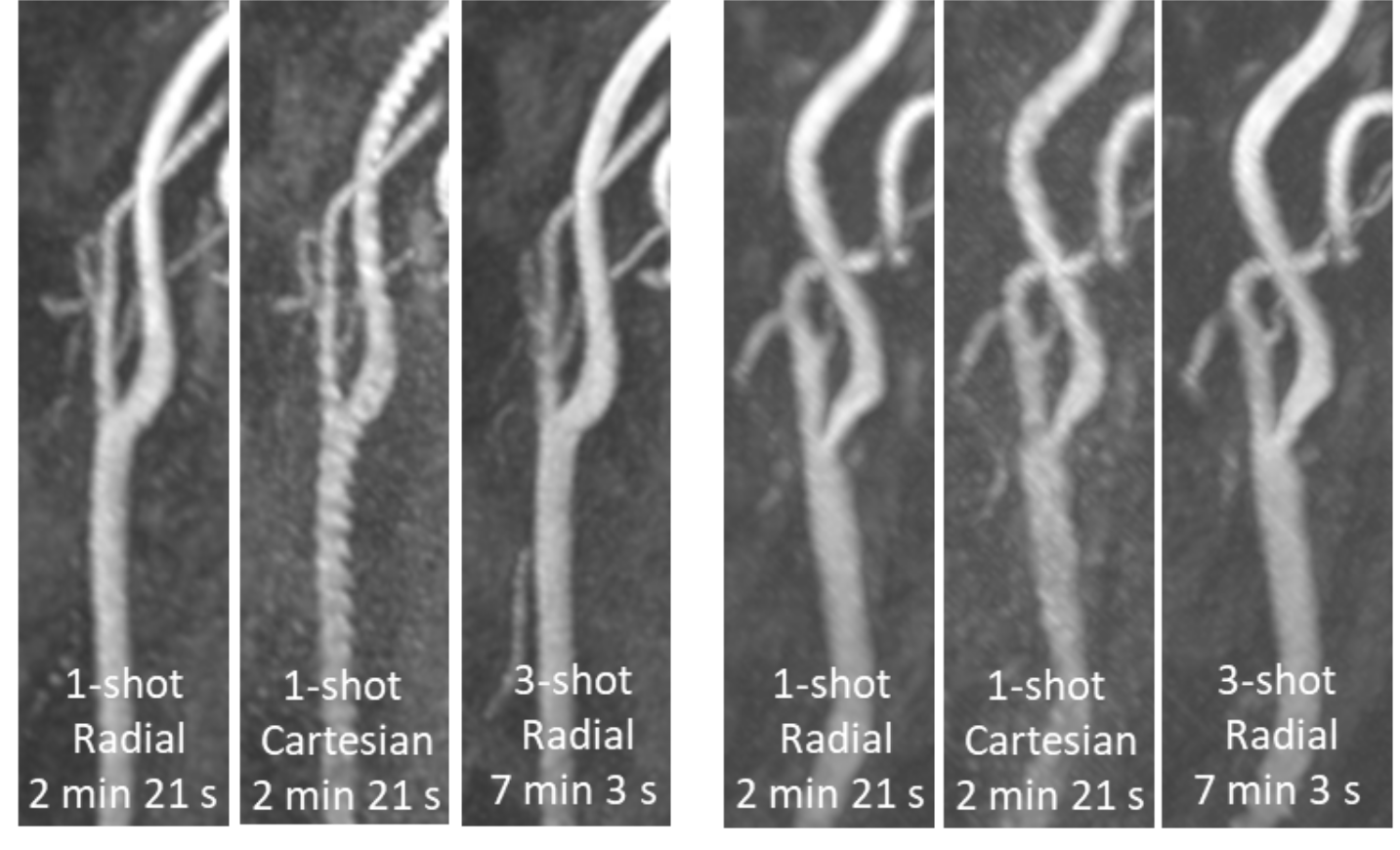
Patient Imaging: Four patients with recent stroke or transient ischemic attack were imaged to probe the feasibility of rapid single-shot QISS for portraying the extracranial carotid arteries in such a patient cohort.
Results
Figure 1 shows the image quality obtained with ungated single-shot QISS MRA of the neck obtained using Cartesian and radial k-space sampling. Compared to the use of Cartesian sampling, ungated single-shot QISS MRA using radial sampling provided improved image quality for portraying the extracranial carotid arteries (mean scores of 2.9 versus 2.7, P<0.05) and 2.2-fold higher temporal signal-to-noise ratio (means of 22.2 versus 10.2, P<0.001). Patient imaging (Figure 2) confirmed the results in volunteers, and demonstrated that rapid single-shot radial QISS similarly portrayed the carotid bifurcation with respect to the much lengthier 3-shot protocol.The application of deep learning-based image reconstruction to single-shot radial QISS produced images (Figure 3) with higher SSIM than those obtained with BM3D denoising (0.870±0.019 versus 0.849±0.013, P<0.001); SSIM values were markedly improved with respect to the baseline single-shot images obtained with standard gridding reconstruction (0.772±0.020, P<0.001). Deep learning-based image reconstruction of prospectively-acquired single-shot data produced source images with 14% increased arterial-to-background contrast (8.4 vs 9.6, P=NS), 210% increased apparent CNR (60.5 vs 19.5, P<0.001), and that were preferred by both radiologists using two-alternative forced choice testing (P<0.001). The use of the machine learning algorithm did not cause loss of image features or introduce false ones.
Discussion
The results of our study suggest that 3-fold faster single-shot QISS of the extracranial carotid arteries is feasible, and that radial k-space sampling outperforms Cartesian k-space sampling. Furthermore, our results show that the use of a patch-based deep learning reconstructive strategy improved image fidelity on the basis of SSIM while outperforming the commonly used BM3D algorithm.Conclusion
In conclusion, we found that rapid single-shot ungated radial QISS MRA of the extracranial carotid arteries is feasible in a short scan time of ≈2 minutes, and benefits from the application of deep learning-based image reconstruction to improve image fidelity.Acknowledgements
Funding Source: NIH grant R01 EB027475References
1. Koktzoglou I, Aherne EA, Walker MT, Meyer JR, Edelman RR. Ungated nonenhanced radial quiescent interval slice-selective (QISS) magnetic resonance angiography of the neck: Evaluation of image quality. J Magn Reson Imaging. 2019 May 11. doi:10.1002/jmri.26781. [Epub ahead of print]
2. Peters S, Huhndorf M, Jensen-Kondering U, Larsen N, Koktzoglou I, Edelman RR, Graessner J, Both M, Jansen O, Salehi Ravesh M. Non-Contrast-Enhanced Carotid MRA: Clinical Evaluation of a Novel Ungated Radial Quiescent-Interval Slice-Selective MRA at 1.5T. AJNR Am J Neuroradiol. 2019 Sep;40(9):1529-1537. doi: 10.3174/ajnr.A6171. Epub 2019 Aug 8.
3. Ronneberger O, Fischer P, Brox T. U-net: Convolutional networks for biomedical image segmentation. In: Lecture Notes in Computer Science (including subseries Lecture Notes in Artificial Intelligence and Lecture Notes in Bioinformatics). ; 2015. doi: 10.1007/978-3-319-24574-4_28.
4. Dabov K, Foi A, Katkovnik V, Egiazarian K. Image Denoising by Sparse 3-D Transform-Domain Collaborative Filtering. IEEE Trans. Image Process. 2007;16:2080–2095
Figures