1061
Imaging Endpoints by Pulse Sequence Type for Intracranial Atherosclerosis using Vessel Wall MR Imaging1Radiology, University of Pennsylvania, Philadelphia, PA, United States, 2Radiology, Cedars Sinai Medical Center, Los Angeles, CA, United States, 3Neurology, University of Pennsylvania, Philadelphia, PA, United States
Synopsis
Intracranial atherosclerosis is a common cause of ischemic stroke. Variability in protocol/pulse sequence design of intracranial vessel wall MR imaging (VWI) has led to different imaging endpoints to detect and characterize atherosclerosis. We systematically reviewed the literature to identify VWI investigations studying atherosclerosis to identify commonly reported imaging endpoints. The most common imaging endpoints using T1-weighting included wall enhancement, thickening, plaque quadrant in cross-section, and stenosis; on T2-weighting, intraplaque T2 signal intensity and wall thickening were common endpoints. Establishing diagnostically accurate imaging endpoints to validate as atherosclerosis biomarkers are critical to understand where efforts for technique optimization should be directed.
Introduction
Intracranial atherosclerosis remains one of the leading causes of ischemic stroke.1 Intracranial vessel wall MR imaging (VWI) is used to help distinguish among different intracranial vasculopathies. However, the protocols and pulse sequences used for VWI vary widely.2 As a result, the reported imaging endpoints to detect and characterize atherosclerosis in many investigations also vary widely leading to challenges in synthesizing the literature and limits the generalizability of the results.3 The most common endpoints by pulse sequence type remain unknown. We systematically reviewed the VWI literature to identify the commonly reported primary and secondary imaging endpoints for atherosclerosis on T1-weighted and T2-weighted by VWI.Methods
PubMed, EMBASE, and MEDLINE databases were searched up to September 2018. Inclusion criteria were investigations on humans, VWI, and intracranial vasculopathies. Single case reports, pediatric investigations and conference abstracts were excluded. A subset of included articles examining intracranial atherosclerosis underwent qualitative review. Data on MR pulse sequences, protocol design, technical parameters and primary and secondary imaging endpoints were collected. Categorical variables are presented as counts and percentages. SPSS, Inc (IBM) was used for data management and analyses. The Preferred Reporting Items for Systematic Reviews and Meta-Analyses guidelines were used.Results
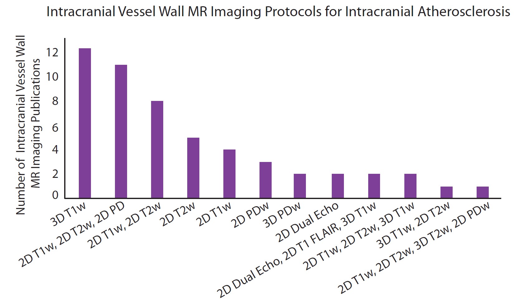
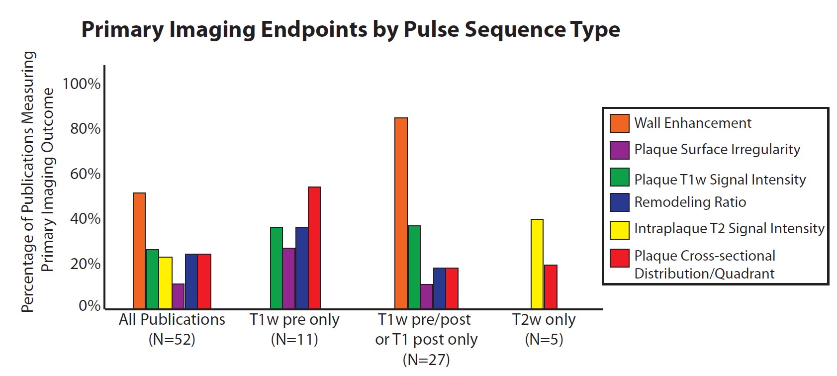
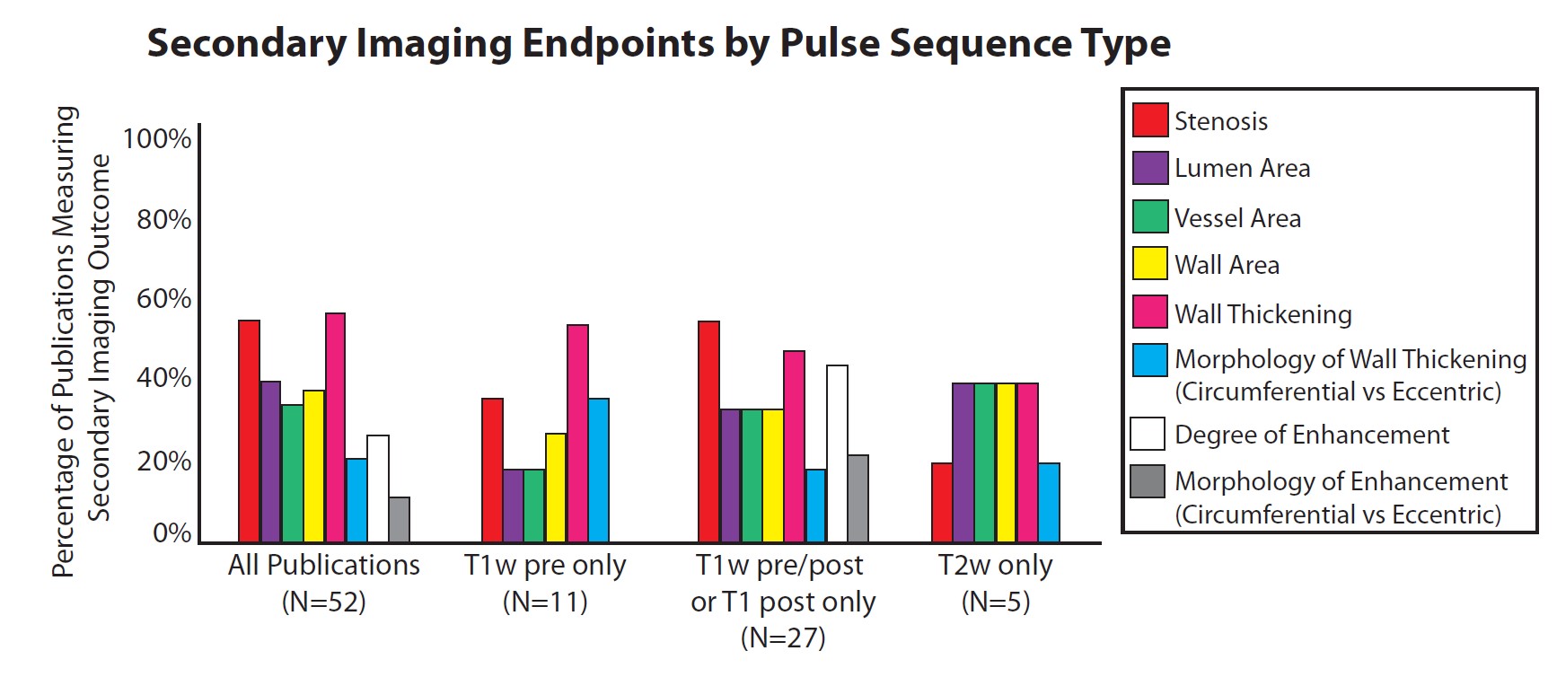
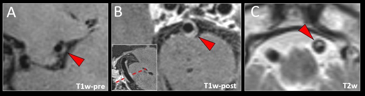
From 2,431 publications, full-text data extraction was performed on 54 articles using intracranial vessel wall MR imaging (VWI) to study intracranial atherosclerosis. VWI protocols to characterize atherosclerosis varied widely with the most common protocol including 3D T1-weighted (T1w) acquisitions (Fig 1). The most common imaging definition of plaque was focal, eccentric vessel wall thickening (40%, n=21). Notably, most publications did not clearly define how plaque was identified on VWI (60%, n=31). To measure imaging endpoints, T1-weighted acquisitions (40%, n=21) were most commonly used followed by T2-weighted acquisitions (T2w) (21%, n=11) (Fig 2). Among all publications, the most common primary and secondary imaging endpoints were wall enhancement (52%) and thickening (58%), respectively (Fig 3). On T1w pre-contrast images, identifying plaque by quadrant on cross-section of an artery to assess whether plaque occluded the ostium of a perforator artery (55%) and wall thickening (55%) were the most common imaging endpoints (Fig 4A). For T1w-postcontrast images, wall enhancement (85%) and thickening (56%) were most frequently assessed (Fig 4B). Finally, for T2w images, intraplaque T2 signal intensity (40%) (Fig 4C) and wall thickening (40%) as well as lumen/vessel/wall areas (40%) were common primary and secondary endpoints, respectively.Discussion
The results reveal a wide spectrum of VWI protocol and pulse sequence designs used to study intracranial atherosclerosis. Primary and secondary imaging endpoints used to define and characterize plaque also vary widely due to these technical differences. Vessel wall enhancement and thickening emerged as the most common endpoints for all studies. Analyses by pulse sequence type showed plaque quadrant distribution, wall enhancement, and intraplaque T2 signal to be the most common primary imaging endpoints for T1w-precontrast, T1w-postcontrast, and T2w acquisitions, respectively. While some reliability and reproducibility investigations align with these common imaging endpoints (e.g., enhancement4), others focus on less commonly used imaging endpoints (e.g., wall and lumen areas5). Moreover, each investigation used different pulse sequences. Consensus on imaging endpoints and pulse sequences would help focus research resources and efforts. Future directions to reach consensus include comparing the diagnostic accuracies of these imaging endpoints by VWI pulse sequence to characterize atherosclerosis.Conclusion
Variability in VWI protocols and pulse sequences result in a number of different imaging endpoints to detect and characterize intracranial atherosclerosis. More studies are warranted to identify which pulse sequences and imaging endpoints are diagnostically accurate and clinically achievable to establish a VWI biomarker for intracranial atherosclerosis.Acknowledgements
No acknowledgement found.References
1. Qureshi, A. I. & Caplan, L. R. Intracranial atherosclerosis. Lancet 383, 984-998 (2014).
2. Mandell, D. M. et al. Intracranial Vessel Wall MRI: Principles and Expert Consensus Recommendations of the American Society of Neuroradiology. AJNR Am. J. Neuroradiol. 38, 218-229 (2017).
3. Lee, H. N., Ryu, C. W. & Yun, S. J. Vessel-Wall Magnetic Resonance Imaging of Intracranial Atherosclerotic Plaque and Ischemic Stroke: A Systematic Review and Meta-Analysis. Front. Neurol. 9, 1032 (2018).
4. Mossa-Basha M, Watase H, Sun J, et al. Inter-rater and scan-rescan reproducibility of the detection of intracranial atherosclerosis on contrast-enhanced 3D vessel wall MRI. Br J Radiol. 92 (2019).
5. Zhang X, Zhu C, Peng W, et al. Scan-rescan reproducibility of high resolution magnetic resonance imaging of atherosclerotic plaque in the middle cerebral artery. PLoS ONE. 10, e0134913 (2015).
Figures