1049
Chemical sHift bAsed pRospectIve k-Space anonyMizAtion (CHARISMA)1Biomedical Magnetic Resonance, Otto-von-Guericke University, Magdeburg, Germany, 2Medicine and Digitalization, Otto-von-Guericke University, Magdeburg, Germany, 3German Center for Neurodegenerative Disease, Magdeburg, Germany, 4Center for Behavioral Brain Sciences, Magdeburg, Germany, 5Leibniz Institute for Neurobiology, Magdeburg, Germany
Synopsis
One key element of open science is to make all data publicly available. In case of neuroscience, reconstructed images can be defaced to prevent data privacy violations, but no strategy to anonymize raw data has been presented to our best knowledge.
Here, chemical shift based prospective k-Space anonymization is presented. The subject wears an oil-filled mask which is superimposed onto the subject’s skin due to chemical shift. This low-cost solution (<15€) is easy to build and applicable for sequences with sufficient chemical shift in the A-P direction.
Introduction
Publicly available data is one keystones of open science. However, open data can conflict with data privacy. Although reconstructed images can be defaced and subject information can be removed from the metadata1, raw data anonymization remains an open challenge. Especially raw data of brain scans are vulnerable because the subject’s face can be reconstructed from it.In this study, Chemical sHift bAsed pRospectIve k-Space anonyMizAtion (CHARISMA) is presented and tested. The subject wears an oil-filled mask. Due to the chemical shift2,3, the oil can be superimposed on the subject’s skin if the protocol is adjusted accordingly. Hence, chemical shift artifacts could be exploited to anonymize data during the acquisition.
Methods
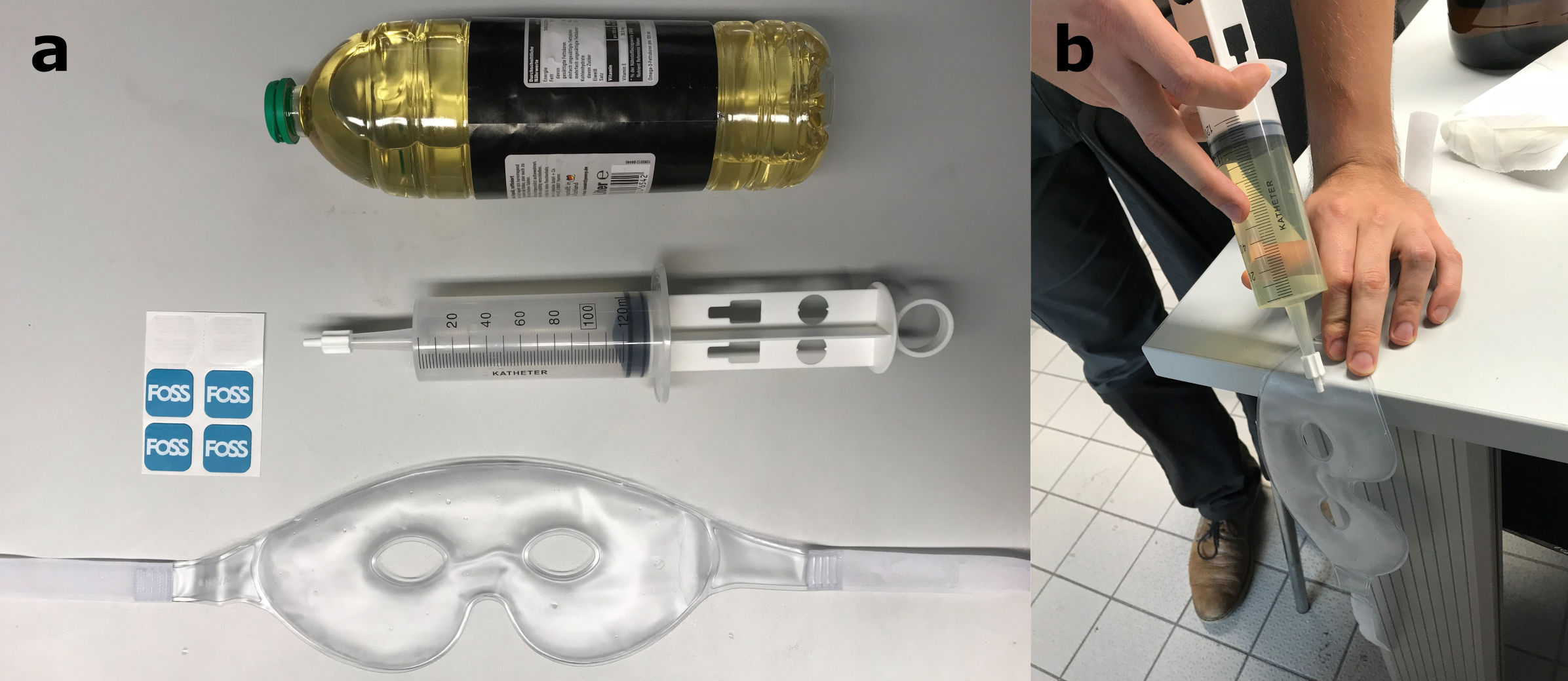
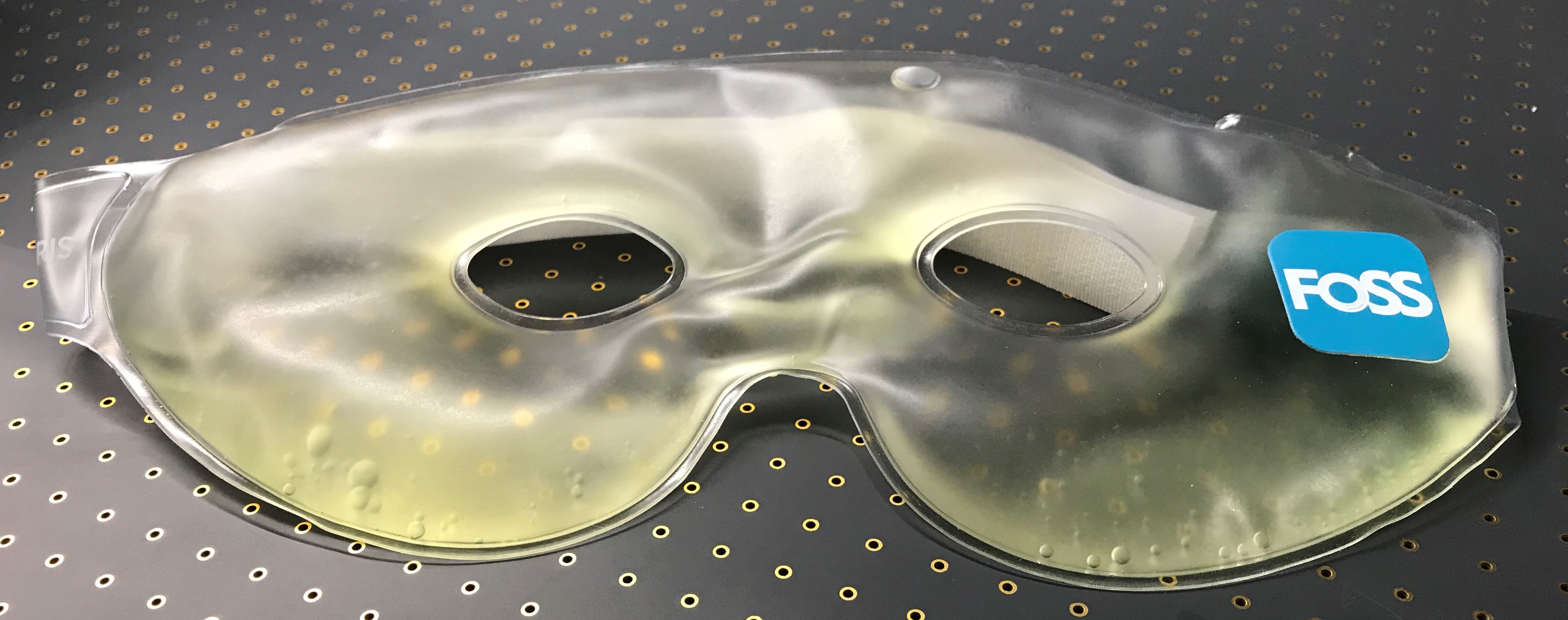
Mask designA commercially available cooling mask with water-based filling was refilled with colza oil (fat shift$$$\approx$$$3.5ppm2). Filling was done through a small hole with a syringe/catheter tip. The hole was sealed using an adhesive patch (bicycle equipment, no vulcanization). Fig.1 shows the materials used and the filling process. The final mask is depicted in Fig.2. Fabrication of a single mask costs less than 15€.
Data acquisition
For proof of principle, a single subject was scanned at 7-Tesla (Siemens Healthineers, Germany) with a 32-channel head coil (Nova Medical, USA) after given written consent.
To superimpose the oil on the skin the read-out direction of all GRE-based sequences were adjusted to A-P. All slab-selective scans were acquired with 1mm isotropic voxel size and 256x224x96mm FOV.
PD-weighted scans with and without the mask were acquired with a bandwidth (BW) of 200 Hz/px (apparent fat shift of 4.9mm) to depict the subject’s skin without dephasing-induced blurring. To achieve the TE of 3.4ms (TR=8.1ms), flow compensation was disabled.
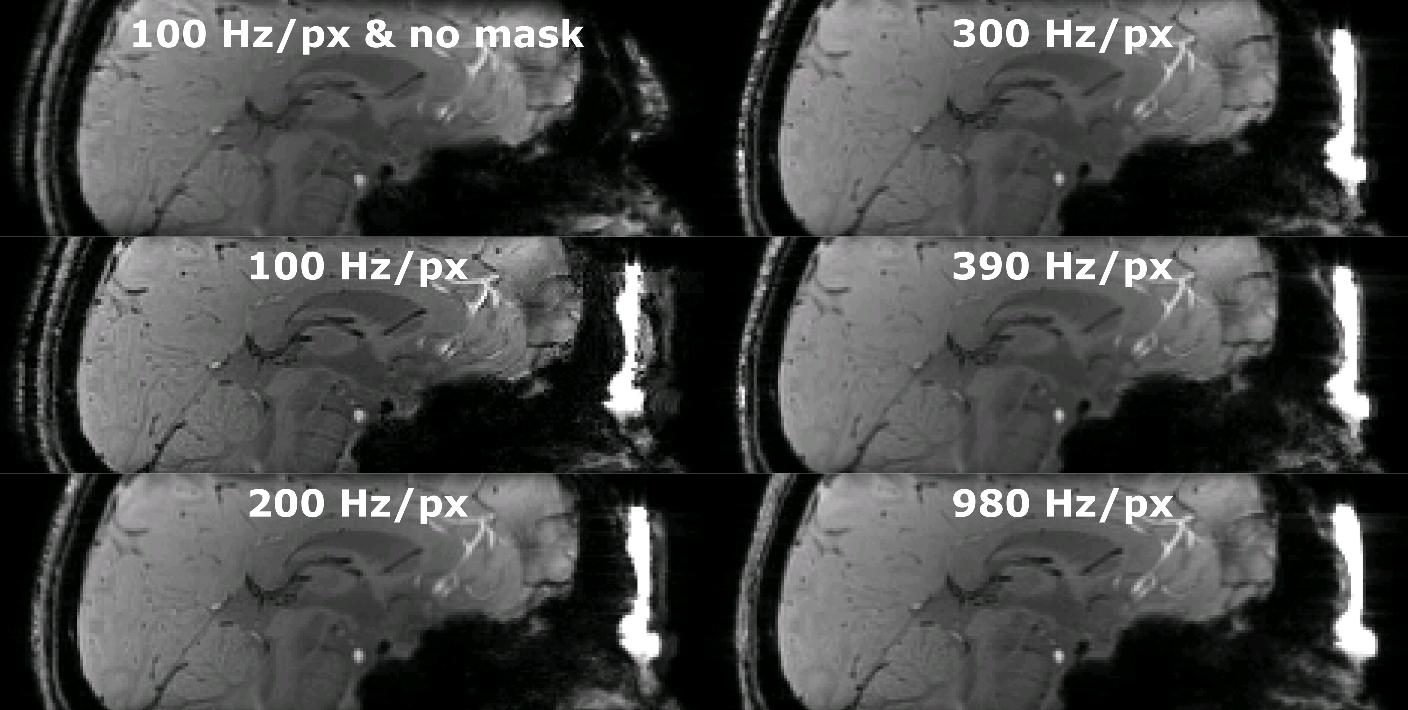
From the PD-weighted scan a T2*-weighted protocol was derived (TE/TR=9/17ms, flow compensation enabled). T2* images were acquired at several BW with the mask: 100Hz/px (9.8mm shift); 200Hz/px (4.9mm shift); 300Hz/px (3.3mm shift); 390Hz/px (2.5mm shift); and 980Hz/px (1.0mm shift). For reference, an un-masked data set (BW=100Hz/px) was acquired.
Post processing
For each contrast, data was co-registered to the masked volume with the lowest BW using FSL FLIRT4. Sagittal images were compared to evaluate qualitatively in superposition of mask and skin. From the masked PD-weighted scan a 3D rendering of the face was reconstructed. The counterexample (without mask) was not rendered to maintain data privacy.
Results
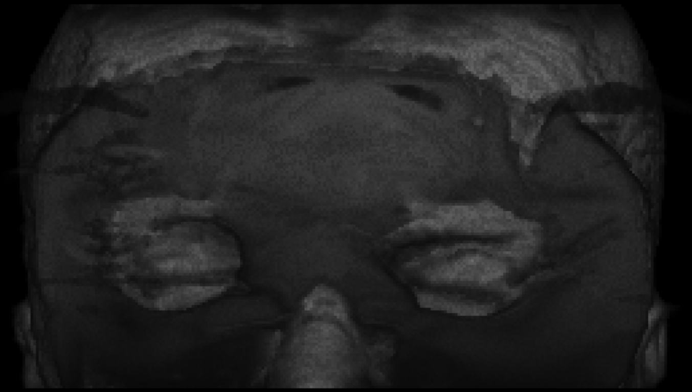
Fig.3 shows the masked and unmasked PD-weighted images. The oil of the mask is successfully superimposed onto the skin. Note that the water content of the mask is also depicted as a shadow at the physical location of the mask. The mask covers most of the subjects face, considerably complicating subject identification, while eyes remain uncovered to improve subject comfort (see 3D rendering in Fig.4).Several BWs for T2*-weighted imaging have been acquired (see Fig.5). If the chemical shift is reduced (i.e. BW=980Hz/px, 1mm apparent shift), the masking mechanism becomes less efficient and separating the mask from the skin could be possible. Based on the data present, a shift of at least 3mm is recommended, but further tests with multiple subjects, protocols, and field strengths are required to give reliable recommendations.
Discussion
CHARISMA is a low-cost solution to anonymize raw data prospectively for sequences with read-out direction in A-P and sufficient chemical shift. Since the apparent chemical shift is a function of BW, voxel size, B0-field strength, and material (mask filling) used3, CHARISMA provides flexibility to be adapted to different imaging scenarios.As with any security mechanism, CHARISMA provides no absolute safety. Currently, nose, mouth, and eyes are not covered by the mask to improve subject comfort, but could be concealed if the mask if extended. Furthermore, with sufficient effort (e.g. segmentation or inpainting5) or access to privileged data repositories (finding matching brain or dental records) the subject’s identity could be uncovered. If multiple gradient echoes are acquired separate fat and water images can be reconstructed6, potentially jeopardizing the masking.
Despite these challenges, CHARISMA is to our best knowledge the first prospective anonymization strategy for MR-raw data acquisition. Alternative approaches could be based on altering the transversal magnetization locally, i.e. dephasing, saturating, or not exciting the magnetization of the subject’s face. Potential implementation of these anonymization strategies included the uses of parallel transmission, non-linear gradients, or additional saturation pulses. Although these approaches could be more effective and general solutions, they are associated with considerable costs and efforts compared to the low-cost, do-it-yourself CHARISMA method and remain topics for future research.
Anonymizing k-space prospectively could help to implement open data repositories of brain exams. These publically available data could then be used in the development of advanced reconstruction methods or to improve reproducibility in MRI research.
Conclusion
To our best knowledge, CHARISMA is the first attempt to anonymize k-space data prospectively. The low-cost solution is easy to build and applicable for sequences with sufficient chemical shift in the A-P direction.Eventually, this abstract could foster the discussion and development of prospective k-space anonymization to enable open data for brain examinations.
Acknowledgements
This work was supported by the NIH, grant number 1R01-DA021146 and received funding from the federal state of Saxony-Anhalt under grant number ‘I 88’.References
1. Bischoff-Grethe A, Ozyurt IB, Busa E et al. A technique for the deidentification of structural brain MR images. Hum Brain Mapp. 2007;28(9):892–903. doi: 10.1002/hbm.20312.
2. Bley TA, Wieben O, François CJ, Brittain JH, Reeder SB. Fat and water magnetic resonance imaging. J Magn Reson Imaging 2010;31(1):4–18. doi: 10.1002/jmri.21895.
3. Parizel PM, van Hasselt BA, van den Hauwe L, van Goethem JW, Schepper AM de. Understanding chemical shift induced boundary artefacts as a function of field strength: influence of imaging parameters (bandwidth, field-of-view, and matrix size). Eur J Radiol. 1994;18(3):158–164.
4. Jenkinson M, Beckmann CF, Behrens TEJ, Woolrich MW, Smith SM. FSL. Neuroimage. 2012;62(2):782–790. eng doi: 10.1016/j.neuroimage.2011.09.015.
5. Abramian D, Eklund A. Refacing: Reconstructing Anonymized Facial Features Using GANS. In: ISBI 2019: 2019 IEEE International Symposium on Biomedical Imaging April 8-11, 2019, Hilton Molino Stucky, Venice, Italy. 2019 IEEE 16th International Symposium on Biomedical Imaging (ISBI); 4/8/2019 - 4/11/2019; Venice, Italy: IEEE; 2019. p. 1104–1108.
6. Berglund J, Rydén H, Avventi E, Norbeck O, Sprenger T, Skare S. Fat/water separation in k-space with real-valued estimates and its combination with POCS. Magn Reson Med. 2019. doi: 10.1002/mrm.27949.
Figures