0966
An enhanced turboPROP+ technique for diffusion weighted imaging1Neuroradiology, Barrow Neurological Institute, Phoenix, AZ, United States, 2Philips Healthcare, Gainesville, FL, United States
Synopsis
Diffusion weighted MRI is a useful technique for the diagnosis of neurological disorders. DW EPI is time efficient but suffer from geometric distortions. DW PROPELLER and its variants, including turboPROP and turboPROP+, have been proposed to generate distortion free images. This project improves the turboPROP+ technique by incorporating LRX RF phase modulation approach to improve SNR and signal stability, and by revising the phase correction algorithm to minimize residual artifacts. Volunteer results demonstrate reduced artifacts compared to the original phase correction algorithm, and increased SNR/image quality compared to original XY2 phase modulation.
Introduction
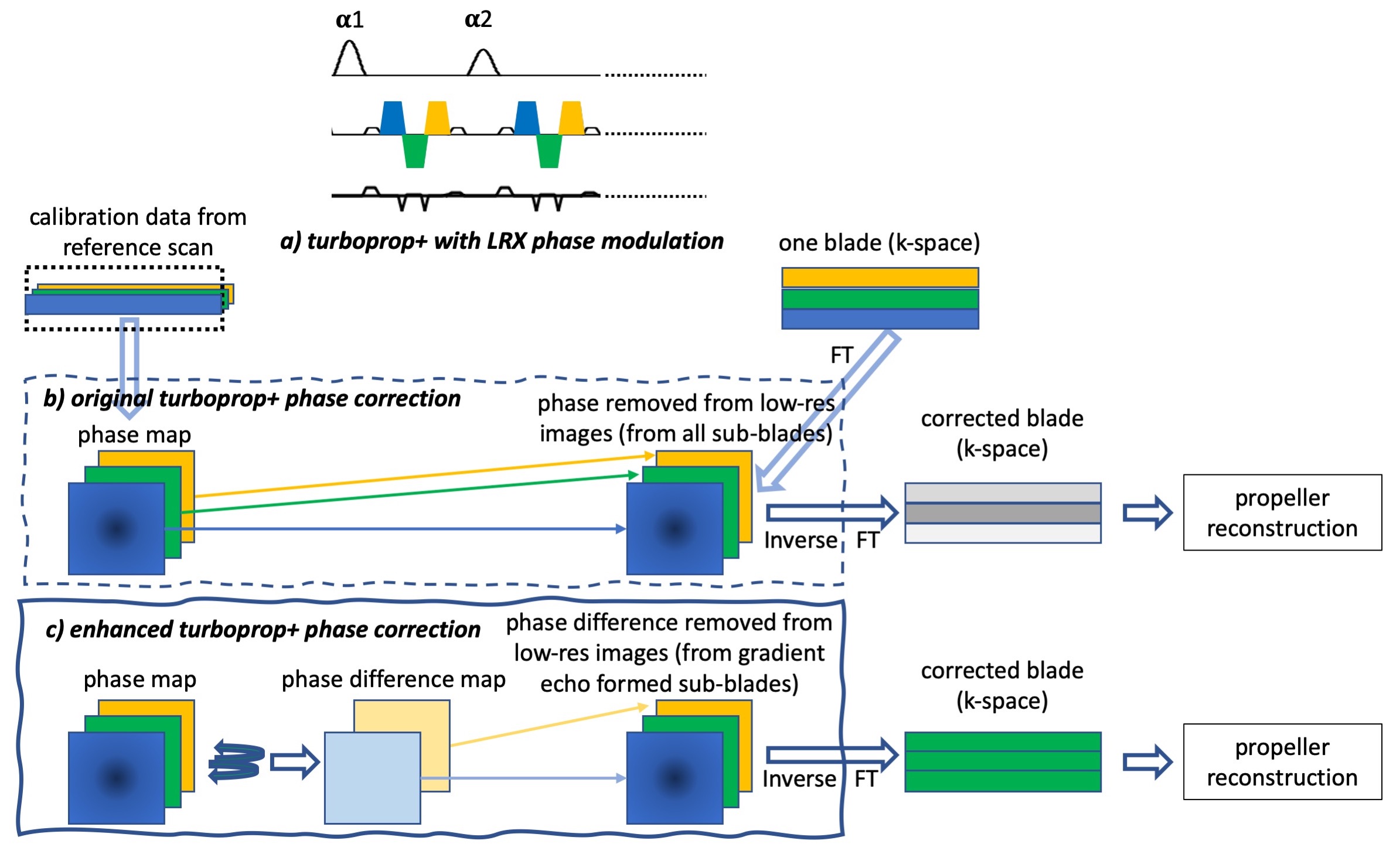
Diffusion weighted MRI is a useful technique for the diagnosis of many neurological disorders. EPI is currently the method of choice for DWI primarily because of its time efficiency. Nonetheless, EPI based DW images suffer from geometric distortions due to field inhomogeneities and susceptibility variations. DW PROPELLER, based on TSE, has been demonstrated to be distortion-free, but less SNR- and SAR-efficient. 1 TurboPROP has been developed to take advantages of both EPI and PROPELLER readouts, while minimizing their drawbacks. 2 One variant, called turboPROP+, applies 2D phase correction on the sub-blades to improve its robustness to field variations. 3 In this work, we further improve the turboPROP+ technique by incorporating a more efficient phase modulation method to increase SNR efficiency, and revising the phase correction algorithm to minimize residual artifacts.Methods
The original turboPROP+ technique uses XY2 phase modulation (i.e., the refocusing RF pulses alternate between X and Y directions) to alleviate the violation of the CPMG condition 1. XY2 phase modulation not only requires high flip angles (~180°), but also leads to fast signal decay along the echo train. A different strategy, called LRX phase modulation, 4 manipulates the phase of the refocusing RF pulses (Fig. 1a) to produce higher and more stable signals than XY2 modulation. 4,5 LRX phase modulation is incorporated into turboPROP+ in this work.In the original 2D phase correction method of turboPROP+ (Fig. 1b), phase maps are generated from the calibration data, and then removed in image space from corresponding sub-blades, each from a gradient or spin echo. These sub-blades are subsequently converted back to k-space to form full composite blades and pass through the regular PROPELLER reconstruction pipeline. However, this may introduce data inconsistency between sub-blades as the phase map may not be accurate enough due to the narrow width of the sub-blades, and therefore lead to image artifacts. To alleviate this issue, the phase difference maps between the gradient echo formed sub-blades and the spin-echo formed sub-blades are generated; the phase differences are then removed from the gradient echo formed sub-blades, while leaving the spin echo formed sub-blades intact (as illustrated in Fig. 1c). These phase difference maps reflect the errors only due to the turbo readout. Additional phase errors are removed in the blade-level phase correction, which is more robust due to the wider blade width, when the coil data are combined in the regular PROPELLER reconstruction pipeline. 1
The sequence was implemented on a 3T Philips Ingenia scanner (Philips Healthcare, Best, the Netherlands) with a 13-channel head array coil. Volunteer data were acquired with both XY2 and LRX phase modulation. Imaging parameters include: FOV = 240x240 mm2, resolution = 1.25x1.25 mm2, slice thickness = 5 mm, 20 slices, flip angle = 180°, one b = 0 and three b = 1000 s/mm2 on orthogonal directions, EPI factor = 5, ETL = 16, TR = 3.2 s. Image reconstruction was done offline.
Results
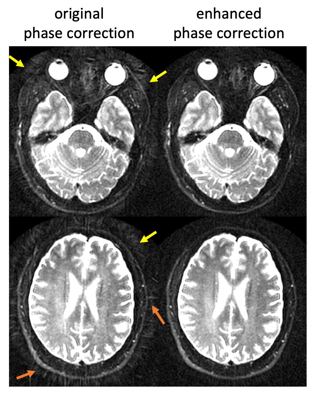
Fig. 2 demonstrates the image quality improvement obtained with the enhanced 2D phase correction algorithm. The window/level were adjusted to better illustrate the incoherent (yellow arrows) and streak-like artifacts (orange arrows) with the original phase correction algorithm, which are minimized with the enhanced algorithm.Fig. 3 compares the overall quality of diffusion trace images obtained with original XY2 and LRX phase modulation approaches, both utilizing the enhanced phase correction algorithm. Images with LRX phase modulation show higher SNR and fewer artifacts.
Discussions and Conclusion
In the current implementation, the two blades from each shot are placed in the same blade angle in k-space. They can be acquired in an orthogonal fashion, as in the conventional DW PROPELLER. This not only makes the scan 2 times faster, also enables mutual calibration 6 to allow for further scan time reduction. LRX phase modulation is also less sensitive to deviation of the flip angles from 180°. 5 This allows for reduction of SAR, especially at high fields.In summary, the preliminary results have shown that turboPROP+ with LRX phase modulation and enhanced phase correction generates high quality DW images and therefore provides a potentially robust tool for neuro imaging.
Acknowledgements
This work is partially supported by Philips Healthcare.References
1. Pipe JG, et al. Multishot diffusion weighted FSE using PROPELLER MRI. Magn Reson Med 2002;47:42-52.
2. Pipe JG, et al. Turboprop: Improved PROPELLER imaging. Magn Reson Med 2006;55:380-385.
3. Lee CY, et al. Turboprop+: Enhanced turboprop diffusion weighted imaging with a new phase correction. Magn Reson Med 2013;70:497-503.
4. Le Roux P. Non-CPMG fast spine echo with full signal. J Magn Reson 2002;155:278-292.
5. Li Z, et al. Robust diffusion weighted imaging using split-blade PROPELLER and LRX phase modulation. ISMRM 2009;p3516.
6. Li Z, et al, A parallel imaging technique using mutual calibration for split-blade diffusion-weighted PROPELLER. Magn Reson Med. 2011;65:638-644.
Figures