0965
Diffusion Imaging with Very High Resolution and Very Short Echo Time1ETH and University of Zürich, Zürich, Switzerland
Synopsis
To achieve high-resolution diffusion imaging with short echo times, single-shot spiral DWI using a recently developed gradient insert (strength=200 mT/m, slew=600 T/m/s) was implemented. The high gradient strength in combination with the spiral readout allowed for an echo time as short as 19 ms at a b-factor of 1000 s/mm2. The high slew rate enabled shortening of the spiral readout duration which reduces sensitivity against static off-resonance and T2* blurring artifacts, and allowed imaging with an in-plane image resolution of only 0.69 mm. First in-vivo results are presented.
Introduction
Diffusion-weighted (DW) imaging is often limited by low spatial resolution, artifacts due to physiological motion, and inherently low SNR that is further degraded by the long echo times required for the diffusion encoding.Single-shot acquisition techniques are most commonly used to acquire DW images, since they are robust against motion and SNR efficient. A combination of single-shot MRI with spiral readouts provides the possibility to achieve shortest echo times for a given diffusion encoding and thereby to improve the achievable SNR (1).
However, an undesirable property of single-shot acquisitions is their sensitivity to static off-resonance and T2* blurring artifacts, in particular when using long readout trains to achieve high image resolution. Moreover, even shorter echo times are often desirable, especially when requiring more SNR to target higher image resolution, stronger diffusion weighting, or the assessment of diffusion properties of tissues with short T2.
To address these needs, we implemented single-shot spiral DWI using a recently developed gradient insert (2) which allows for a gradient strength of 200 mT/m on each axis and a slew rate of 600 T/m/s. The high gradient strength in combination with the spiral readout allowed for an echo time as short as 19 ms for a b-factor of 1000 s/mm2. The high slew rate enabled shortening of the spiral readout duration which reduces the sensitivity against static off-resonance and T2* blurring artifacts, and allowed imaging with an in-plane image resolution of 0.69 mm. First in-vivo results are presented.
Methods
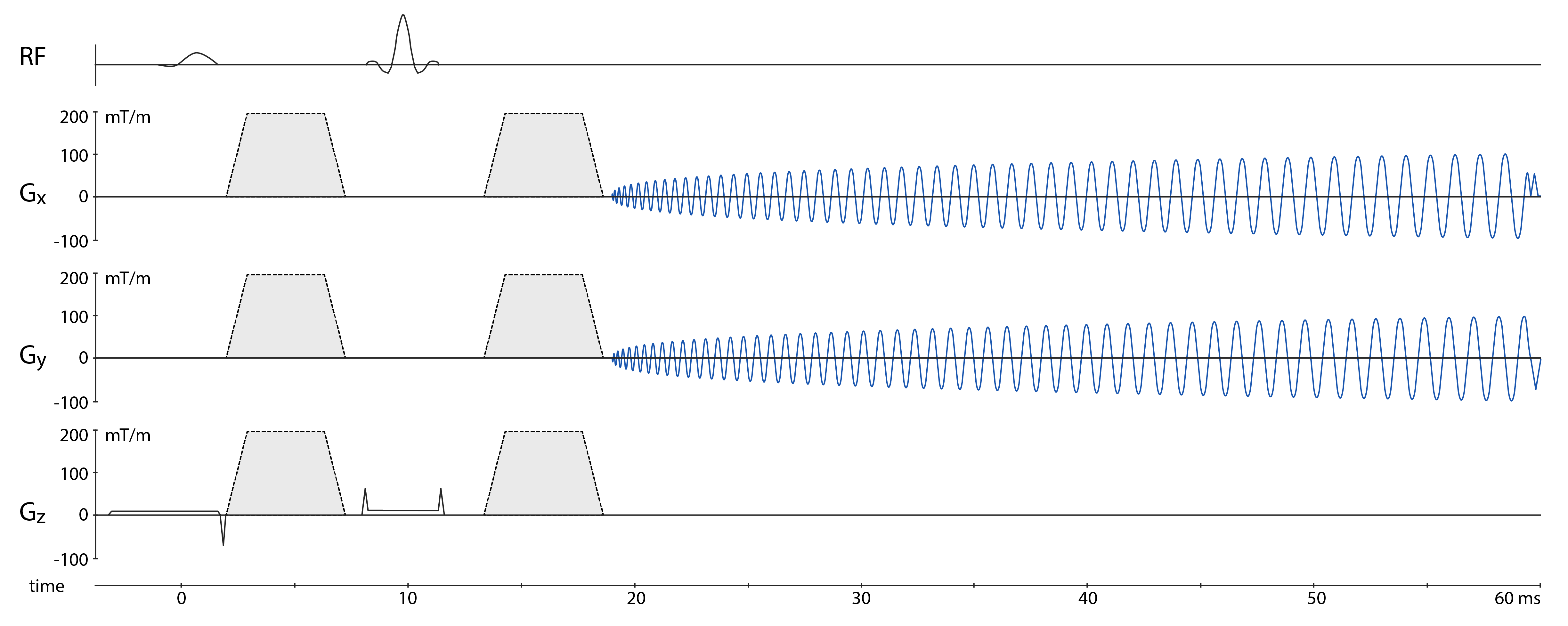
MR scanning was performed on a 3T Achieva (Philips Healthcare, Best, The Netherlands) using an 8-channel transmit-receive coil array (3) and a custom-built gradient insert (2).A diffusion-weighted spin-echo single-shot spiral sequence (Fig. 1) (FOV: 22 cm, in-plane resolution: 0.68 mm, slice thickness: 3 mm, averages = 60, SENSE: R=3, DW: b=0 and b=1000 s/mm2 in 3 directions) was applied in a healthy subject. The gradient insert was operated with 200 mT/m and 600 T/m/s, opting for the shortest echo time; the readout duration was 40 ms. Coil sensitivities as well as static B0 maps were obtained from a multi-echo gradient echo scan on the same geometry.
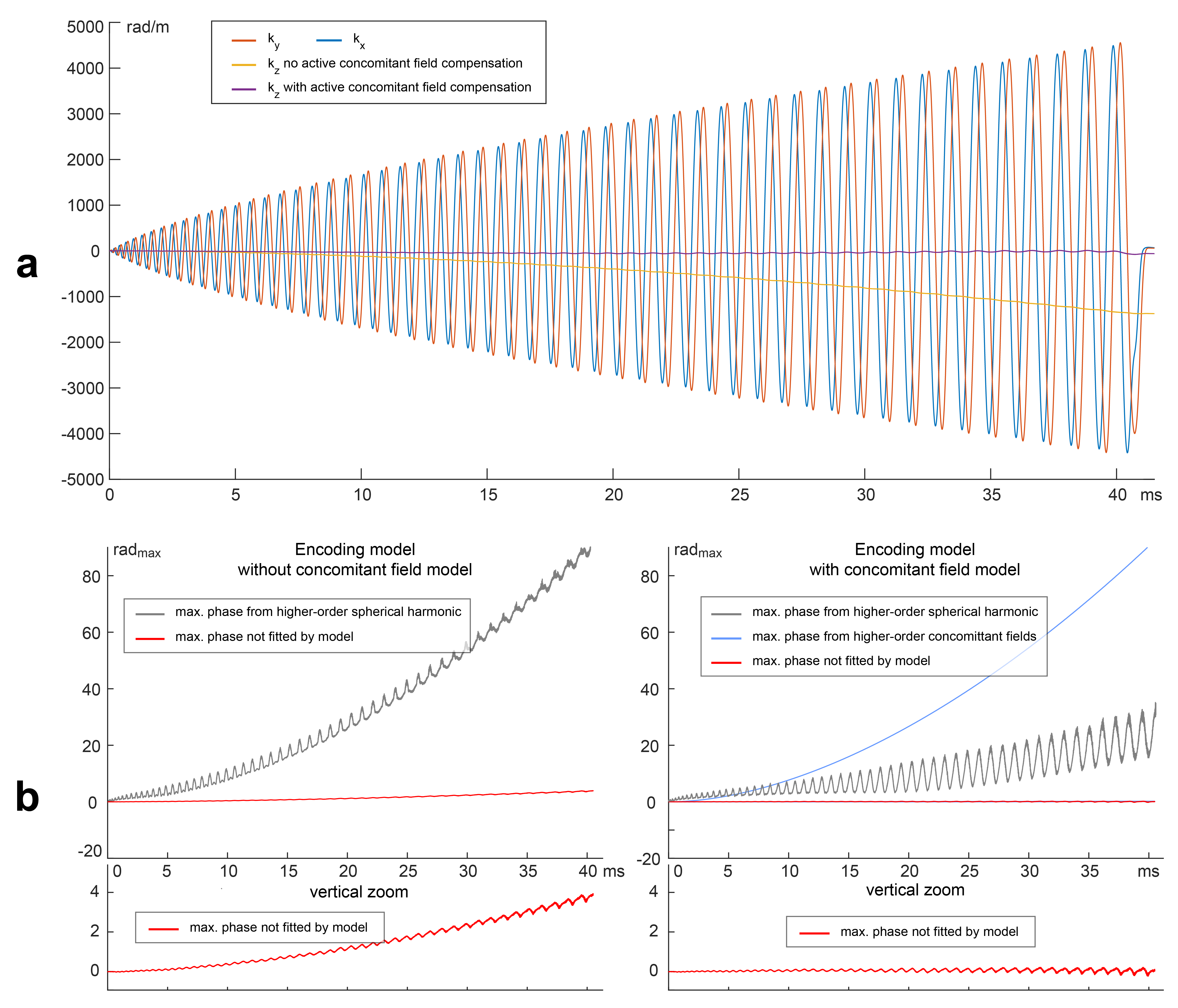
Operating gradient systems at such strength and slew rates imply increased eddy currents which need to be accounted for. Moreover concomitant (Maxwell) fields are also amplified at higher gradient strength. To find an accurate encoding model, the spiral readout was recorded using a Dynamic Field Camera (Skope MRT, Zurich, Switzerland) in a repeated scan. Field monitoring was performed with three different field camera positions. From the combined data, a 5th order spherical harmonic model was fitted. Higher-order concomitant fields are commonly modelled separately based on an analytical description (5). Due to the spatial non-linearity of the gradient system, it is however unclear how well the concomitant fields are described by this model. To judge the validity of the concomitant field model, the fit error of the 5th order encoding model (using 48 field probes and 36 unknown coefficients) was evaluated with and without incorporation of the analytical concomitant field model.
In addition to higher-order concomitant fields, 0th order and linear concomitant field components arise as a results of z-gradient asymmetry (4). The linear components of the concomitant fields were actively compensated (6) during the readout to avoid related signal dephasing.
All images were reconstructed accounting for receive coil sensitivities, B0 map and measured (higher-order) k-space coefficients (7).
Results and Discussion
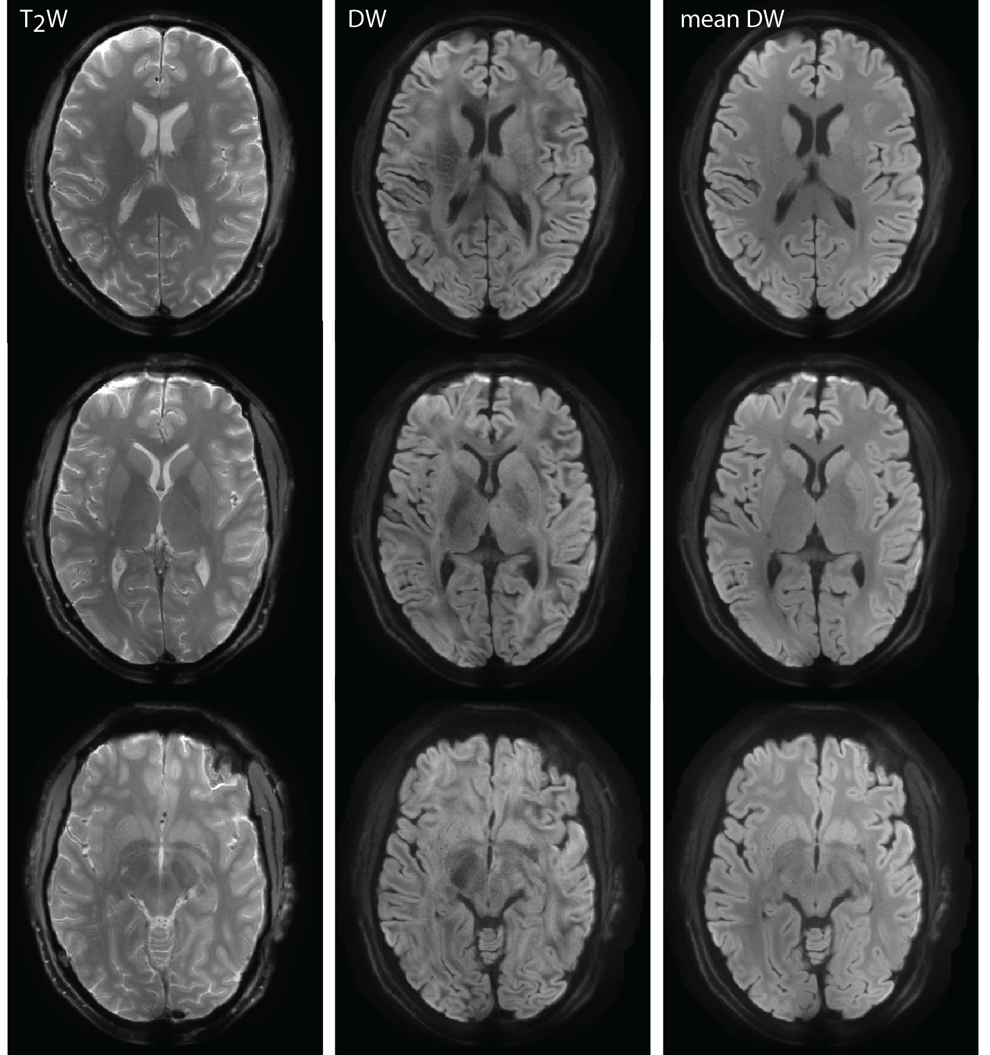
Employing such strong gradients for the diffusion encoding in conjunction with the spiral readout allowed for an echo time of only 19 ms - as compared to around 60-100 ms (depending on resolution and parallel-imaging acceleration) for single-shot EPI with typical gradient specifications (50 mT/m, 200 T/m/s). The field monitoring data showed that the encoded image resolution was π/kmax = 0.685 mm (Fig. 2a), which is only slightly less than the targeted 0.68 mm - presumably due to eddy currents. The active compensation of linear concomitant fields to avoid unwanted dephasing was confirmed by the field monitoring results (Fig. 2a). Dynamic fields of higher order in space had a substantial contribution to the image encoding (Fig 2b). Including the analytical concomitant field model (5) into the encoding model (Fig 2b) diminished non-fitted phase cumulating over the readout to less than 0.5 rad (down from 4 rad), which, given the acquisition bandwidth per pixel, can be regarded as sufficient.The reconstructed images (Fig. 3) show fine anatomical details, with little effects from static B0 off-resonance and T2* blurring, probably owing to the accuracy of the encoding model and the high bandwidth of the acquisition.
Conclusion
This is the first presentation of a setup and sequence that allows for in-vivo brain DWI with such short echo time and high image resolution. The achieved short echo time increases the sensitivity to diffusion properties of tissues with short T2 such as myelin water (8) by an order of magnitude or more. The enabled resolution and improved sensitivity may readily be employed for advanced studies of brain microstructure in-vivo.Acknowledgements
No acknowledgement found.References
(1) Lee Y, Wilm B, Nagy Z, Pruessmann K. High-Resolution Diffusion MRI: In-Vivo Demonstration of the SNR Benefit of Single-Shot Spiral Acquisition vs. EPI. In Proceedings of 27th Annual Meeting of ISMRM, Montreal, Canada, 2019 n.d.:767.
(2) Weiger M, Overweg J, Rösler MB, Froidevaux R, Hennel F, Wilm BJ, Penn A, Sturzenegger U, Schuth W, Mathlener M, Borgo M, Börnert P, Leussler C, Luechinger R, Dietrich BE, Reber J, Brunner DO, Schmid T, Vionnet L, Pruessmann KP. A High-Performance Gradient Insert for Rapid and Short-T2 Imaging at Full Duty Cycle. Magn Reson Med 2018;79:3256–3266.
(3) Roesler MB, Leussler C, Brunner DO, Schmid T, Weiger M, Hennel F, Luechinger R, Pruessmann KP. A Head Transmit-Receive Array for a High Performance Gradient Insert. In Proceedings of 27th Annual Meeting of ISMRM, Montreal, Canada, 2019 n.d.:80.
(4) Meier C, Zwanger M, Feiweier T, Porter D. Concomitant Field Terms for Asymmetric Gradient Coils: Consequences for Diffusion, Flow, and Echo-Planar Imaging. Magnetic Resonance in Medicine 2008;60:128–134.
(5) Bernstein MA, Zhou XJ, Polzin JA, King KF, Ganin A, Pelc NJ, Glover GH. Concomitant Gradient Terms in Phase Contrast MR: Analysis and Correction. Magn Reson Med 1998;39:300–308. (6) Tao S, Weavers PT, Trzasko JD, Shu Y, Huston J, Lee S-K, Frigo LM, Bernstein MA. Gradient Pre-Emphasis to Counteract First-Order Concomitant Fields on Asymmetric MRI Gradient Systems. Magnetic Resonance in Medicine 2017;77:2250–2262.
(7) Wilm BJ, Barmet C, Pavan M, Pruessmann KP. Higher Order Reconstruction for MRI in the Presence of Spatiotemporal Field Perturbations. Magn Reson Med 2011;65:1690–1701.
(8) Tax C, Rudrapatna U, Mueller L, Jones T. Characterizing Diffusion of Myelin Water in the Living Human Brain Using Ultra-Strong Gradients and Spiral Readout. In Proceedings of 27th Annual Meeting of ISMRM, Montreal, Canada, 2019 n.d.:1115.
Figures