0958
A TSE BLADE based distortion-free diffusion-weighted imaging method with Dixon water-fat separation1Siemens Shenzhen Magnetic Resonance Ltd., Shenzhen, China, 2Siemens Medical Solutions USA, Boston, MA, United States
Synopsis
Diffusion weighted imaging with EPI can suffer from image distortions due to sensitivity to B0 inhomogeneity and chemical shift related artifacts induced by incomplete fat suppression. In this study, we propose a TSE BLADE sequence with Dixon water-fat separation for DWI. With this technique, distortion-free DWI with robust fat suppression was shown to be feasible, even in body regions with strong B0 inhomogeneity.
INTRODUCTION
Diffusion weighted imaging (DWI) is commonly achieved by single-shot EPI (SS-EPI) technique, due to its high acquisition efficiency and insensitivity to bulk motion and physiological movements. However, SS-EPI can suffer from off-resonance related artifacts accumulated along the phase encoding direction, e.g. image distortions due to B0 inhomogeneity or chemical shift artifacts due to incomplete fat suppressions.On the other hand, the DW-PROPELLER technique [1], which combines the Fast Spin Echo (or Turbo Spin Echo, TSE) sequence and the PROPELLER (BLADE) trajectory, has intrinsic advantages over EPI for reduced sensitivity to B0 inhomogeneity. With this method, residue artifacts due to incomplete fat suppression does not manifest as the chemical shift artifacts. However, especially at high b-value in DWI, such residue fat signals may still obscure critical structures and lesions.
Regardless of the k-space acquisition strategy, the Dixon’s method [2] can also provide advantages for insensitivity to B0 inhomogeneity over other fat suppression techniques, which reconstructs chemical shift encoded data and produces water-only and fat-only images, respectively.
Furthermore, the DW-PROPELLER acquisition can be speed up with recent developments of the multi-blade readout (X-Prop) techniques [3]. As the blades acquired in one shot at different echo times are chemical shift encoded, the Dixon’s method can be applied to these data for more robust fat suppression.
Therefore, based on these existing methods, we propose a technique, which combines the Dixon reconstruction and the BLADE DWI sequence with phase insensitive preparation and multi-blade acquisition(PIP-MULTI-BLADE) [4-5], to produce distortion-free diffusion weighted images with water-fat separation.
METHODS
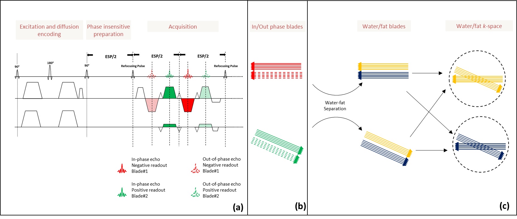
The sequence diagram of PIP-MULTI-BLADE is shown in Figure 1a [6]. Two pairs of chemical-shift encoded echoes (red and green lines) were acquired in each spin echo interval: Each pair of in-phase echoes (solid-line) and out-of-phase echoes (dashed-line) were collected at the same k-space location, with same readout direction but difference echo time. Such acquisition was repeated throughout the whole echo train with different phase encoding steps, producing all k-space lines for each blade. The k-space locations of these blades are shown in Figure 1b.The reconstruction pipeline is shown in Figure 1c. Dual-echo Dixon reconstruction [2] was performed on each pair of the chemical-shift encoded blades, with water and fat signals generated for each blade. The water and fat blades were loaded into the standard BLADE reconstruction to generate water and fat images with full k-space coverage, respectively.
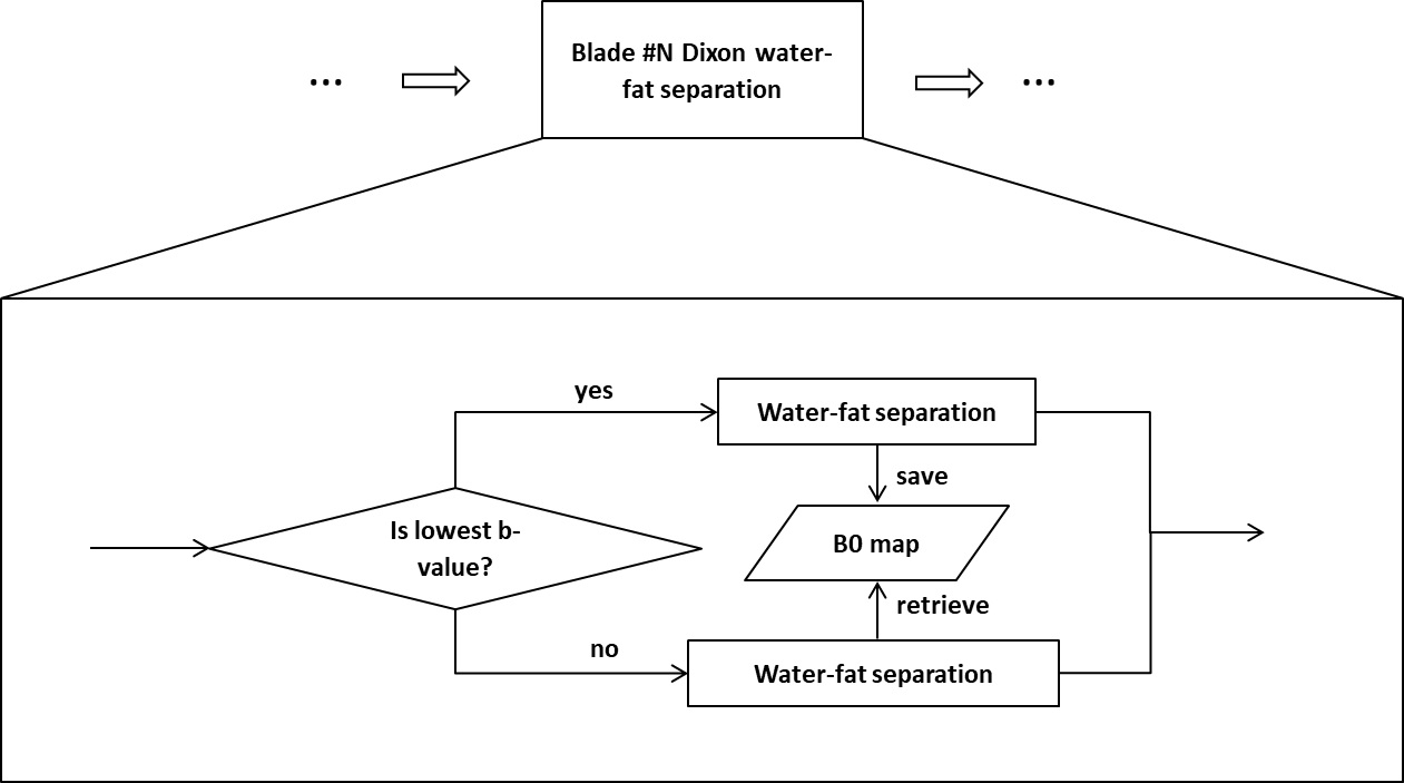
The water-fat separation can be compromised by inaccurate B0 map estimation due to low SNR at high b-value. The B0 map obtained from the Dixon reconstructed with the lowest b-value was stored for calculations at high b-values, as shown in figure 2.
The proposed prototyping sequence was tested in the neck region of a healthy volunteer at a Siemens 1.5T MAGNETOM Aera scanner (Siemens Healthcare Gmbh, Erlangen, Germany). The following parameters were used in the proposed sequence: FoV = 260 x 260 mm2, slice thickness = 5 mm, 20 slices, bandwidth = 955 Hz/Pixel, TE/TR = 43/5000ms, acceleration factor in phase encoding direction = 3, 4-scan trace diffusion mode, b value = 50, 800 s/mm2, 1 average for b = 50 and 2 average for b = 800, measurement time = 4’08’’. For comparison, SS-EPI with short term inversion recovery (IR-SS-EPI) was acquired with matching geometric and diffusion parameters. The different parameters are: FoV = 260 x 178 mm2, bandwidth = 1565 Hz/Pixel, TE/TR = 62/5230ms, 3 average for b = 50 and 8 average for b = 800, measurement time = 4’06’’. A TSE Dixon with matching geometric parameters was also acquired.
RESULTS
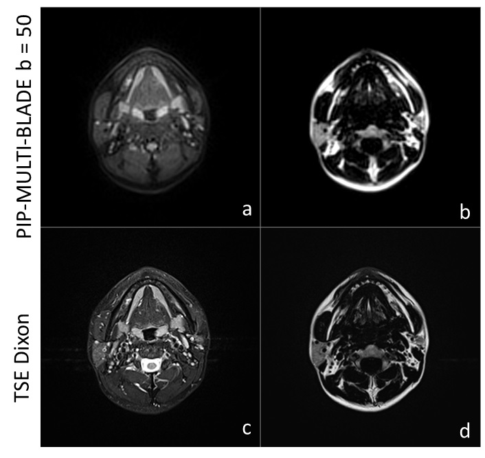
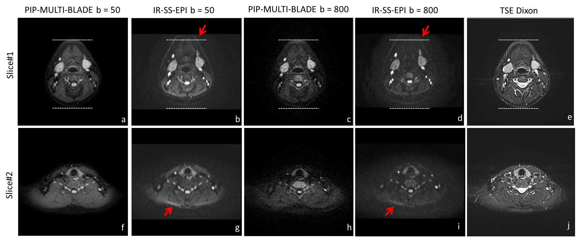
The first slice acquired with the proposed PIP-MULTI-BLADE sequence with b=50 s/mm2 (a-b) and TSE Dixon sequence (c-d), respectively, were shown in Figure 3. The water-fat separation is successful by the proposed sequence with Dixon’s method, indicated by the comparison of the water/fat images to the TSE Dixon images. In Figure 4, the PIP-MULTI-BLADE DWI water images (a, c, f, h), IR-SS-EPI images (b, d, g, i) and TSE Dixon water images (e, j) from two exemplary slices were presented, respectively. As shown in Figure 4b and Figure 4d, the distortion can be observed on the IR-SS-EPI images, but is absent in the BLADE DWI image, which has its geometry closely matched with the TSE images.In the second slice, due to severe field inhomogeneity, susceptibility artifacts can be found in IR-SS-EPI images, as indicated by red arrow in Figure 3g and Figure 3i. As adiabatic inversion recovery pulse was used, the fat suppression by the IR-SS-EPI is comparable to the PIP-MULTI-BLADE method, but comes a tradeoff of SNR loss.
DISCUSSION AND CONCLUSION
In diffusion imaging, shot-dependent phase variations induced by microscopic motion can be problematic. As a multi-shot DWI sequence, the PIP-MULTI-BLADE avoids this problem as the in-phase and out-of-phase blades used for water-fat separation are acquired within the same shot.In this work, Dixon water-fat separation was combined with the distortion-free BLADE DWI sequence by taking advantages of the inherent chemical-shift encoding in the multi-blade readout. Our experimental results show its promising potential as an alternative technique to SS-EPI for distortion-free DWI with robust fat suppression, especially from the successful experiments on the neck regions with strong B0 inhomogeneity.
Acknowledgements
The authors thank Dr. Shi Cheng and Dr. Rui Tian for help with the preparation of the abstract.References
1. Pipe JG. Split-blade PROPELLER DWI. In Proceedings of the 11th annual meeting of ISMRM, Toronto, Canada, 2003. p 2126.
2. Eggers H. Dual-echo Dixon imaging with flexible choice of echo times. Magnetic resonance in medicine. 2011 Jan;65(1):96-107.
3. Li Z, Pipe JG, Lee CY, Debbins JP, Karis JP, Huo D. X-PROP: a fast and robust diffusion-weighted propeller technique. Magn Reson Med 2011;66:341–347.
4. Zhou K. Multi-Blade Acquisition of Split Turbo Spin Echoes: A Robust and Fast Diffusion Imaging Technique. Proc. Intl. Soc. Mag. Reson. Med.24 (2016).
5. Zhou K. Non-CPMG PROPELLER diffusion imaging: comparison of phase insensitive preparation with split acquisition. Proc. Intl. Soc. Mag. Reson. Med.26 (2018).
6. Zhou K. A novel method combining Dixon water-fat separation and BLADE imaging. Proc. Intl. Soc. Mag. Reson. Med.27 (2019).
Figures