0955
Simultaneous acquisition of diffusion weighted images and conductivity maps using a balanced double echo steady state (DESS) sequence1Philips Research, Hamburg, Germany
Synopsis
A combined acquisition of distortion-free diffusion-weighted images and tissue conductivity maps is explored using a fully balanced double echo steady state (DESS) sequence. Banding artifacts are avoided using sufficiently high gradient moments of the diffusion gradient, such that the banding is contained within single voxels. The stability of the B1 transceive phase measurement by the balanced DESS sequence allows the derivation of quantitative tissue conductivity based second derivative using standard EPT (electrical properties tomography) methods. Feasibility of simultaneous DWI and EPT is shown on a 3T MRI system in phantom and volunteer experiments (head).
Introduction
Diffusivity and tissue conductivity are physiologic parameters with manifold applications e.g. in tumor characterization, typically assessed by Diffusion Weighted Imaging (DWI) and Electrical Properties Tomography (EPT) in separate sequences. EPI based DWI sequences often suffer from geometric distortions (magnetic field inhomogeneity). Diffusion weighted dual-echo steady-state (DW-DESS) MRI using unipolar gradients provides a distortion free alternative1, but is inherently sensitive to motion and does not exploit the steady-state signal, because of non-balanced gradients. In this study, a balanced DW-DESS sequence was developed using bipolar DW gradients while avoiding dark-band artefacts. EPT is based on the transceive phase φ purely related to B1 (not impacted by B0) as in spin-echo (SE) based sequences as well as in balanced steady state sequences2. This study investigates the use of φ from balanced DW-DESS as basis for EPT, which would synergistically allow to assess two relevant physiological parameters from a single MR acquisition.Methods
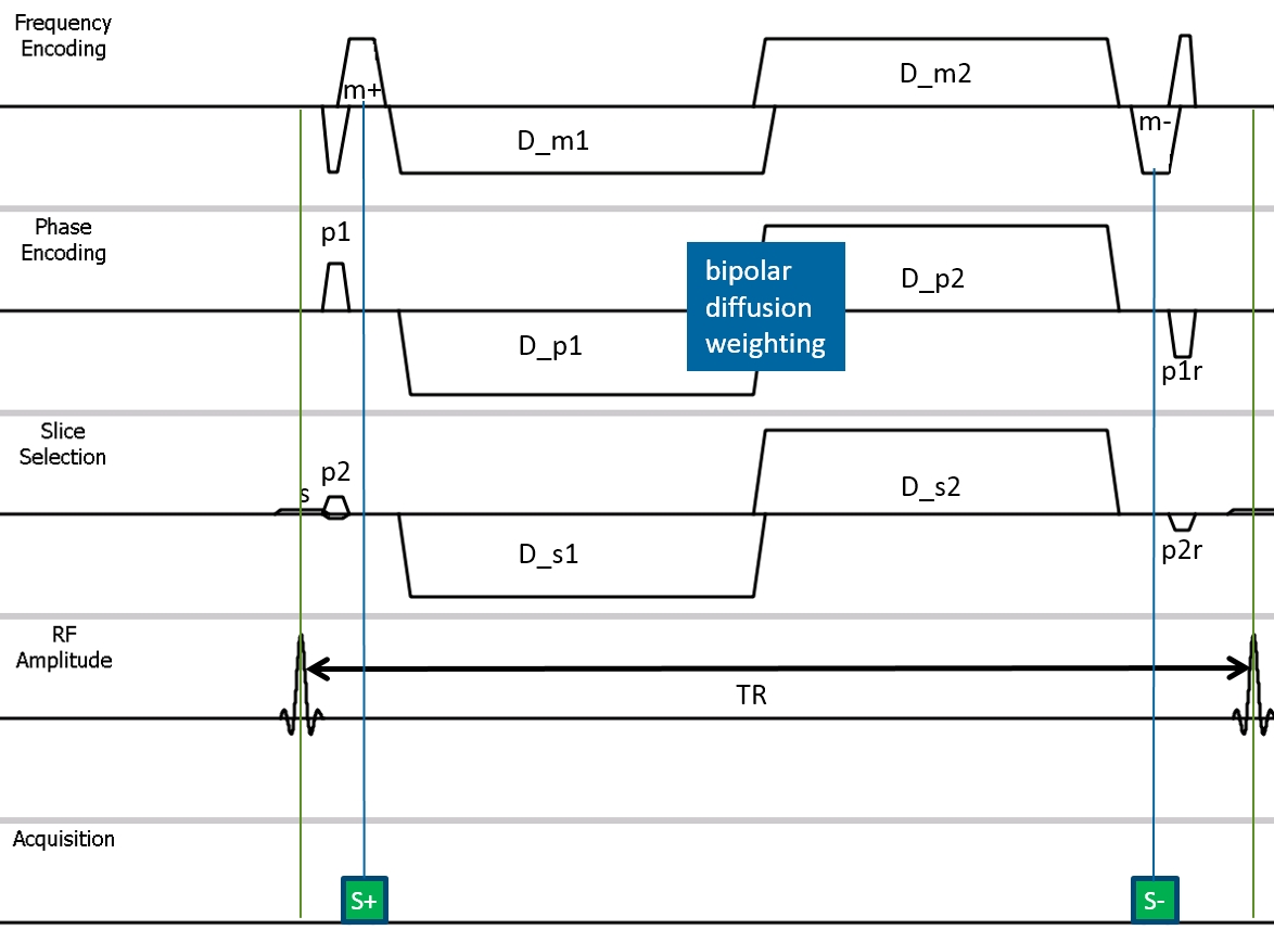
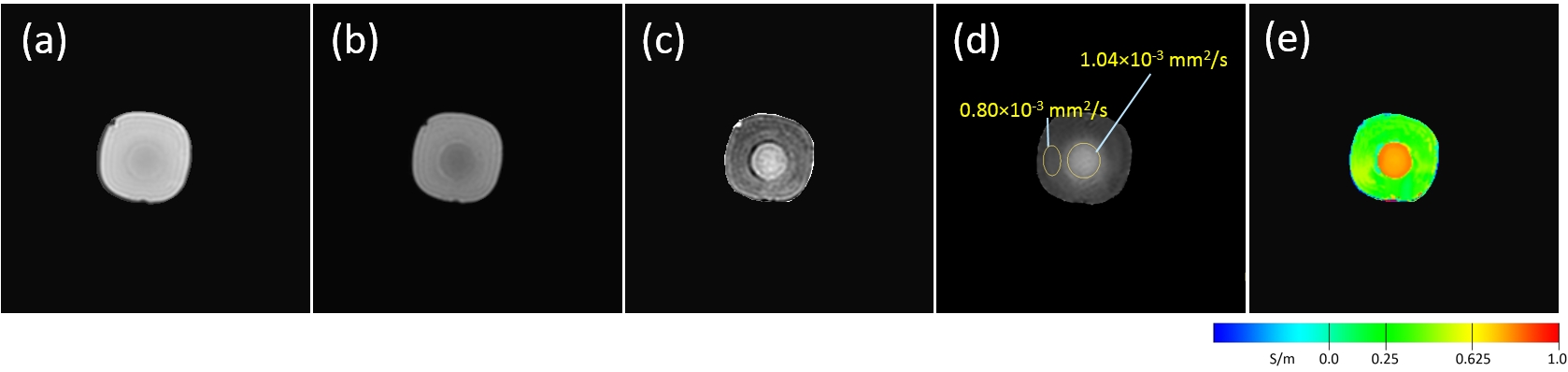
A fully-balanced DW-DESS sequence is used in combination with bipolar DW gradients (example pulse sequence diagram in Figure 1). For (single echo) balanced SSFP, the dark band spacing only depends on TR and off-resonances (frequency spacing 1/TR), as the echo is fully refocused at TE=TR/2. The dark-bands of S+ and S- in fully-balanced DESS also depend on the moments of the diffusion-weighting gradient lobes, which appear as an additional off-resonance effect in the gradient direction. At high gradient moments, the orientation and spacing of the dark bands is dominated by the gradient effect and less influenced by actual off-resonances. This study applies sufficiently high gradient moments of the bipolar gradient lobes such that the spatial distance of the dark band artefacts is reduced to a value smaller than the imaging voxel size. Dark bands within a voxel partly reduce the overall signal intensity but are not visible as artefacts. Thus, a strong diffusion weighting can be induced at high SNR efficiency by combining all coherence pathways for the overall FID (echo1, S+) and ECHO (echo2, S-) of the DESS acquisition.From the acquired DESS signals, conductivity σ was calculated via $$$σ = \nabla^{2}φ/(2μω)$$$ (with vacuum permeability μ and Larmor frequency ω) in combination with a bilateral denoising filter2. A phantom was composed of polyvinylpyrolidon (P), gelatine (G), NaCl (S) and H2O (W) with different diffusion and conductivity values in an outer and inner compartment (inner: D=1.04×10⁻³ mm2/s, σ=0.66 S/m, P/G/S/W=5/3/0.5/91.5 mass%; outer: D=0.8×10⁻³ mm2/s, σ=0.42 S/m, P/G/S/W=25/3/0.3/71.7 m%).
Combined DW-DESS and EPT acquisition was tested on a 3T MRI system (Achieva TX, Philips, NL) on the phantom and in a volunteer head examination (male, age 50 yrs), with written consent obtained, using the following imaging parameters: 3D balanced dual-echo SSFP, 8-channel head coil, TR/TE1/TE2=31/1.8/26 ms (phantom: 53/1.85/50.8 ms) , FOV 224×224×120 mm3, pixel 1.8×1.8 mm2 , reconstruction 224×224, 24 slices (5mm in vivo, 1.8mm phantom), pixel bandwidth 1.3 kHz, bipolar or unipolar diffusion gradients (3 simultaneous directions, duration 2×11 ms (phantom: 2×22 ms), slopes 0.4 ms, strength 18 mT/m), two signal averages (phantom: 6), total scan duration 2min 55s (phantom: 7min). Diffusion weighted images were computed as ratio S+/S-.
Results
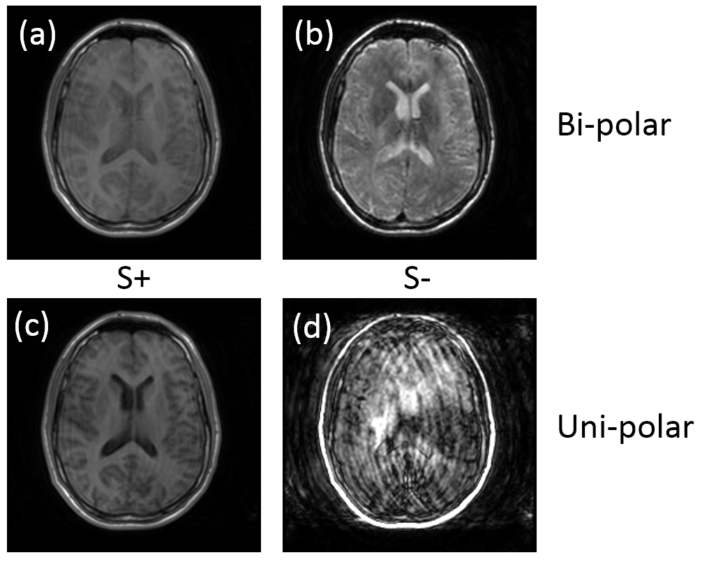
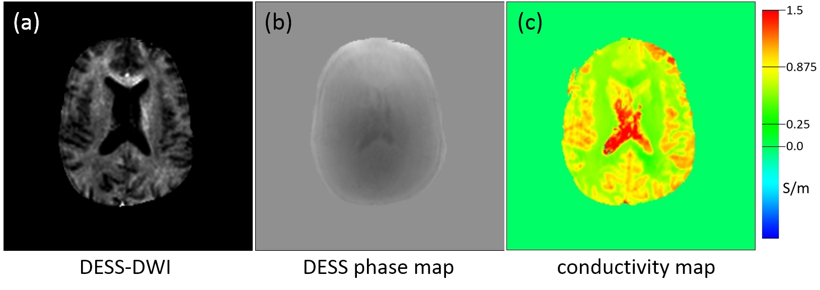
Phantom results are shown in Fig.2. While the first echo (S+, FID, Fig.2a) shows minimal contrast between the two compartments, the second echo (S-, Fig.2b) has overall a lower signal and shows further lowered signal from in the inner compartment with larger diffusivity. The resulting image (S+/S-, Fig.2c) confirms the diffusion weighting and can be compared with and ADC map obtained using a standard DWI sequence (EPI, 8 b-values 0…1400, Fig.2d). Fig.2e shows the conductivity map obtained from S+ and the measured σ values, inner/outer=(0.77±0.02)/(0.31±0.06)S/m, correspond to the phantom preparation.Volunteer results are shown in Fig.3 and Fig.4. Using bipolar (Fig.3 a/b) instead of unipolar diffusion weighting gradients (Fig3. c/d) is much more robust to motion effects, in particular for the second echo (S-). In Fig. 4, a DW-DESS image of the volunteer is shown together with the corresponding phase map and the resulting conductivity map, yielding conductivity values as expected from literature4.
Discussion & Conclusion
A balanced DW-DESS acquisition could be successfully implemented using large bipolar DW gradients that avoid banding artifacts and show low motion sensitivity. Although the SNR is lowered by dark band contents within the voxels, image quality was clearly improved as compared to unipolar gradients. A drawback of bipolar DW-DESS is given by its limitation of the achievable b-values. In this initial demonstration, the diffusion weighted images also include a considerable T2 weighting, because of the long second echo time (26 or 51 ms). Multiple b-values with the same echo time could be used to reduce the T2 weighting (b=0 cannot be used because of banding artifacts). The transceive phase of DW-DESS can be used for EPT, yielding conductivity maps with comparable quality as previously obtained in the brain5. Because of the lower overall SNR in S- images (DW and long TE), EPT reconstructions were preferably calculated from the first echo S+.In conclusion, DW-DESS is able to produce distortion-free diffusion weighted images and conductivity maps simultaneously. It is thus expected to be a valuable sequence particularly for tumor characterization.
Acknowledgements
Cordial thanks to Christian Stehning and Philipp Karkowski for technical help.References
1. Granlund KL et al., High-resolution, three-dimensional diffusion-weighted breast imaging using DESS. Magn Reson Imaging. 2014; 32:330
2. Stehning C et al., Real-time conductivity mapping using balanced SSFP and phase-based reconstruction. ISMRM 2011; 19: 128
3. Katscher U, van den Berg CAT. Electric properties tomography: Biochemical, physical and technical background, evaluation and clinical applications. NMR Biomed. 2017; 30:3729
4. Gabriel S et al., The dielectric properties of biological tissues II. Measurements in the frequency range 10Hz to 20GHz. Phys Med Biol. 1996;41: 2251
5. Tha KK et al., Noninvasive electrical conductivity measurement by MRI: a test of its validity and the electrical conductivity characteristics of glioma. Eur Radiol. 2018; 28:348
Figures