0937
Quantifying Hippocampal Dentation in Epilepsy: a comparison of absolute mean curvature versus visual inspection and their memory correlates.1Neurology, University of Alabama at Birmingham, Birmingham, AL, United States, 2Birmingham VA Medical Center, Birmingham, AL, United States, 3University of Alabama at Birmingham, Birmingham, AL, United States
Synopsis
Hippocampal dentation (HD) is a morphologic feature of the human hippocampus that has been recently described and has been shown to correlate with aspects of verbal and visual memory. It varies dramatically across healthy individuals and can be affected by diseases such as epilepsy. We demonstrate a method to extract ultra-high-resolution surface contours from common MPRAGE images. We also propose a method based on absolute mean curvature to quantify HD and compare that to visual inspection in a cohort of temporal lobe epilepsy patients. Finally, we examine correlations between HD and measures of verbal and visual memory across methods.
Introduction
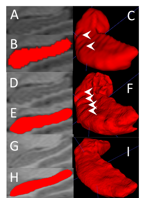
Hippocampal dentation (HD) is morphologic feature of the human hippocampus characterized by the dentated (“tooth-like”) contour of the inferior aspect of the subiculum and CA1 subregions. The degree of HD has been shown to vary dramatically across healthy individuals from prominently dentated to virtually no dentation.1 Furthermore, it has been shown that the degree of HD correlates with memory performance.1 HD can be seen visually on common T1w MPRAGE images interpolated to a higher resolution, but standard hippocampal segmentation approaches do not render HD well (Fig.1).2 This is due to the fact that segmentations are conventionally done in native resolution of the image (~1mm3 isotropic), which is rather large compared to the contours of HD. Previous work has demonstrated the ability to extract ultra-high-resolution segmentations that depict dentation well from common T1w images but required proprietary software that was not robust across computing platforms,2 which limits its use. Furthermore, until now quantification of HD has been limited to visual assessment of the quantity or quality of HD that is inherently subjective. Objective quantitative measures of HD are needed to study its significance in various diseases affecting the hippocampus such as temporal lobe epilepsy (TLE), Alzheimer’s disease, and schizophrenia.Significance
In this work we demonstrate 4 key findings:- We demonstrate a simple, efficient method to perform super-resolution hippocampal segmentation that allows visualization of HD using freely available software.
- We demonstrate an objective method based on absolute mean curvature (AMC) to quantify HD and compare it with simple visual assessment of number of dentes.
- We compare the degree of HD in TLE patients and demonstrate that HD is asymmetrically lost in TLE.
- Visual assessment of HD showed significant correlation measures of verbal and visual memory, but the quantitative measure did not, indicating that additional work is needed to objectively quantify HD.
Methods
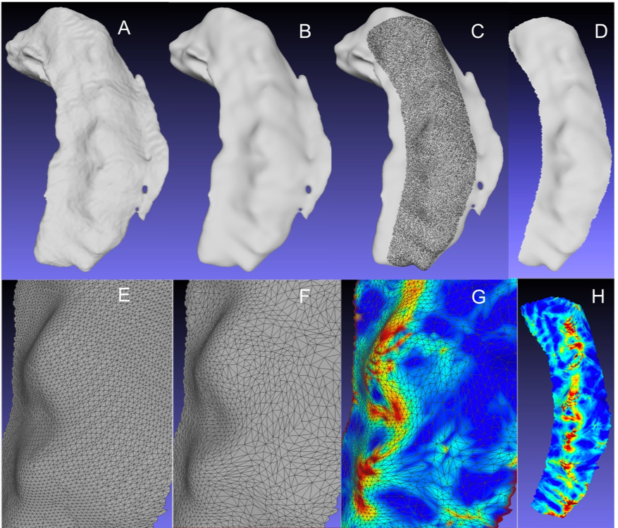
Fifty-nine patients with MRI evidence of TLE/hippocampal sclerosis were retrospectively identified. Clinical T1w MPRAGE scans with a voxel volume of 1.0-1.2 mm3 were upsampled with cubic spline interpolation to a resolution of 0.25 mm in three dimensions. The upsampled images and source images were processed with Automated Segmentation of Hippocampal Subfields (ASHS) using the Penn TLE atlas,3 which provides whole hippocampal segmentation instead of subfield segmentation. ASHS can be run in a linux, windows, or OSX environment and has the advantage that the segmentation is in the resolution of the target image (in this case 0.25 mm) not that of the source atlas. The hippocampal segmentations were exported as .stl surface mesh files using ITK-SNAP4 and imported into MeshLab.5 Figure 2 illustrates the following steps. A Taubin smoothing filter6 (λ=0.5/μ=-0.51/steps=500) was applied to remove surface irregularities. The inferior aspect of the whole hippocampal surface mesh (closed) was trimmed such that only the region that typically contains dentation remained as an open surface. Remeshing using the Quadric Edge Collapse Decimation filter reduced the number of faces by 60%. This step regularizes the spacing of the vertices and simplifies the mesh. AMC was calculated7 at each vertex using MeshLab. Observation of numerous examples showed us that AMC values related to dentation were in the range of 0.5-1.2, while higher values came from sources not related to HD, such as artifactually extreme values on the free edge of the open surface. The summary measure of curvature was calculated as the number of vertices with AMC values in the range of 0.5-1.2 multiplied by the average AMC value of those vertices. As a comparison, the number of dentes on each side was counted visually using the 3D view and 3-plane views of the upsampled image. Visual and objective measures of HD for each hippocampus in both left- and right-sided TLE cases were correlated with long-delay free recall (LDFR), a measure of verbal memory that is commonly impaired in left TLE, and the Rey Recognition Score (RRS), a measure of visuospatial memory that is commonly impaired in right TLE.Results
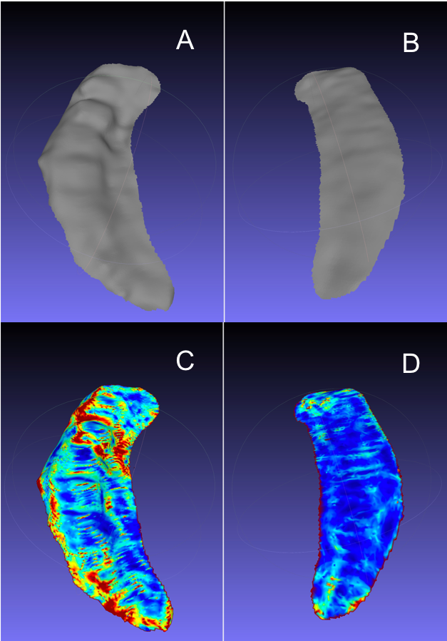
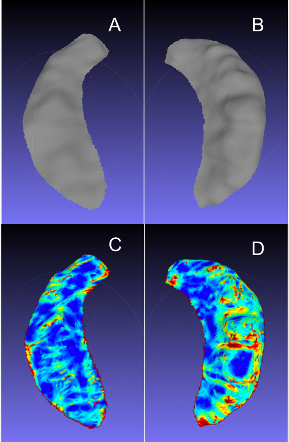
Surfaces and AMC maps show HD well (Fig.3). Both AMC and visual counts of dentes showed significantly less HD in the hippocampus affected by TLE (ipsilateral) versus the contralateral normal hippocampus (p=0.0013, p<0.0001 respectively) (Fig.4). There was a moderate but significant correlation between the AMC measure and visual counting (r=0.52, p<0.0001). Visual counts from the right hippocampus showed a significant correlation with LDFR in left TLE cases (r=0.42, p=0.0274), but the AMC measure did not. Similarly, visual counts from the left hippocampus showed a significant correlation with RRS in right TLE cases (r=0.49, p=0.037), but the AMC measure did not. No other comparisons were significant.Discussion/Conclusions
These results show that surface features of HD can be extracted from common T1w volumes with moderate resolution allowing clear visualization and potential for quantification using mesh-based metrics. Our objective, quantitative AMC-based measure of HD can show a difference between epileptic and contralateral non-epileptic hippocampi, the former having less HD. This suggests that the pathology of TLE reduces the morphologic complexity of the hippocampus. However, the objective measure was not able to demonstrate correlations with measures of memory performance, while visual counting of the number of dentes was significant. Further development of objective measures of HD is needed to surpass subjective visual assessment.Acknowledgements
This work was supported by NIH/NINDS R01NS094743References
1. Beattie JF, Martin RC, Kana RK, et al. Hippocampal dentation: Structural variation and its association with episodic memory in healthy adults. Neuropsychologia. 2017; 101: 65-75.
2. Chang C, Huang C, Zhou N, et al. The bumps under the hippocampus. Hum Brain Mapp. 2018; 39(1): 472-90.
3. Hadar PN, Kini LG, Coto C, et al. Clinical validation of automated hippocampal segmentation in temporal lobe epilepsy. Neuroimage Clin. 2018; 20: 1139-47.
4. Pluta J, Yushkevich P, Das S, et al. In vivo analysis of hippocampal subfield atrophy in mild cognitive impairment via semi-automatic segmentation of T2-weighted MRI. J Alzheimers Dis. 2012; 31(1): 85-99.
5. Cignoni P, Callieri M, Corsini M, et al. Meshlab: an open-source mesh processing tool. Eurographics Italian conference, proceedings, 2008.
6. Taubin G. A signal processing approach to fair surface design. ACM SIGGRAPH, 1995.
7. Surazhsky T, Magid E, Soldea O, et al. A comparison of gaussian and mean curvatures estimation methods on triangular meshes. ICRA, 2003.
Figures