0916
Gait - related white matter tracts damage in idiopathic normal pressure hydrocephalus
shuai xu1, ye yao2, jing ding3, and he wang*4,5
1Fudan University, Shanghai, China, 2School of Public Health, Fudan University, Shanghai, China, 3Department of Neurology, Zhongshan Hospital, Fudan University, Shanghai, China, 4Institute of Science and Technology for Brain-Inspired Intelligence, Fudan University, Shanghai, China, 5Human Phenome Institute, Fudan University, shanghai, China
1Fudan University, Shanghai, China, 2School of Public Health, Fudan University, Shanghai, China, 3Department of Neurology, Zhongshan Hospital, Fudan University, Shanghai, China, 4Institute of Science and Technology for Brain-Inspired Intelligence, Fudan University, Shanghai, China, 5Human Phenome Institute, Fudan University, shanghai, China
Synopsis
Grouping based on white matter hyperactivities (WMH) of each white matter tract, 15 idiopathic normal pressure hydrocephalus (iNPH) patients’ 10 gait index were compared by double sample t test. The results showed some white matter tracts with strongest gait index relationships located in motor and sensory pathways including middle cerebellar peduncle (MCP), left medial lemniscus, left posterior limb of internal capsule and right posterior limb of internal capsule.
Introduction
Idiopathic normal pressure hydrocephalus (iNPH) is a common hydrocephalus in the elderly adults. The iNPH patients are usually characterized by progressive gait impairment, cognitive deficits and urinary urgency and/or incontinence1. More radiographic studies manifested iNPH patients have enlarged ventricle and altered brain morphology, but few of study focus on the relationships between changed brain structure and gait dysfunctions due to iNPH. Thus, the aims of this study were to evaluate the abnormalities of white matter (WM) correlated with gait impairment in iNPH patients and to have a better understanding of its pathology.Materials and Methods
Fifteen iNPH patients (5 females, 10 males) were enrolled in this study and each patients’ demographic and gait index were collected, see table 1. The images were acquired using T2-weight TSE sequence with the following parameters: TR=6000ms, TE=93ms, slice thickness=3mm. First, we proformed correlation analysis between demographic and gait index. Then, grouping based on white matter hyperactivities (WMH, means white matter tract damage) extracted from different WM tracts (JHU white matter tractography atlas), all ten gait index compared by double sample t test and synthesized through Fisher's method.Result
Removing effects of age, gender and illness duration, correlation analysis showed negative correlation between step length(r=-0.73, p=0.007), pace(r=-0.74, p=0.005) and clinical grade score and positive correlation between 3m round trip time(r=0.66, p=0.021), rising time(r=0.76, p=0.004) and clinical grade score, see figure 1. Based on WMH of every white matter tracts, gait index showed significant differences (p<0.05/48, corrected by bonferroni) between fewer WMH patients and more WMH in middle cerebellar peduncle (MCP), left medial lemniscus, left posterior limb of internal capsule and right posterior limb of internal capsule, see figure 2,3.Discussion and Conclude
As the disease progresses, iNPH patients represented severer gait performance. The MCP, as the biggest afferent fiber of cerebellum, receives information from contralateral motor cortex. The posterior limb of internal capsule is located in the motor conduction pathway as part of corticospinal tract (CST) whose damage may cause motor dysfunction. Meanwhile, the medial colliculus is the proprioceptive ascending pathway of the extremities that serves as processing of sensory information. Therefore, these white matter tracts damage may have an important role at abnormal gait of iNPH.Acknowledgements
This work was supported by Shanghai Municipal Science and Technology Major Project (No.2017SHZDZX01), Shanghai Municipal Science and Technology Major Project (No.2018SHZDZX01) and ZJLab, Shanghai Natural Science Foundation (No. 17ZR1401600) and the National Natural Science Foundation of China (No. 81971583).References
Adams RD, Fisher CM, Hakim S, Ojemann RG, Sweet WH. Symptomatic Occult Hydrocephalus with "Normal" Cerebrospinal-Fluid Pressure.A Treatable Syndrome. N Engl J Med 1965;273:117-126.Figures
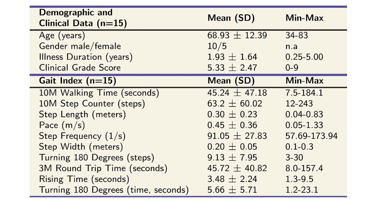
Table 1 Demographic, Clincal and Gait data of Patients
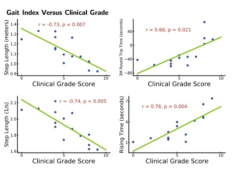
Figure 1 Gait Index Versus Clinical
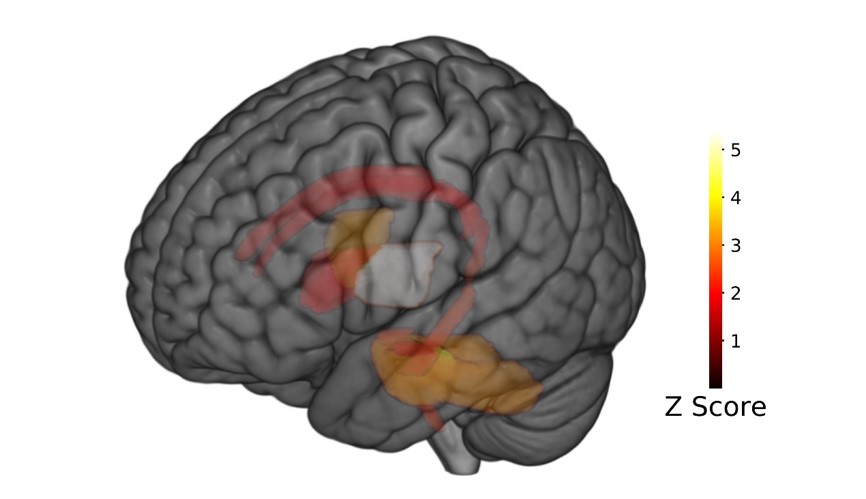
Figure 2 White Matter Tracts Related with Gait Indexes
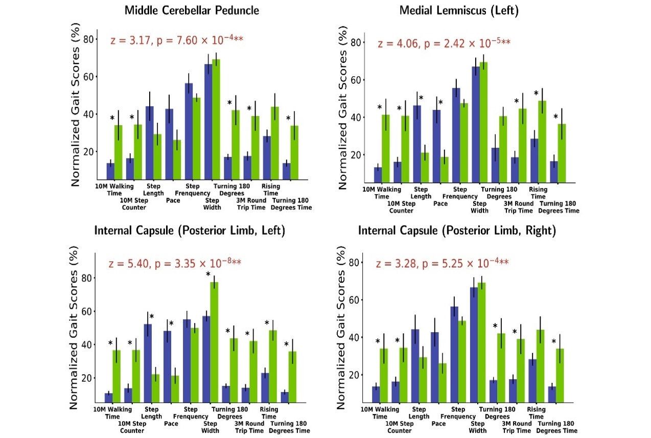
Figure 3 White Matter Tracts with Strongest Gait Index Relationships
blue means patients with fewer WMH, green means patients with more WMH