0873
Evaluation of transmit sensitivity (B1+) encoding in MR fingerprinting at 7T1Center for Advanced Imaging Innovation and Research, Department of Radiology, New York University School of Medicine, New York, NY, United States, 2Center for Biomedical Imaging, Department of Radiology, New York University School of Medicine, New York, NY, United States, 3Sackler Institute of Graduate Biomedical Sciences, New York University School of Medicine, New York, NY, United States
Synopsis
We evaluated the ability of three reported MR fingerprinting methods to mitigate the effect of B1+ inhomogeneity at 7T. Results from each method were compared with gold standard results. All methods provided relatively accurate T1 quantification. We show that T2 cannot be accurately quantified at 7T without accounting for B1+ in the MR fingerprinting dictionary. The Inversion-Recovery-FISP-FLASH (IRFF) method provided the most accurate T2 values. We conclude that the use of both FISP and FLASH segments best encodes B1+ into the fingerprint.
Introduction
MR fingerprinting (MRF)1 can simultaneously quantify multiple tissue parameters in a single scan with relatively short acquisition time. Transmit radiofrequency (RF) field (B1+) inhomogeneity is prominent at ultra-high magnetic field strengths (≥7T) where the RF wavelength becomes comparable to subject size. It has been shown early on that MRF results could be greatly affected by B1+ imperfections if they are not encoded in the dictionary2,3. Since then only a few studies4-6 have reported methods to implicitly mitigate the effect of B1+. Here, we evaluated the ability of these methods to mitigate the effect of B1+ inhomogeneity in a phantom study at 7T.Methods
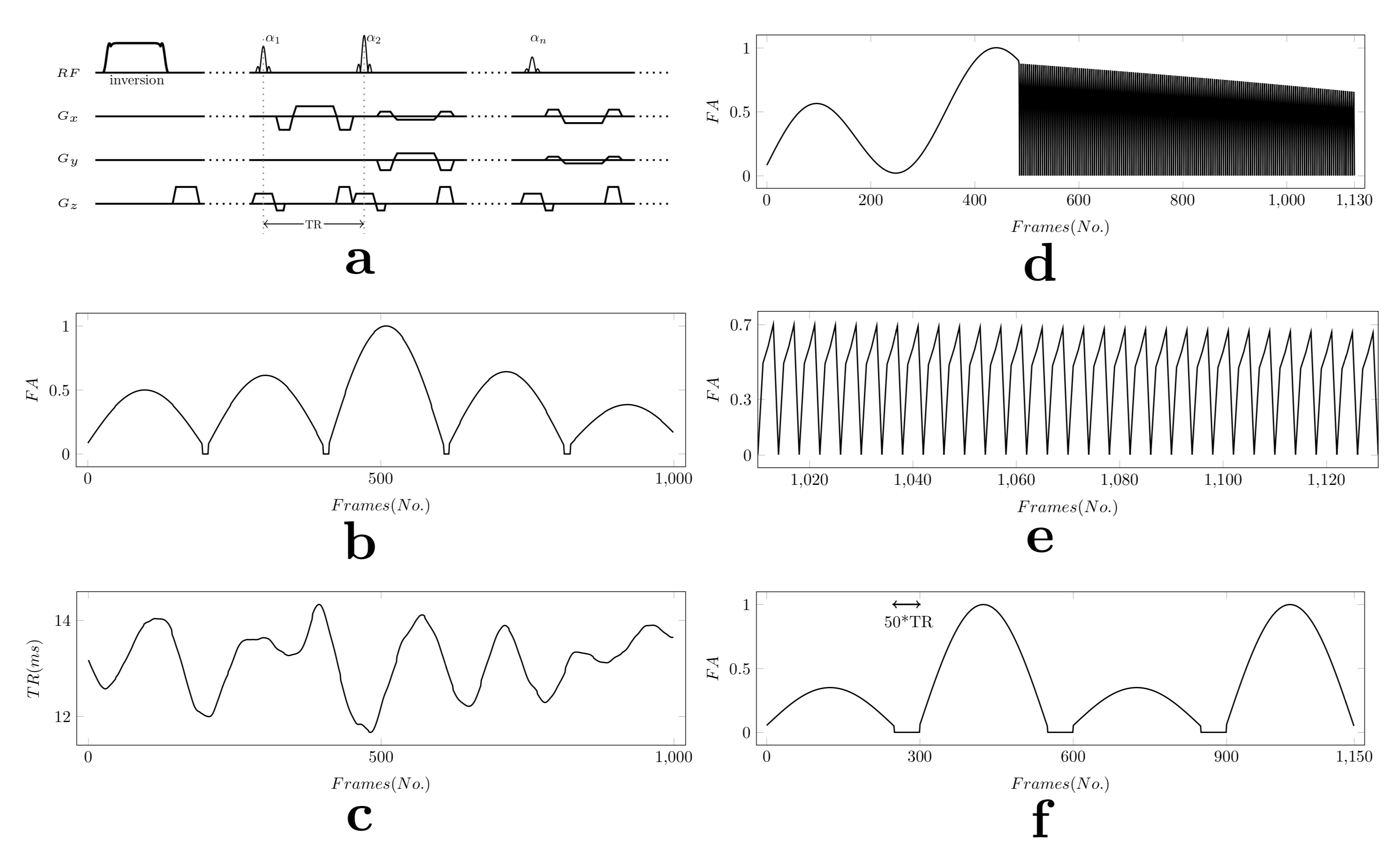
We implemented three different methods (Figure 1): the MRF-FISP approach (without & with external B1+)5,7, the renewed scheme proposed by Buonincontri et al.6 (referred here as Sawtooth-B1), and our Inversion-Recovery-FISP-FLASH (IRFF) method. All sequences start with an adiabatic inversion (IR), followed by a 2D golden-angle radial readout (Figure 1a). The excitation pulse is a 2ms Hanning-filtered Sinc waveform with TBW=3, a spoiler is applied in the slice direction to produce 8pi phase dispersion across a 5mm slice, TE=3.4ms, and 10s delay between each radial spoke. The FA train (maxFA=70°) and the variable TR train (11.5-14.5ms) of MRF-FISP are shown in Figure 1b-c. Sawtooth-B1 is based on MRF-FISP, but contains a segment with abrupt “sawtooth” FA changes (maxFA=80°) to encode B1+ and uses a fixed TR=11ms (Figure 1d-e). IRFF (Figure 1f) consisted of 4xSegments of 250xExcitations separated by 50xTRs delays, maxFA=60° and fixed TR=7.5ms. The last two segments employ FLASH, which predominantly encodes B1+ and T1.Experiments were performed on a 7T MRI system (Magnetom, Siemens) with a 1-channel transmit/32-channel receive head coil (Nova Medical). The phantom consisted of 7 glass tubes with different T1/T2 values immersed in saline solution. The three MRF sequences were tested, with 6 and 10 radial spokes per time-point, for a 5mm slice (256x256, 0.5x0.5mm2 in-plane resolution). A reference B1+ map was acquired with a method that uses TurboFLASH readout (tfl_b1map)11. Spin-echo experiments were performed for the same slice to obtain gold standard T1 (TI={25,50,100,200,400,800,1600,3200,6400}ms) and T2 (TE={12,24,36,48,60,72,84,96,144,192,278,384}ms) maps, using TR=8s to minimize saturation effects.
Parameter maps were reconstructed with an algorithm8 described before, and the EPG-X framework9 was employed to simulate the fingerprints in the dictionary. To improve the accuracy of the dictionary, a 16-bin slice profile simulation10 was included in the dictionary generation. Each dictionary for the three MRF methods contained 40076 entries (30xT1 ranging from 110-1745ms, 32xT2 ranging from 11-210ms and 43xB1+ ranging from 2-86°, where entries with T2>T1 were excluded). For MRF-FISP, three sets of results were generated: assuming homogeneous B1+ across the slice (MRF-FISP w/o B1)7, using the measured fingerprints to also match for B1+ (MRF-FISP w/ B1), adopting the B1+ map from the separate scan (tfl_b1map) to correct biases in the estimated parameters (MRF-FISP ext B1)5.
Results & Discussion
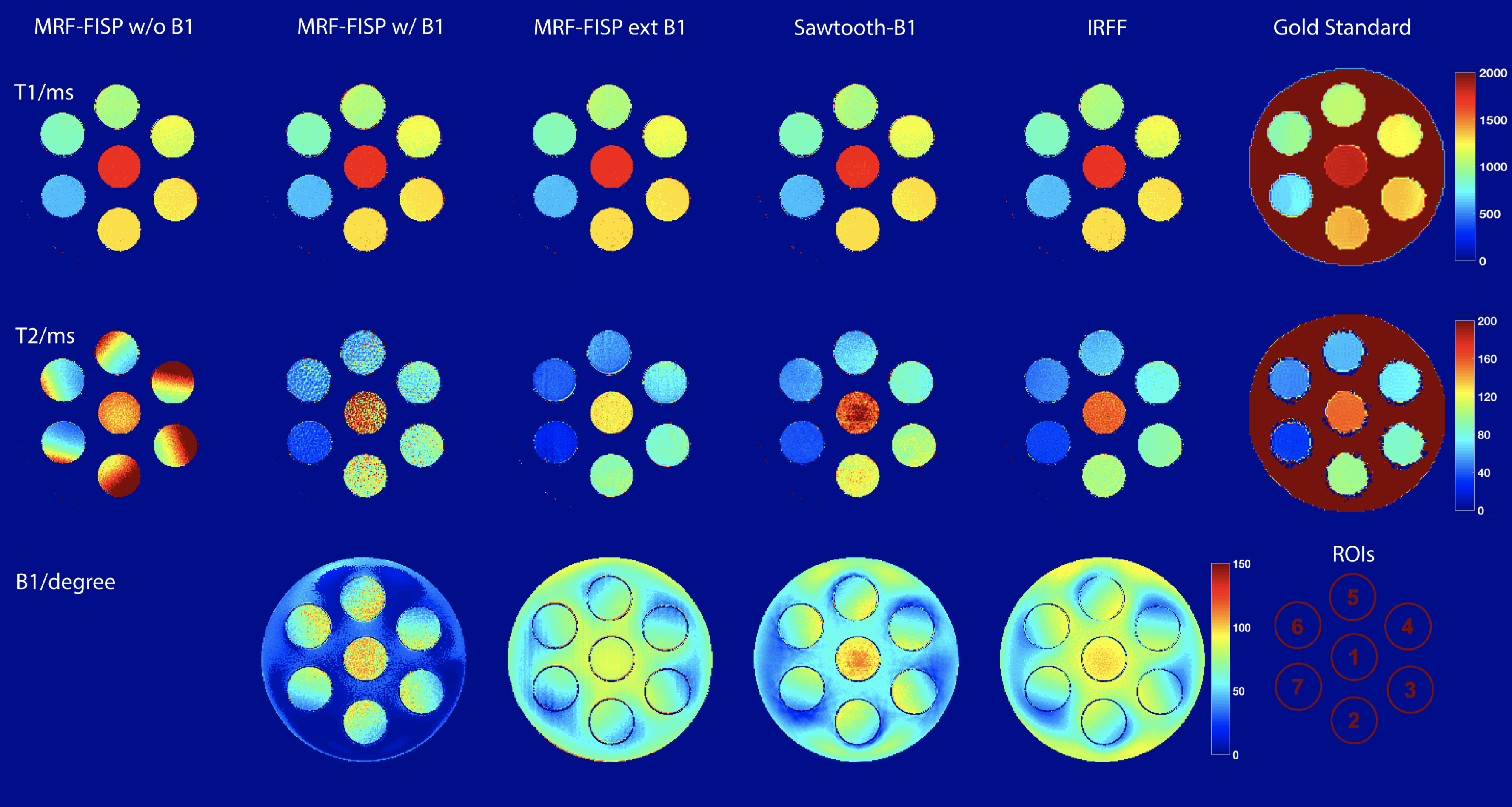
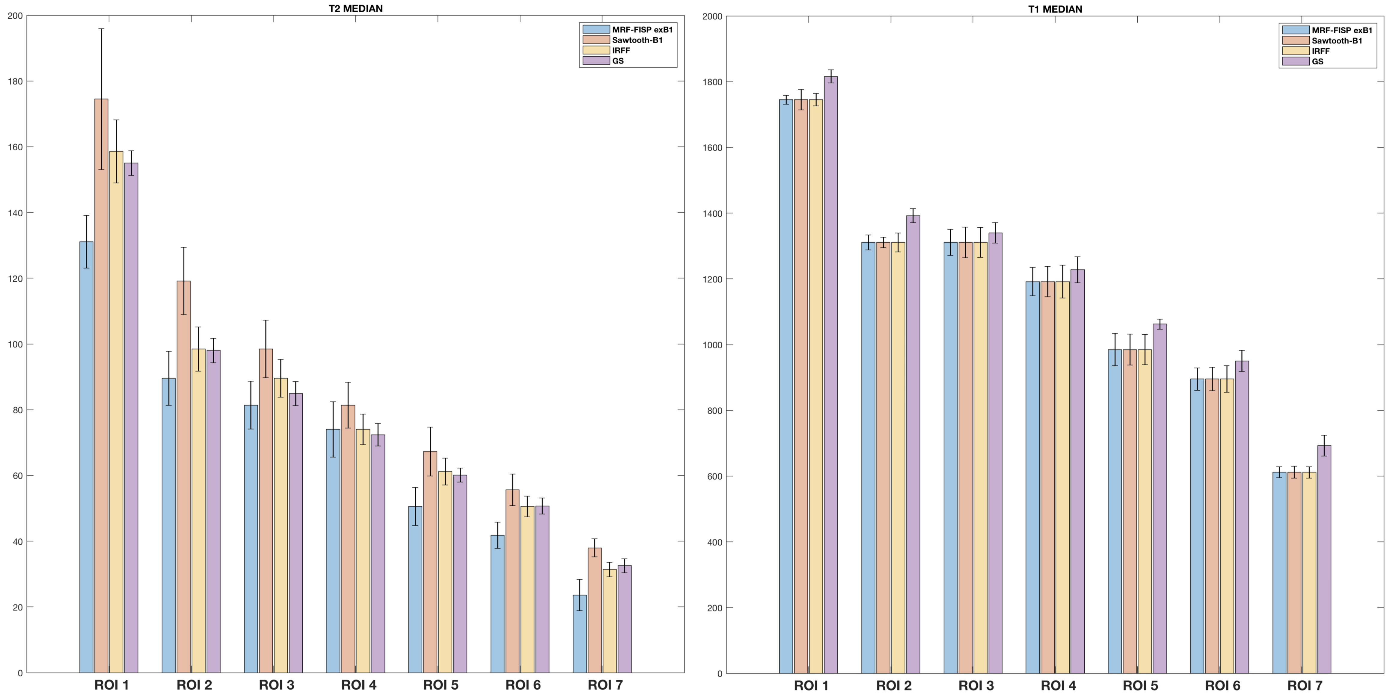
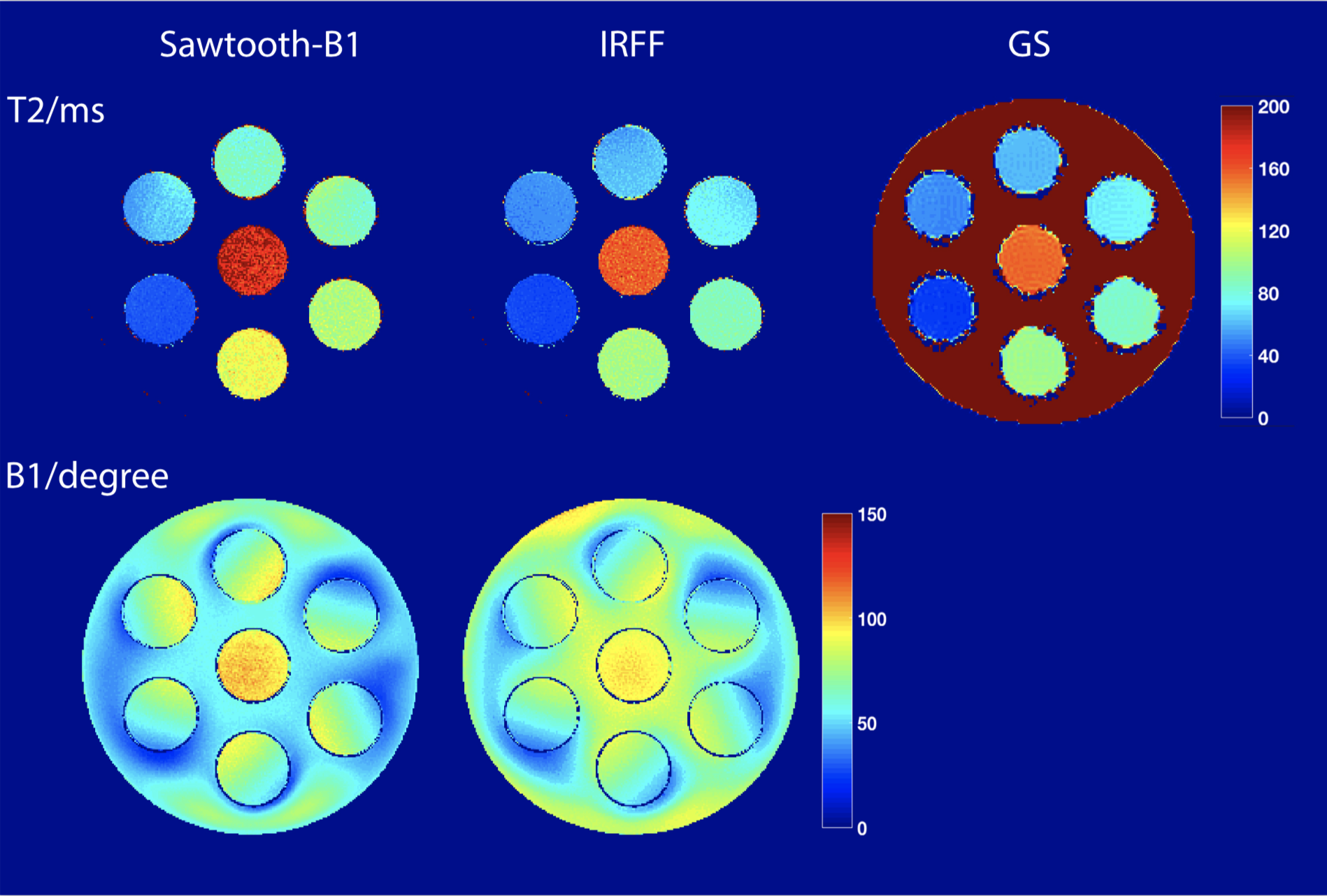
Results from the phantom experiment are shown in Figure 2 for the case of 6 radial spokes per timepoint. As expected, the T2 values were distorted due to variations in B1+. Simply extending the dictionary to include B1+ could not fully remove the bias either. The FISP segments only provided a relatively weak B1+ information, thus yielding only small improvement. The use of an external B1+ calibration in combination with the MRF-FISP sequence produced improved T2 values, however an artifact pattern from the reference B1+ map is visible in the T2 map. When compared with the gold standard T2 map, results from our IRFF showed best agreement (Figure 3). Results of MRF-FISP ext B1 appeared to underestimate T2, possibly due to inaccuracies in the external B1+ map. One the other hand, the Sawtooth-B1 results showed an overestimation of T2, which may be linked to an overestimation in B1+. The comparison between Sawtooth-B1 and IRFF for 10 radial spokes in Figure 4 suggests that the overestimation of B1+ and T2 was probably not due to under-sampling artifacts. Overall, T1 values were stable due to the use of the same adiabatic IR at the beginning of all MRF sequences, but they all showed a slight underestimation when compared to the gold standard (Figure 3). This could be attributed to the adiabatic IR not being strong enough to achieve a full inversion; or to the bias in the gold standard T1 map due to the non-selective IR experiencing certain degrees of B1+ inhomogeneity, as visible for some of tubes in the gold standard T1 map (Figure 2).Conclusion
After evaluating different methods to mitigate the effect of B1+ in MR fingerprinting, we concluded that IRFF with the use of both FISP and FLASH segments best encodes B1+ into the fingerprint. Since the IRFF implementation has not yet been optimized with respect to noise and under-sampling artifacts, it may be improved even further.Acknowledgements
This work was supported in part by NIH R01 AR070297 and was performed under the rubric of the Center for Advanced Imaging Innovation and Research (CAI2R, www.cai2r.net), an NIBIB Biomedical Technology Resource Center (NIH P41 EB017183).References
1. Ma, D., Gulani, V., Seiberlich, N., Liu, K., Sunshine, J. L., Duerk, J. L., & Griswold, M. A. (2013). Magnetic resonance fingerprinting. Nature, 495(7440), 187.
2. Cloos, M. A., Alon, L., Grppert, C., Sodickson, D. K., & Lattanzi, R. (2015). Rapid T1 and T2 mapping of the hip articular cartilage with radial MR fingerprinting. In Proc Int Soc Magn Reson Med (Vol. 23, p. 113).
3. Cloos, M. A., Knoll, F., Zhao, T., Block, K. T., Bruno, M., Wiggins, G. C., & Sodickson, D. K. (2016). Multiparametric imaging with heterogeneous radiofrequency fields. Nature communications, 7, 12445.
4. Buonincontri, G., & Sawiak, S. J. (2016). MR fingerprinting with simultaneous B1 estimation. Magnetic resonance in medicine, 76(4), 1127-1135.
5. Ma, D., Coppo, S., Chen, Y., McGivney, D. F., Jiang, Y., Pahwa, S., ... & Griswold, M. A. (2017). Slice profile and B1 corrections in 2D magnetic resonance fingerprinting. Magnetic resonance in medicine, 78(5), 1781-1789.
6. Buonincontri, G., Schulte, R. F., Cosottini, M., & Tosetti, M. (2017). Spiral MR fingerprinting at 7 T with simultaneous B1 estimation. Magnetic resonance imaging, 41, 1-6.
7. Jiang, Y., Ma, D., Seiberlich, N., Gulani, V., & Griswold, M. A. (2015). MR fingerprinting using fast imaging with steady state precession (FISP) with spiral readout. Magnetic resonance in medicine, 74(6), 1621-1631.
8. Cloos, M. A., Assländer, J., Abbas, B., Fishbaugh, J., Babb, J. S., Gerig, G., & Lattanzi, R. (2019). Rapid radial T1 and T2 mapping of the hip articular cartilage with magnetic resonance fingerprinting. Journal of Magnetic Resonance Imaging, 50(3), 810-815.
9. Malik, S. J., Teixeira, R. P. A., & Hajnal, J. V. (2018). Extended phase graph formalism for systems with magnetization transfer and exchange. Magnetic resonance in medicine, 80(2), 767-779.
10. Hilbert, T., Xia, D., Block, K. T., Yu, Z., Lattanzi, R., Sodickson, D. K., ... & Cloos, M. A. (2019). Magnetization Transfer in Magnetic Resonance Fingerprinting. Magnetic resonance in medicine, In press. arXiv preprint arXiv:1907.13262.
11. Chung, S., Kim, D., Breton, E., & Axel, L. (2010). Rapid B1+ mapping using a preconditioning RF pulse with TurboFLASH readout. Magnetic resonance in medicine, 64(2), 439-446.
Figures